Ⅰ. 서 론
간장(liver), 지라(spleen), 위장(stomach), 작은창자 (small bowel), 큰창자(large bowel) 등 여러 복부장기의 질환 환자에서 종양이나 출혈성 질환 및 혈관질환이 있는 환자에게 해부학적 혈관 구조를 파악하는 것이 중요한데,
과거에는 고식적 혈관촬영술이 표준적 검사로 여겨져 왔 다. 그러나 고식적 혈관조영술은 침습적 시술이며, 검사 비용이 비싸고, 감염의 위험성이 있는 이차원적 검사라는 단점과 복부혈관의 다양한 변위 때문에 정확한 혈관의 구 조를 파악할 수 없는 단점이 있다. 이에 3차원 CT 혈관 조영술(Three Dimensional Computed Tomography Angio−graphy, 이하 3D CTA)을 이용하여 고식적 혈관 조영술을 대체하고자 하는 다양한 연구들이 이루어지고 있다1~3). 1990년대 후반 Multidetector row Computed Tomo−graphy (이하 MDCT)의 출현은 고식적 나선형 CT 에 비해 검사시간의 단축과 고해상능의 얇은 단면을 이용 한 재구성 영상을 제공함으로 특히 동맥조영 CT 영상을 교신저자: 장영일, (545-703) 전남 광양시 한려대길 111
광양보건대학교 방사선과
Tel : 061-760-1452, Fax : 061-763-1505 E-mail : radpacs@hanmail.net
* 접수일(2013년 3월 20일), 1차 심사일(2013년 5월 6일), 확정일(2013년 6월 18일)
MDCT의 3차원 볼륨렌더링을 이용한 복강축과 위창자간막동맥의 변위 형태에 관한연구
- A Study on Variation Types in Celiac Axis and Superior Mesenteric Artery using 3D Volume Rendering of MDCT -
동신대학교 보건복지대학 방사선학과 ․ 광양보건대학교 방사선과1) 이정근 ․ 장영일1)․ 장성주
― 국문초록 ―
복부 전산화단층촬영 후 3차원 볼륨렌더링으로 재구성한 영상을 분석하여 복부대동맥에서 분지하는 복강축 과 위창자간막동맥을 기준으로 해부학적 변위를 분류하여 평가하고자 하였다.
복부 전산화단층혈관촬영을 시행한 613명 환자의 3차원 볼륨렌더링 영상을 이용하여 해부학적 변위를 형태 별로 분류한 결과 552명(Type Ⅰ, Ⅱ)은 정상 구조에 속하였고, 61명(Type Ⅲ∼Ⅻ)은 변위로 분류하였다.
Type Ⅰ이 339명 (55.31%), Type Ⅱ가 213명(34.74%)으로 나타났으며 변위로 분류된 경우는 Type Ⅲ은 18명 (2.93%), Type Ⅳ는 12명 (1.95%), Type Ⅴ는 11명 (1.79%), Type Ⅵ는 9명 (1.46%), Type Ⅶ는 6명 (0.97%) 으로 나타났으며, Type Ⅷ∼Ⅻ는 각각 1명 (0.16%)으로 전혀 새로운 변위형태로 분류되었다.
결론적으로 복강축과 위창자간막동맥을 기준으로 변위를 분류한 결과 그동안 간동맥 중심의 해부학적 변위 분류에서는 관찰되지 않았던 9가지의 새로운 변위형태를 파악할 수 있었다. 이는 새로운 혈류지도를 만드는 중요한 자료로 이용될 수 있을 것으로 사료된다.
중심 단어 : 변위, 복강축, 위창자간막동맥, 3차원 볼륨렌더링
얻을 수 있는 스캔속도가 10배 이상 빨라졌으며 동맥기 영상도 초기와 후기의 두 가지 영상을 얻을 수 있게 되어 고식적 나선형 CT보다 공간분해능이 우수하고 고화질의 3D CTA을 얻을 수 있게 되었다. 이로 인해 복부혈관의 정확한 해부학적 구조를 알 수 있게 되어 수술이나 방사 선학적중재적시술(Interventional Radiology, 이하 IVR) 의 성공여부를 좌우할 수 있는 중요한 검사법으로 간주되 었다. 특히 복부장기의 혈관분포는 대단히 복잡하고 해부 학적 변위가 많아 혈관질환 환자의 수술 및 IVR시 고식 적 혈관촬영술을 시행하기 전에 3D CTA로 혈관의 정확 한 해부학적 구조를 먼저 파악하는 것이 매우 중요하다고 볼 수 있다. 정상적인 혈관구조로 복부대동맥에서 기원하 는 복강축(Caliac Axis, 이하 CA)과 위창자간막동맥 (Superior Mesenteric Artery, 이하 SMA)의 혈관들이 많은 예에서 변위4,13)를 보인다고 알려져 있고, 이에 대한 많은 해부학적 혹은 외과학적 연구15)나 혈관조영술, 3D CTA의 소견을 중심으로 변위에 관한 연구가 진행되어 왔 으나 이는 주로 간동맥을 중심으로 연구결과4,12~14)가 발 표되었다. 그러나 MDCT기술의 발달로 복부스캔 후 3D 체적렌더링기술(Volume Rendering Technique, 이하 VRT)5)을 이용한 혈관영상재구성으로 복부대동맥에서 분 지하는 복부혈관의 전반적인 해부학적 구조를 관찰하여 CA와 SMA를 중심으로 한국인에 대한 혈관의 해부학적 구조와 변위에 관한 연구가 미약하여 본 연구자는 CTA에 의해 얻어진 2차원적인 영상정보를 3D VRT로 재구성하 여 복부대동맥에서 분지하는 CA와 SMA의 혈관영상 형태 를 분석하여 정상적인 해부학적 구조와 변위를 가지고 있 는 혈관의 해부학적 형태의 유형을 알아보고자 하였다.
Ⅱ. 대상 및 방법
1. 연구대상
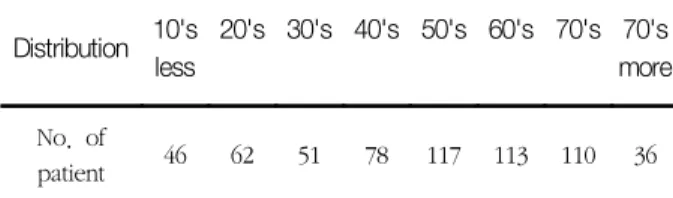
연구윤리심의위원회 승인을 얻었으며 환자 참여 동의서는 적용되지 않았다. 광주광역시소재 C-대학병원에서 2011년 7월부터 2011년 12월까지 6개월 동안 CTA을 시행한 환자 중 총 613명의 환자의 스캔영상을 전산망을 이용하여 Terarecon Workstation(Aquarlus intuition Edition, forster city, USA)에서 3D VRT로 재구성하여 복부대동 맥에서 분지한 CA와 SMA를 중심으로 변위 형태를 분석 하였다. 이들의 연령분포는 8∼95세로 평균 53.0세로 남 자 환자 401명과 여자 환자가 212명으로 남자 환자가 약 2배정도 많았으며 연령별 나이분포는 Table 1과 같다.
Table 1. Distribution according to age (n=613) Distribution 10's
less
20's 30's 40's 50's 60's 70's 70's more No. of
patient 46 62 51 78 117 113 110 36
2. 영상 획득 방법
검사에 사용된 CT 장비로는 MDCT SOMATOM Sensation Cardiac 64(Siemens Healthcare, Forchheim, Germany)과 Dual Source SOMATOM Definition Flash (Siemens Healthcare, Forchheim, Germany) single - energy mode를 사용하여, 전형적인 심전도 동기화 high-pitch spiral 방식을 적용하였다. 사용된 영상변수 는 rotation time 0.5초, acquisition 64×0.6 mm, reconstruction selection thickness 3 mm, pitch factor 1 mm, field of view 300~400 mm, Matrix size 512×512 pixels, 조영제는 Ultravist 370 (Schering, Berlin, Germany) 130∼150 ml를 상완정맥 을 통해 20 gauge catheter line을 확보하고 초당 3.0~4.0ml로 조영제 자동 주입장치(Optivantage;
Liebel-Flarsheim, USA)를 사용하여 아래대동맥에 100HU 도달 후 15초에 동맥상을 획득하고, 40초 후에 문맥상을 100초 후에 지연상을 각각 획득하였다. 영상재 구성은 Bolus Tracking of abdominal aorta(Fig. 1)와 Bolus Tracking Result(Fig. 2)로 Terarecon Workstation (Aquarlus intuition Edition, forster city, USA)을 이 용하여 절편두께 1.0 mm, 절편간격 1 mm 영상을 최대 투시강도(maximum intensity projection, 이하 MIP)기 법을 이용하여 삼차원 CTA의 횡단면(transverse), 사위 관상면(oblique coronal), 그리고 사위시상면(oblique sagittal)의 MIP 기법을 이용한VRT로 재구성한 후 복부 대동맥에서 기시하는 CA와 SMA의 영상을 분석 후 통계 분석 처리는 SPSS version 18.0(statistical package for the social sciences, SPSS INC. Chicagi, IL, U.S.A)을 이용하였다.
Fig. 1 Bolus tracking of abdominal aorta
Fig. 2 Bolus tracking result
Ⅲ. 결 과
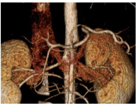
CA에서 왼위동맥(left gastric artery), 지라동맥 (splenic artery), 온간동맥(common hepatic artery)이 분지하고 SMA에서 작은창자(small bowel)와 큰창자 (large bowel) 일부에 혈액을 공급하는 동맥분지 혈관구 조를 정상해부학(normal anatomy)으로 분류하였으며, 그 외에 다른 구조로 분지된 형태는 해부학적 변위(anatomic variation)로 분류하였다. 전체 613명의 복부전산화단층 촬영상의 3D 혈관영상을 분석한 결과 정상형태인 CA의
한 곳에서 3개의 혈관이 분지한 경우가 552명인 90.05%
로 나타났으며 해부학적 변위로 분류되는 경우는 10가지 의 서로 다른 형태로 총 61명인 9.95%로 조사되었다 (Table 2).
정상해부학적 형태인 CA에서 3개의 혈관이 한꺼번에 분지하는 경우를 Type Ⅰ으로 분류하여 339명 55.31%로 나타났고(Fig. 3), CA에서 좌위동맥이 먼저 분지하고 나 중에 온간동맥과 지라동맥이 함께 분지하는 경우를 Type
Ⅱ로 분류하여 213명으로 34.74%로 조사되었다(Fig. 4).
또한 해부학적 변위로 분류된 경우는 총 10가지 형태로 총 64명에 10.44%로 나타났는데, 형태별로 변위의 유형 을 살펴보면 Type Ⅲ는 복부대동맥에서 CA와 SMA가 각 각 기원하면서 CA에서 좌위동맥과 왼간동맥, 지라동맥이 분지하고 SMA에서 오른간동맥이 분지되는 경우로 18명 2.93%(Fig. 5), Type Ⅳ는 복부대동맥에서 하나의 CA가 기원하면서 좌위동맥, 지라동맥, 온간동맥, SMA가 하나의 축을 이루면서 분지되는 형태로 12명 1.95%(Fig. 6), Type Ⅴ는 복부대동맥에서 왼간동맥, CA, SMA가 각각 따로 기원하고 CA에서 좌위동맥이 먼저 분지하고 바로 아 래에서 온간동맥과 지라동맥이 분지되는 형태로 11명 1.79%(Fig. 7), Type Ⅵ는 복부대동맥에서 CA과 SMA가 각각 기원하고 CA에서 좌위동맥과 지라동맥이 분지되며 SMA에서 온간동맥이 분지되는 형태로 9명 1.46%(Fig.
8), Type Ⅶ는 복부대동맥에서 좌위동맥, CA, SMA가 각 각 따로 기원하고 CA에서 온간동맥과 지라동맥이 분지되 는 형태로 6명 0.97%(Fig. 9), Type Ⅷ는 복부대동맥에서 지라동맥과 CA가 기원하고 CA에서 온간동맥과 SMA가 분 지되는 형태로 1명 0.16%(Fig. 10), Type Ⅸ는 복부대동 맥에서 좌위동맥, 지라동맥, CA가 각각 기원하고 CA에서 지라동맥이 분지되며 SMA에서 온간동맥이 분지되는 형 태로 1명 0.16%(Fig. 11), Type Ⅹ는 복부대동맥에서 좌 위동맥, CA, SMA가 각각 기원하고 CA에서 온간동맥과 지라동맥이 분지되는 형태는 1명 0.16%(Fig. 12), Type
Ⅺ는 복부대동맥에서 좌위동맥과 CA이 따로 기원하고 좌 위동맥에서 왼간동맥이 분지되고 CA에서 지라동맥, 온간 동맥, SMA가 분지되는 형태는 1명 0.16%(Fig. 13), Type
Ⅻ는 복부대동맥에서 CA과 SMA가 각각 기원하고, CA에 서 좌위동맥과 지라동맥이 분지되며 SMA에서 온간동맥이 분지되는 형태는 1명 0.16%(Fig. 14)로 조사되었다. 이중 Type Ⅳ∼Ⅻ는 다른 연구 결과에서 나타나지 않은 특별한 경우로 3D VRT의 영상을 이용하여 분류한 본 연구 결과 에서 나타났다(Table 2).
Table 2. Types of celiac axis and SMA in 613 patients
Type Ⅰ Ⅱ Ⅲ Ⅳ Ⅴ Ⅵ Ⅶ Ⅷ Ⅸ Ⅹ Ⅺ Ⅻ
No. of Patients 339 213 18 12 11 9 6 1 1 1 1 1
% 55.31 34.74 2.93 1.95 1.79 1.46 0.97 0.16 0.16 0.16 0.16 0.16
Fig. 3. Type I Celiac axis and SMA each originates from abdominal aorta with left gastric artery, common hepatic artery, and splenic artery branching from celiac axis at one point and SMA branching right below.
Fig. 4. Type Ⅱ Celiac axis and SMA each originates from abdominal aorta with left gastric artery branching from celiac axis first followed by common hepatic artery and splenic artery branching below.
Fig. 5. Type-Ⅲ Celiac axis and SMA each originates from abdominal aorta with left gastric artery, left hepatic artery, and splenic artery branching from celiac axis and right hepatic artery branching from SMA.
Fig. 6. Type Ⅳ One celiac axis originating from abdominal aorta with left gastric artery, splenic artery, common hepatic artery, and SMA branching form from it's one axis.
Fig. 7. Type Ⅴ Left hepatic artery, celiac axis, and SMA each originating from abdominal aorta with ahead left gastric artery branching from the celiac axis and common hepatic artery and splenic artery branching right below.
Fig. 8. Type Ⅵ Celiac axis and SMA each originating from abdominal aorta with left gastric artery and splenic artery branching from celiac axis and common hepatic artery originating from SMA.
Ⅳ. 고 찰
복부대동맥에서 기원하는 CA와 SMA는 정상적인 해부 학적 형태와 다양한 변이를 갖는다고 알려져 있으며 이러 한 해부학적 변이 유무는 혈관질환의 수술이나 IVR시 정
확한 혈관의 해부학적 구조의 파악이 매우 중요하다11,12). 과거 간동맥을 중심으로 Michels Classification에 의하 면 Table 3과 같이 12가지 형태로 분류하였는데, 제 1형 은 고유간동맥에서 좌우간동맥이 분지하는 표준기본형태 이고 제 2형은 좌위동맥에서 왼간동맥이 분지하는 경우로 Fig. 9. Type Ⅶ Left gastric artery, celiac axis, and SMA
each originating separately from abdominal aorta with common hepatic artery and splenic artery branching from celiac axis.
Fig. 10. Type Ⅷ Splenic artery and celiac axis originating from abdominal aorta with common hepatic artery and SMA branching from celiac axis.
Fig. 11. Type Ⅸ Left gastric artery, splenic artery, celiac axis each originating from abdominal aorta with splenic artery branching from celiac axis and common hepatic artery branching from SMA.
Fig. 12. Type Ⅹ Left gastric artery, celiac axis, and SMA each originating from abdominal aorta with common hepatic artery and splenic artery branching from celiac axis.
Fig. 13. Type Ⅺ Left gastric artery and celiac axis originating separately from abdominal aorta with left hepatic artery branching from left gastric artery and splenic artery, common hepatic artery, and SMA branching from celiac axis.
Fig. 14. Type Ⅻ Celiac axis and SMA each originating from abdominal aorta with left gastric artery and splenic artery branching from celiac axis and common hepatic artery branching from SMA.
제 3형은 SMA에서 오른간동맥이 분지하는 경우로 제 4 형은 좌우간동맥이 바뀐 경우로 제 5형은 부가적인 왼간 동맥이 좌위동맥으로 대치되는 경우로 제 6형은 부가적으 로 오른간동맥이 SMA으로부터 기시하는 경우로 제 7형 은 부수적인 왼간동맥과 오른간동맥이 존재하는 경우이며 제 8a형은 오른간동맥이 존재하고 부가적인 왼간동맥이 존재하는 경우로 제 8b형은 왼간동맥이 놓여있고 부가적 인 오른간동맥이 존재하는 경우로 제 9형은 고유간동맥이 SMA에서 기시하는 경우로 제 10형은 모든 간동맥이 왼 위동맥에서 분지하는 경우로 제 11형은 어느 동맥변위 형 태가 앞에 열거한 11가지의 형태로 분류할 수 없는 경우 로 나타내었다. 또한 나선형 CT와 계수적감산혈관조영술
(Digital Subtraction Angiography, 이하 DSA)을 이용 한 간동맥중심의 변위의 형태를 분류하여 발표한 송순용4) 등은 Michels Classification을 기준으로 12개의 가능한 CA 변이를 총 67예의 환자에서 10가지의 해부학적 변위 로 분류하였는데, 간동맥 중심으로 해부학적 변위 형태를 발생 빈도순으로 Table 4와 같이 분류하였다. 제 1형으로 정상 간동맥 유형 30예(67%)에서 관찰되었고, 다음으로 왼위동맥에서 왼간동맥이 기시하는 변위가 6예(13%), 제 9형인 간동맥 전체가 SMA에서 기시하는 변위가 4예(9%) 순으로 나타난 연구 결과를 발표하였다. 한문희13) 분류에 의하면 총 311명의 환자의 복부간동맥혈관촬영상의 분류 에서 CA 및 간동맥을 관찰하여 혈관구조 및 정상변위를
Table 3. Michels classification of hepatic artery anatomy
Type Michels anatomy
Ⅰ Standard anatomy
Ⅱ Left HA replaced to left GA
Ⅲ Right HA replaced to SMA
Ⅳ Both right and left hepatic arteries replaced
Ⅴ Accessory left HA replaced to left GA
Ⅵ Accessory right HA arising from SMA
Ⅶ Accessory left HA and right HA
Ⅷa Replaced right HA and accessory left HA
Ⅷb Replaced left HA and accessory right HA
Ⅸ Proper HA arising from left GA
Ⅹ Entire HA arising from left GA
Ⅺ Designation of any arterial variant not described for types Ⅰ-Ⅺ Note. GA: HA, HA: SMA, SMA: superor mesenteric artery
Table 4. Findings on conventional angiography based on Michels classification
Type Michels anatomy No. of Case(%)
Ⅰ Conventional anatomy 30(67)
Ⅱ Replaced left HA 6(13)
Ⅲ Replaced right HA 1(2)
Ⅳ Replaced left HA and replaced right HA 0(0)
Ⅴ Accessory left HA 3(7)
Ⅵ Accessory right HA 0(0)
Ⅶ Accessory right and left HA 0(0)
Ⅷ Replaced right and accessory left HA or accessory right and replaced left HA 1(2)
Ⅸ Entire hepatic trunk that arises from SMA 4(9)
Ⅹ Entire hepatic trunk that arises from left GA 0(0)
Note. GA: gastric artery, HA: hepatic artery, SMA: superor mesenteric artery
9가지로 분류하였다. 총 311례 중 CA에서 기시하는 정상 온간동맥은 제 1형은 고유간동맥에서 좌우간동맥이 분지 하는 경우, 제 2형은 총간동맥에서 왼간동맥이 분지하고 고유간동맥에서 오른 간동맥이 분지하는 경우, 제 5형은 SMA에서 오른간동맥의 일부가 분지하는 경우, 제 7형은 SMA에서 지라동맥이 분지하는 경우, 제 8형은 CA에서 왼위동맥이 먼저 분지하고 총간동맥에서 왼․오른 간동맥 이 분지하는 경우, 제 9형은 CA에서 왼위동맥이 먼저 분 지하고 고유간동맥에서 오른간동맥의 일부가 분지되면서 고유간동맥에서 오른․왼간동맥이 분지하는 경우 등으로 89.8%에서 관찰되었다.
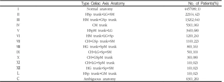
송순영4) 등이 발표한 연구결과에서 총 14가지의 해부학 적 변위에 따른 간동맥의 해부학적 변위 형태의 빈도순은 Table 5과 같다. 이는 5002명의 환자 중 정상 CA은 4457명(89.1%)에서 확인되었고, CA 변이의 12가지 형태 가 482명(9.64%)의 환자에서 확인되었으며 나머지 63명 (1.26%)의 환자에서는 간동맥과 위십이지장 동맥이 서로 다른 기원을 가지고 있어서 온간동맥(common hepatic artery)이 존재하지 않기 때문에(n=55), 또는 연속적인 문합의 경로로 인해 총간동맥의 기원이 결정될 수 없었기 때문에(n=8) CA는 ‘모호함’으로 분류되었다. 정상 CA 을 기원으로 하는 총간동맥은 문정맥(n=6) 또는 이자관통 (n=1) 경로를 가지고 있었다. 좌측 위동맥을 기원으로 하 는 8개의 총간동맥 모두 정맥관인대의 절개를 통과했다.
위창자간동맥을 기원으로 하는 148개의 총간동맥(창자위, 창자아래, 창자관통) 및 위창자간-문정맥축과 다양한 관
계를 보였다. 대동맥을 기원으로 하는 20개의 총간동맥은 정상 췌장위 전문 경로를 가지고 있었다.
또한 정상 간동맥에 대하여 Koops는14) 93.6%, Daseler15) 는 83%, Browne16)은 92.8%에서 관찰된다고 보고하여 서 로 약간의 차이를 보이나 유형에 있어 큰 차이는 없었다.
총간동맥이 다른 곳에서 전치되어 기시하는 경우가 제6형 으로 1.9%인 6례로 나타났고, 모두 SMA에서 기시하였다.
전체 총간동맥은 SMA에서 기시하는 경우가 가장 많으며 Michels17)는 8예(4%) 중 5예가, Daseler15)는 23예(4.6%) 중 22예가 SMA에서 기시하였다고 보고하였다. 또한 총 간동맥이 기원하고 모든 간동맥이 온간동맥에서 기원하는 고식적인 구조는 55%로 가장 흔하며 다음으로 왼위동맥 에서 왼간동맥이 기원하는 변이가 10%, 오른간동맥이 SMA에서 기원하는변이가 11%라고 하였다. 과다혈관상 종양의 진단과 여러 가지 삼차원 기법을 이용한 재구성 영상으로 여러 간질환의 주변 장기 침범 뿐만 아니라 3D CTA로 얻을 수 있다.
CT를 이용한 동맥기 영상은 복부 CT에서 과다혈관상 종양의 진단과 여러 가지 삼차원 기법을 이용한 재구성 영상으로 CA과 SMA에서 분지하는 3D CTA을 얻을 수
있다2,4,7,8). 동맥의 해부학적 구조 및 여러 장기와의 관계
를 잘 나타내기 위해서 널리 쓰이는 3D CTA 기법에는 앞서 언급했던 것처럼 SSD, MIT, VRT, 등이 있다4,6~10). VRT은 겹쳐진 구조물을 삼차원적으로 구분해서 볼 수 있 고 특히 큰 혈관이나 겹쳐진 혈관을 보는데 유용하며 혈 관직경과 혈관의 내부를 볼 수 있는 장점이 있다. 단점으
Table 5. Celiac axis variation in 5002 patients
Type Celiac Axis Anatomy No. of Patients(%)
Ⅰ Normal anatomy 4457(89.1)
Ⅱ HSp trunk+LG+SM 221(4.42)
Ⅲ HM trunk+GSp trunk 132(2.64)
Ⅳ CM trunk 53(1.06)
Ⅴ HSpM trunk+LG 34(0.68)
Ⅵ HM trunk+LG+Sp 12(0.24)
Ⅶ CH+GSp trunk+SM 11(0.22)
Ⅷ HG trunk+SpM trunk 8(0.16)
Ⅸ CH+LG+Sp+SM 5(0.10)
Ⅹ CH+GSpM trunk 3(0.08)
Ⅺ CH+LG+SpM trunk 1(0.02)
Ⅻ HG trunk+Sp+SM 1(0.02)
Ⅼ HSp trunk+GM trunk 1(0.02)
Ⅽ Ambiguous anatomy 63(1.26)
Note. Numbers in parentheses an percentages
Normal anatomy refers to hepatogastrosplenic trunk plus SMA. GSp=gasrtosplenic.
로는 영상재구성에 약간의 시간소요와 술자 의존성의 문 제가 있다. SSD는 분명한 경계를 가진 삼차원적영상을 보여주는 장점이 있으나 장기 내부의 혈관을 볼 수 없다 는 단점이 있다5,7,9,10). MIP 기법은 고식적 혈관조영술과 유사한 영상을 얻을 수 있는 장점을 가지고 있다. 따라서 본 저자는 복부대동맥에서 분지하는 CA과 SMA의 변위 관찰이 목적이었기 때문에 VRT을 이용하였다.
따라서 본 연구자는 CA와 SMA 위주로 변위를 관찰했기 때문에 CA에서 간으로 혈류를 공급하는 형태가 90.05%로 조사되었고, 해부학적 정상변위가 있는 경우로 CA의 분지 가 아닌 SMA나 다른 곳에서 간혈류를 공급하는 경우가 10 개의 서로 다른 형태에서 9.95%로 조사되었다.
Ⅴ. 결 론
복부대동맥에서 분지하는 CA와 SMA에서 간으로 혈류 를 공급하는 것이 정상적인 해부학적 구조 형태가 대부분 이지만 약 10% 정도에서 변위를 가지고 있고, 또한 9가지 형태의 새로운 변위의 결과로 이는 침습적인 혈관촬영을 시행하기 전에 3D CTA를 통하여 파악된다면 좀 더 쉽게 수술 및 중재적 시술을 시행할 수 있을 것이다. 관찰된 변 이 및 그들의 해부학적 경로를 통합해 구축된 가설상의 해 부학 모형은 이를 다루는 분야의 전문가들에게 다양한 변 이들의 기본적인 발달상의 기초를 이해하는 데에 도움이 될 수 있을 것으로 사료된다.
본 연구는 몇 가지 한계를 가지고 있다. 첫째, 본 연구는 후향적으로 수행되었다. 둘째, 본 연구는 MDCT을 이용하 여 CTA의 3D VRT 영상에 대한 분석을 바탕으로 했기 때 문에 미세한 동맥네트워크에 대한 묘사가 누락될 수 있으며 또한 CT 검사의 역량을 벗어나는 것이기도 했다. 셋째, 본 연구는 연구자의 주관적인 판단에 의해 수행되는 영상 해석 을 바탕으로 했다. 마지막으로 본 연구자가 제안한 새로운 형태가 중재 절차와 수술 절차에 미치는 영향은 고려하지 않았다. 그러나 새로운 명명체계가 복잡해 보일지라도 이 체계는 우리가 여러 미보고 변이를 포함해 관찰되는 모든 변이의 상세한 3D 해부학을 기술할 수 있도록 해준다.
참고문헌
1. In Ho Go, Dae Chul Kwon, et al. Textbook of
Computed Tomography. Chung-Ku Publisher, 14-283, 2009
2. Moon-Chan Kim. Doses of Coronary Study in 64 Channel Multi-Detector Computed Tomography : Reduced Radiation Doses According to Varify of Examination Protocols. The Korean Society of Radioligical Science, 300; 1~8, 2009
3. Tacell Ni, Remy-Jardin M, Flohr T, Delannoy V, Duhamel A, Remy J, Dual-souece chest CT an- giography with high temporal resolution and high pitch modes : evaluation of image quality in 140 patients. European Society of Radiology, 1188-96, 2009
4. Soon-Young Song,M.D. Jin Wook Chung,M.D.
Yong Hu Yin,M.D. et al. Celiac Axis and Common Hepatic Artery Variations in 5002 Patients: Systematic Analysis with Spiral CT and DSA. Vascular and Interventional Radiology.
RSNA, 278-288, 2010
5. Dong-Won Kang, Hyun-Soo Kim et al. Computed Tomography. Daihak Publishing Company, 14-156, 2007
6. Hideo Adachi, Jun Nagai, Three-dimensional CT angiography, Journal of Joint Surgery, 5:647-652, 2005
7. Mathias Prokop. General principles of MDCT.
European Journal of Radiology, 45: 4-10, 2003 8. A. Calzado, S. Ruiz, M. Melchor and E. Vano. A
comparison of measured and claculated organ doses from CT examinations. Radiation Protection Dosimetry, 57; 381-385, 1995
9. Kalender, WA, Schmitt B, Zankl M, et al. A PC program for estimating organ dose and effective dose values in computed tomography. Eur Radiol, 9(3): 555-562, 1999
10. Clarke, J., Cranley, K., Robinson, J. et al.
Application of draft European Commission refer- ence levels to a regional CT dose survey. Br J Radiol, 73; 43-50, 200
11. Suzuki T, Nakayasu A, Kawabe K, Takede H, Honjo I, Surgical significance of anatomic varia- tion of the hepatic artery. The American Journal of Surgery, 122: 505-512, 1971
12. Jae-Young Lee, Jain-Ok, Jung et al. Diagnosis of Normal Variation of Hepatic artery on Axial Image of Spiral CT : Importance of a Vascular Structure in a Portocaval space and Fissure of Ligamentum Venosum, 37: 473-478, 1997
13. Moon Hee Han,M.D. Yup Yoon,M.D Man Chung Han,M.D. A Radiological study on Normal Variation of Abdominal Aorta and its Major Branches. The Korean Radioligical Society, Vol.
XVII, No. 1. 69-75, 1981
14. A. Koops. B, Wojchowski. D.C. Broering. G.
Adam. G, Krupski-Berdien. Anatomic Variation
of the hepatic artery in 604 selective celiac and superior mesenteric angiographies. Surg Radiol Anat, 26:239-244, 2004
15. Daseler, EH Anson, B J : The cystic artery con- stituents of the hepatic pedicles, Surg Gyn and Obst, 8: 45-63, 1947
16. Browne, EZ : Variation in origin and course of the hepatic artery and its banches, Surgery, 8:
424-445, 1940
17. Michels, NA: Blood supply and anatomy of the upper abdominal organs. Pitman Medical Publishing Co., 1955
∙Abstract
A Study on Variation Types in Celiac Axis and Superior Mesenteric Artery using 3D Volume Rendering of MDCT
Jeong-Keun Lee·Young-Ill Jang1)·Seong-Joo Jang
Department of Radiological physics Graduate School of DongShin University
1)Department of Radiological Technology of Kwangyang Health College
The aim of this study was to evaluate the variation which based on Celiac axis and SMA using by CT volume rendering images. 613 patients underwent abdominal CTA, there were 552 patients (99.05%, TypeⅠ,
Ⅱ ) with normal anatomical form and 61 (9.95%, Type Ⅲ~Ⅻ) with variation. TypeⅠ was 339(55.31%), Type Ⅱ was 213 (34.74%), Type Ⅲ was 18 (2.93%), Type Ⅳ was 12 patients (1.95%), Type Ⅴ was 11 pa- tient (1.79%), Type Ⅵ was 9 patients (1.46%), Type Ⅶ was 6 patients (0.97%), Type Ⅷ was 1 patient (0.16%), Type Ⅸ was 1 patient (0.16%), Type Ⅹ was 1 patient (0.16%), Type Ⅺ was 1 patient (0.16%), and Type Ⅻ was 1 patient (0.16%) into totally new types of variation. In conclusion, we could found 9 new types of variation by classifying based on celiac axis and superior mesenteric artery. These results were considered to be an important opportunity for a new vessel map.
Key Words : variation, celiac axis, superior mesenteric artery, 3D-volume rendering