Ⅰ. INTRODUCTION
18
F-FDG PET/CT is sensitive and specific in the diagnosis and staging of several types of cancers.
FDG is a radio-labeled sugar able to detect region
The Value of Delayed 18 F-FDG PET/CT Imaging for Differentiating Axillary Lymph Nodes in Breast Cancers
- 유방암 환자에서 액와 림프절 진단을 위한 18 F-FDG PET/CT 지연 검사의 유용성 -
Dept. of Nuclear Medicine, Dongnam Institute of Radiological & Medical Sciences Cancer Center
1)
Dept. of Radiological Technology Dong-Eui Institute of Technology Young-Sik Ji ․ Ju-Cheol Son ․ Cheol-Woo Park
1)― Abstract ―
Positron emission tomography/computed tomography (PET/CT) imaging with fluorodeoxyglucose (FDG) have been used as a powerful fusion modality in nuclear medicine not only for detecting cancer but also for staging and therapy monitoring. Nevertheless, there are various causes of FDG uptake in normal and/or benign tissues. The purpose of present study was to investigate whether additional delayed imaging can improve the diagnosis to differentiate the rates of FDG uptake at axillary lymph nodes (ALN) between ma- lignant and benign in breast cancer patients. 180 PET/CT images were obtained for 27 patients with ALN uptake.
The patients who had radiotherapy and chemotherapy were excluded from the study.
18F-FDG PET/CT scan at 50 min (early phase) and 90 min (delayed phase) after
18F-FDG injection were included in this ret- rospective study. The staging of cancers was confirmed by final clinical according to radiologic follow-up and pathologic findings. The standardized uptake value (SUV) of ALN was measured at the Syngo Acquisition Workplace by Siemens.
The 27 patients included 18 malignant and 9 ALN benign groups and the 18 malignant groups were classified into the 3 groups according to number of metastatic ALN in each patient. ALNs were categorized less than or equal 3 as N1, between 4 to 9 as N2 and more than 10 as N3 group.
Results are expressed as the mean ± standard deviation (S.D.) and statistically analyzed by SPSS. As a result, Retention index (RI-SUV max) in metastasis was significantly higher than that in non-metastasis about 5 fold increased. On the other hand, RI-SUV max in N group tended to decrease gradually from N1 to N3.
However, we could not prove significance statistically in malignant group with ANOVA. As a consequence, RI-SUV max was good indicator for differentiating ALN positive group from node negative group in breast cancer patients. These results show that dual-time-point scan appears to be useful in distinguishing malig- nant from benign.
Key Words : PET/CT, FDG, ALN and SUV.
교신저자: 박철우, (614-715) 부산광역시 부산진구 양지로 54 동의과학대학교 방사선학과
Tel : 051-860-3532, Fax : 051-860-3351 E-mail : cwpark@dit.ac.kr
* 접수일(2013년 9월 20일), 1차 심사일(2013년 11월 8일), 확정일(2013
년 12월 5일)
of abnormal glucose metabolism and localize many cancers
1). The degree of FDG accumulation is related to the cellular metabolism and the number of glucose transporters. Increased FDG accumulation in cancers is mainly due to an increased number of glucose transporters
2), enzyme levels of hexokinase and phosphofructokinase promoting glycolysis in cancer cells
3).
However, FDG is not specific to cancers. There are many causes of FDG accumulation in benign lesions
4). Many inflammation and infection have elevated FDG accumulation, leading to false positive results when a patient is managed for a potential malignant disorder. Moreover, there is significant overlap between SUV of metastatic lymph node and benign lymph node, causing difficulty in diagnosing
18
F-FDG PET/CT data
5,6).
ALN involvement is the important prognostic factor in breast cancer patients
7). Therefore, accurate assessment of ALN status is essential not only for predicting outcome but also selecting a therapeutic plan. The most reliable procedure for the examination of ALN is axillary lymph node dissection (ALND). However, the complications related to ALND, such as lymphedema, limitation of shoulder movement and numbness of the skin of the upper arm, lead to decreased quality of life.
Therefore, it is important to investigate methods to assess accurate staging of ALNs without unnecessary ALND.
Several researches have shown that FDG accumulation by malignant tumors increase time dependent manner after injection. Thus, we hypothesized that whether delayed
18F-FDG PET/CT may improve the diagnostic accuracy of staging in breast cancer patients.
Ⅱ. MATERIALS AND METHODS
1. Patient population
This retrospective study was performed on 180 breast cancer patients who underwent
18F-FDG
PET/CT between August 2010 and December 2012.
The patients who had radiotherapy and chemotherapy were excluded from the study.
27 women (mean age 50.6 ± 10.5 years) of them, there were ALN FDG uptake that included 18 metastatic regions and 9 benign regions. The staging of cancers was confirmed from final clinical and radiologic follow-up and pathologic findings.
2.
18F-FDG PET/CT scan
18
F-FDG PET/CT images were acquired by Biograph 6 True Point; (Siemens Medical Solution, Knoxville, TN) from breast cancer patients.
All patients had fasted at least 5 hr before
18
F-FDG injection and their blood sugar levels were restricted less than 130 mg/dl for PET/CT scan.
We performed PET/CT scan twice with each patient. The early scan was acquired from the upper thigh to the mid cranium at 50 min after injection of 7.03MBq/kg of
18F-FDG and also second scan was acquired from thorax region for delayed PET/CT scan at 90 min. The patients were positioned supine with arm raised and they were recommended drinking water and void during the intervening time between scan.
The early and delayed scan protocols were consisted of 6 to 8 and 2 to 3 beds respectively. The image acquisition time was 2 min per bed. Both of early and delayed scan protocol was the same except for the field of view.
3. Image analysis
All PET/CT images were reviewed in the trans axial, coronal and sagittal planes reconstructed at the Syngo Acquisition Workplace by Siemens.
We evaluated both of early(50 min) and delayed(90
min) PET/CT images by drawing a region of interest
(ROI) over the perceptible
18F-FDG uptake at the
ALN which included the largest amount of
radioactivity for semi-quantitative analysis and
then, standardized uptake value (SUV) of
18F-FDG
uptake was measured. If the
18F-FDG uptakes
appeared multiply at ALN, the highest SUV max was measured.
The SUV was calculated as following formula:
SUV = mean ROI activity(MBq/g)
Injected 18F - FDG dose ( MBq/g) - Body weight ( g)
We also estimated the D-SUV max and RI-SUV max for accurate changed SUV max between early SUV max and delayed SUV max at each region.
The D-SUV max and RI-SUV max were calculated as the following formula:
D - SUV max = D elayed SUV max -Early SUV max
RI - SUV max = D elayed SUV max -Early SUV max Early SUV max
4. Statistical analysis
We analyzed all semi-quantitative data by using SPSS (V.18 Inc., USA) and those data were divided as 18 malignant and 9 benign groups depending on each patient’s final diagnosis.
Furthermore, 18 malignant groups were classified in different 3 groups according to the number of accumulated
18F-FDG uptake in axillary lymph nodes(ALN).
The number of ALN were categorized less than or equal 3 as N1, between 4 to 9 as N2 and more than 10 as N3.
All semi-quantitative data appeared as mean ± standard deviation (S.D.). The independent t-test was used to measure semi-quantitative data (Early SUV max, Delayed SUV max, D-SUV max and RI-SUV max) in between benign and malignant groups respectively. P-values of less than 0.05 were considered statically significant for all analysis.
Furthermore, we performed statistically RI-SUV max in N1, N2 and N3 with ANOVA and compare
differences between benign and malignant groups.
Ⅲ. RESULTS
We evaluated 27 breast cancer patients who had perceptible
18F-FDG uptake at ALN between August 2010 and December 2012.
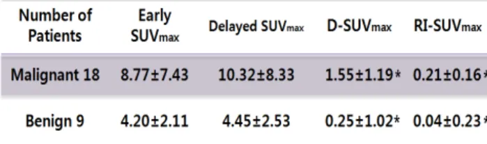
Table.1 shows the mean value of Early SUV max, Delayed SUV max, D-SUV max and RI-SUV max in between benign and malignant group.
The mean values of Early SUV max, Delayed SUV max, D-SUV max and RI-SUV max were 8.77±7.43, 10.32±8.33, 1.55±1.19 and 0.21±0.16 in benign group, and 4.20±2.11, 4.45±2.53, 0.25±1.02 and 0.04±0.23 in malignant group, respectively.
Table 1. Comparison of semi-quantitative analysis as mean
± S.D. in benign and malignant group
* p 〈 0.05 (Independent sample t-test)
Early SUV max and Delayed SUV max have no statistical significance (p=0.085 compared with Early SUV max, p=0.051 compared with Delayed SUV max) in the t-test.
There were statistically significant between benign and malignant group in D-SUV max (p=0.009) and RI-SUV max in the t-test. In particular, RI-SUV max (p=0.044) in malignant group was significantly higher than that of benign group of about 5 fold increase (0.21±0.16 versus 0.04±0.23).
Furthermore, we were able to confirm improved visualization through delayed PET/CT images.
Figure.1 shows an example of distinguishable
visualization about
18F-FDG uptake in malignant and benign group. Malignant focus became more apparent in later images and SUV max increased from 7.33(a) to 10.82(b) whereas benign focus became faint in later images and SUV max decreased from 2.06(c) to 1.59(d).
And also we could identify even 3 additional lesions through delayed PET/CT images from 2 patients.
Fig. 1.
18F-FDG uptake left axilla on 50min (a), 90min (b) of malignant ALN and right axilla on 50min (c), 90min (d) of benign ALN in PET/CT
Table. 2 shows the mean value of Early SUV max, Delayed SUV max, D-SUV max and RI-SUV max among N stages.
The mean values of Early SUV max, Delayed SUV max, D-SUV max and RI-SUV max were 7.74±6.43, 9.28±7.21, 1.54±1.21 and 0.21±0.16 in N1, 4.69±3.94, 5.40±4.24, 0.71±0.32 and 0.18±0.06 in N2 and 14.68±9.87, 16.89±11.06, 2.20±1.33 and 0.15±0.05 in N3 respectively.
Table 2. Comparison of semi-quantitative analysis as mean
± S.D. among N stages
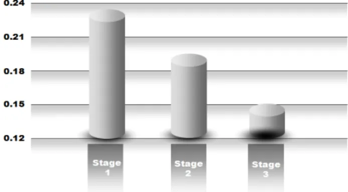
In this case, we could not prove statistically significance with One way ANOVA. There are no statistically significant differences in N1, N2 and N3 in ML group for RI-SUV max (p>0.05). Despite of that reason, RI-SUV max in N stage tended to decrease gradually from N1 to N3 in ML (Figure.2).
Fig. 2. Correlation between RI-SUV max and stage of ML
Ⅳ. DISCUSSION AND CONCLUSION
Based on our study, it informed that metastatic ALN lesion with large increased SUVs over time seemed to be had a malignancy. In contrast, ALN lesion with decreased or slightly increased SUVs over time was likely to have benign etiology.
Although there are no significant differences between N stage and RI-SUV max, the changed
18
F-FDG uptake value in malignant group was very distinguishable from that in benign group.
We regarded that the reason we could not find out the correlation among N stages was small subjects included in this study. However, despite this limitation, this result showed that using delayed PET/CT scan was potentially useful to distinguish metastatic lymphadenopathy (LAP) from benign LAP in breast cancer patients.
The usefulness of delayed PET/CT has already been proven by several studies. A study with
18
F-FDG PET that showed promising results
reported sensitivity and specificity of over 90% in
detection of ALN metastasis from breast cancer
8). Kumar et al
9)also reported that the change of SUVs over time was helpful in differentiating even inflammatory lesions from malignant lesions.
However, we have to consider additional radiation exposure from delayed
18F-FDG PET/CT scan. Even if the radiation exposure cause low radiation dose, meticulous care is required depending on comprehensive state of patients to reduce radiation dose. In the case of children and patients who are undergoing frequent follow-up radiation scan, more invasive test such as biopsy will be helpful for the evaluation of breast pathologies.
18