Received: December 7, 2018 / Revised: December 17, 2018 / Accepted: December 19, 2018
Correspnding author : Yun-Sang Lee, Department of Nuclear Medicine, Seoul National University Hospital, 101, Daehak-ro, Jongno- gu, Seoul 03080, Republic of korea, Tel: +82-2-3668-8906, Fax: +82-745-7690, E-mail: [email protected].
ω-[18F]-Fluorohexadecanoic acid (FHA) has been used for imaging of fatty acid metabolism of myocardium. To increase retention of radiolabeled fatty acid by blocking β-oxidation, methyl branched analogues have been used. In this experiment, we tried to synthesize 18F-labeled α-, β- and ω-FHA for imaging of the myocardial fatty acid metabolism. We synthesized α-, β- and ω-mesylated methyl hexadecanoates and labeled with 18F by reacting with [18F]TBAF in acetonitrile at 80ºC for 10 min. Methyl ester group was removed by 1 M NaOH at 80ºC for 5 min. The yields of α-[18F] and ω-[18F]FHA were 25.5 and 45.5%, respectively [EOS]. However, β-[18F]
FHA was not labeled at all due to a fast elimination reaction. The biodistribution study in ICR-mice showed that ω-[18F]FHA has higher myocardial uptake and lower liver uptake than α-[18F]FHA. The electron-withdrawing effect of fluorine at α- position is believed to be the major factor affecting the biodistribution.
ABSTRACT
Key Word: Myocardial imaging, Positron emission tomography, Radiolabeled fatty acid
Synthesis and biodistribution of
18F-labeled α-, β- and ω-fluorohexadecanoic acid
Yun-Sang Lee*, Young Joo Kim, Gi Jeong Cheon, Jae Min Jeong
Department of Nuclear Medicine, Seoul National University College of Medicine, Seoul, Republic of Korea https://doi.org/10.22643/JRMP.2018.4.2.57
Introduction
Positron emission tomography (PET) of myocardial fatty acid metabolism has been performed mainly using 11C-labeled fatty acid (1, 2). However, it has a limitation due to a short half-life of 11C (T1/2 = 20 min) and high positron range (11C = 4.4 mm vs. 18F = 2.6 mm). To overcome the problem, 18F-labeled fatty acid derivatives have been developed, which have longer physical half-life of 18F (T1/2 = 110 min) (3-5).
For single photon emission computed tomography (SPECT), 123I-labeled fatty acids were developed (6, 7).
Myocardial PET using the labeled fatty acids was suffered from rapid clearance from the myocardium.
To increase retention of the labeled fatty acids in the myocardium, branched derivatives were synthesized for blocking β-oxidation and consequent increase of retention. For example β-methyl fatty acid (8) and β-dimethyl fatty acid (9) were developed for 11C-labeled fatty acids, β-methyl fluorofatty acid (10)and 5-methyl fluorofatty acid 11 were developed for 18F-labeled fatty acids, and α- or β-methyl-p-iodophenyl fatty acids (6, 12, 13) were developed for 123I-labeled fatty acids.
Another trial for blocking β-oxidation was to introduce a thioether group in the middle of fatty acid.
Accordingly, [18F]fluoro-4-thia-heptadecanoic acid or [18F]fluoro-6-thia-heptadecanoic acid have been synthesized and been found increased myocardial
Copyright©2018 The Korean Society of Radiopharmaceuticals and Molecular Probes
retention (14, 15).
In case of 11C-labeled fatty acids, most 11C were labeled mainly at C1 carboxyl position, which would be removed at the early β-oxidation stage. So, the radioactivity was cleared out from the myocardium rapidly, which made imaging difficult. To make longer retention, labeling at the ω-position has been tried (16, 17).
Here, we tried to synthesize 18F-labeled α-, β- and ω-fluorohexadecanoic acid (FHA) and compared their biodistribution for evaluation of feasibility of myocardial imaging agent. (Fig. 1.)
Materials and Methods
General
1H NMR spectroscopy was performed using the AL300 model from the JEOL (Japan). The samples prepared for NMR analysis were dissolved in CDCl3 with TMS as a internal reference or D2O, purchased from Sigma-Aldrich Korea. Mass spectra were obtained with a Hewlett Packard GC(5980)-MSD(5970). A Bio-Scan System 200 imaging scanner was used for Radio-TLC. The ion exchange resin AG1-X8 was purchased from the Bio-Rad Laboratories.
C18 Sep-Pak cartridges were provided by the Waters Division of the Millipore Corporation. For TLC, the glass- backed silica gel 60 F254 (layer thickness 0.25 mm) was purchased from the Merck Company. All reagents and solvents, if not specified, were purchased from Sigma- Aldrich Korea and used without further purification.
1. Synthesis of 18F-labeling precursors
The 18F-labeling precursors, methyl 2-methansulfon yloxyhexadecanoate, methyl 3-methansulfonyloxyhex adecanoate, and methyl 16-methansulfonyloxyhexadec anoate, were synthesized by simply modified methods of the previously described (11) (Fig. 2.).
1.1) Methyl 2-hydroxyhexadecanoate
2-Hydroxyhexadecanoic acid (20 mg, 0.072 mmol) was dissolved in methanol (3 mL), and then 4-M hydrochloric acid in 1,4-dioxane (0.056 mL, 0.04 mmol) was added with stirring. The mixture was heated 100ºC with reflux for 2 hr. After removal of solvent under reduced pressure, dichloromethane (3 mL) was added, washed with saturated sodium chloride solution (1 mL), and dried over anhydrous sodium sulfate. The solvent was removed under reduced pressure to give methyl 2-hydroxyhexadecanoate (21 mg, 0.072 mmol, 99%) as a yellowish oil: 1H NMR (CDCl3) õ 0.87 (3H, t), 1.23-1.70 (26H, m), 3.78 (3H, s), 4.19 (1H, m); MS m/e : 237 (M+-49), 194, 125, 98, 74, 55 (base peak).
1.2) Methyl 2-methansulfonyloxyhexadecanoate To a solution of methyl 2-hydroxyhexadecanoate (21 mg, 0.072 mmol) in dichloromethane (3 mL), methanesulfonyl chloride (0.056 mL, 0.7 mmol) was added slowly with stirring. To this mixture, the solution of triethylamine (0.102 mL, 0.7 mmol) in dichloromethane (1 mL) was added dropwise under
Figure 1. Chemical structures of 18F-labeled α-, β- and ω-FHA
Figure 2. Synthesis of 18F-labeling precursors
cooling with ice-water. After stirring at room temperature for 30 min, the reaction mixture was evaporated. The residue was extracted with dichloromethane (3 mL), and the extract was washed with 1-M hydrochloric acid solution (2 mL), saturated sodium chloride solution (2 mL), and dried over anhydrous sodium sulfate. After removal of dichloromethane, the residue was purified by preparative thin layer chromatography (Merck, 1.13895, ethyl acetate : n-hexane = 1 : 3) to give methyl 2-methansulfonyloxyhexadecanoate (41 mg, 0.071 mmol, 98%) as a white power: 1H NMR (CDCl3) õ 0.87 (3H, t), 1.23-1.70 (26H, m), 3.13 (3H, s), 3.78 (3H, s), 5.01 (1H, m); MS m/e : 285 (M+-79), 253, 236, 225 (base peak), 218, 207, 194, 185, 171, 152, 139, 125, 111, 97, 83, 69, 55.
1.3) Methyl 3-methansulfonyloxyhexadecanoate To a solution of methyl 3-hydroxyhexadecanoate (20 mg, 0.070 mmol) in dichloromethane (3 mL), methanesulfonyl chloride (0.056 mL, 0.7 mmol) was added slowly with stirring. To this mixture, the solution of triethylamine (0.102 mL, 0.7 mmol) in dichloromethane (1 mL) was added dropwise under cooling with ice-water. After stirring at room temperature for 30 min, the reaction mixture was evaporated. The residue was extracted with dichloromethane (3 mL), and the extract was washed with 1-M hydrochloric acid solution (2 mL), saturated sodium chloride solution (2 mL), and dried over anhydrous sodium sulfate. After removal of dichloromethane, the residue was purified by preparative thin layer chromatography (Merck, 1.13895, ethyl acetate : n-hexane = 1 : 3) to give methyl 3-methansulfonyloxyhexadecanoate (25.8 mg, 0.070 mmol, 99%) as a white power: 1H NMR (CDCl3) õ 0.87-0.93 (3H, t), 1.27-1.56 (23H, m), 2.29 (2H, m), 3.05 (3H, s), 3.74 (3H, s), 5.12 (2H, s); MS m/e : 268
(M+-96), 237, 194, 113, 87 (base peak), 55.
1.4) Methyl 16-hydroxyhexadecanoate
16-Hydroxyhexadecanoic acid (200 mg, 0.72 mmol) was dissolved in ethanol (10 mL), and then 4-M hydrochloric acid in 1,4-dioxane (0.1 mL, 0.4 mmol) was added with stirring. The mixture was heated 100ºC with reflux for 2 hr. After removal of solvent under reduced pressure, dichloromethane (5 mL) was added, washed with saturated sodium chloride solution (3 mL), and dried over anhydrous sodium sulfate. Solvent was removed under reduced pressure to give ethyl 16-hydroxyhexadecanoate (216 mg, 0.72 mmol, 99%) as a yellowish oil: 1H NMR (CDCl3) õ 1.23-1.27 (22H, br s), 1.52-1.61 (5H, m), 2.29 (2H, t, J=7.2Hz), 3.64 (2H, t, J=6.6Hz), 3.67 (3H, s); MS m/e : 268 (M+-18), 256, 143, 112, 98, 74 (base peak), 55.
1.5) Methyl 16-methansulfonyloxyhexadecanoate To a solution of ethyl 16-hydroxyhexadecanoate (216 mg, 0.72 mmol) in dichloromethane (10 mL), methanesulfonyl chloride (0.112 mL, 1.4 mmol) was added slowly with stirring. To this mixture, the solution of triethylamine (0.202 mL, 1.4 mmol) in dichloromethane (5 mL) was added dropwise under cooling with ice-water. After stirring at room temperature for 30 min, the reaction mixture was evaporated. The residue was extracted with dichloromethane (10 mL), and the extract was washed with 1-N hydrochloric acid solution (5 mL), saturated sodium chloride solution (5 mL), and dried over anhydrous sodium sulfate. After removal of dichloromethane, the residue was purified by preparative thin layer chromatography (Merck, 1.13895, ethyl acetate : n-hexane = 1 : 3) to give ethyl 16-methansulfonyloxyhexadecanoate (269 mg, 0.71 mmol, 98%) as a white power: 1H NMR (CDCl3) õ
1.26-1.39 (22H, br t), 1.55-1.79 (4H, m), 2.30 (2H, t, J=7.7Hz), 3.00 (3H, s), 3.67 (3H, s), 4.22 (2H, t, J=6.8Hz); MS m/e : 332 (M+-32), 304, 276, 253, 237, 225, 207, 194, 185, 171, 152, 140, 123, 112, 98.
2.
18F labeling
[18F]Fluoride was produced by bombardment of
18O-enriched (>95%) water with 13-MeV proton beam using a TR13 cyclotron (EBCO Technologies).
The [18F]fluoride produced was captured by an AG1-X8 microcolumn and eluted with 1-mL of tetrabutylammonium bicarbonate solution in acetonitrile.
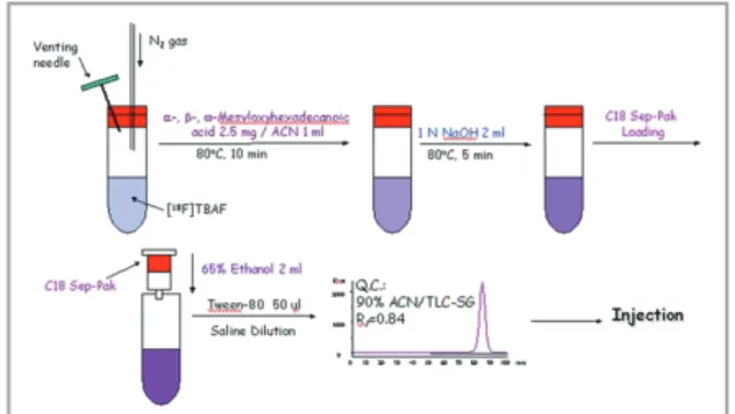
The eluted [18F]fluoride was dried by purging with nitrogen gas at 80°C. After drying, acetonitrile (2 mL) was added, and dried again to remove water. A solution of methyl 2- or 16-methansulfonyloxyhexadecanoate (8 mg, 0.02 mmol) in acetonitrile (1 mL) was added to the dried [18F]fluoride, and the mixture was heated at 80°C for 10 min. To this reaction mixture, 1 M sodium hydroxide solution was added (Fig. 3.). After being heated at 80°C for 10 min, the reaction mixture was passed through a C18 Sep-Pak cartridge, and then washed with distilled water (3 mL) and 0.1-M phosphoric acid solution (3 mL). The radioactive product was eluted with 80% ethanol (2 mL), and the purity was checked by TLC (ethyl acetate : n-hexane = 1 : 1) (Fig. 4).
3. The cold form synthesis and GC-MS analysis
To make the cold form of methyl α-, β- and ω-fluorohexadecanoate, methyl 2-methansulfonyloxy hexadecanoate, methyl 3-methansulfonyloxyhexadeca noate, and methyl 16-methansulfonyloxyhexadecanoa te were mixed with fluoride ion in the basic solution.
The reaction mixtures were analyzed by GC-MSD (Hewlett Packard, USA, GC-5980, MSD-5972) or LC- MS (LC-HP1100, USA, MS-Perkin Elmer, API3000, USA) with no further purification. The GC column was a high performance capillary column (Hewlett Packard, HP-5 Trace Analysis, 5% PH ME Siloxane, film thickness-0.33 μm, column ID-0.20 mm, length-50 m) used for mass analysis. The injection port temperature was set to 150°C to prevent the thermal dissociation of labile compounds.
3.1) Methyl 2-fluorohexadecanoate (α-FHA) Acetonitrile (1 mL) was added to a mixture solution of tetrabutylammonium bicarbonate in acetonitrile solution (320 μL) and tetrabutylammonium fluoride in tetrahydrofuran solution (0.5 mL). The solution was dried by purging with nitrogen gas at 80°C. After drying, acetonitrile (1 mL) was added, and dried again to remove water., A solution of methyl 2-methansulfon
Figure 3. 18F-labeling
Figure 4. Purification of α- and ω-[18F]FHA
yloxyhexadecanoate (2.5 mg, 6.25 μmol) in acetonitrile (1 mL) was added to this solution, and the mixture was heated at 80°C for 10 min. The reaction mixture was analyzed by GC-MSD with no further purification.
3.2) Methyl 3-fluorohexadecanoate (β-FHA) Acetonitrile (1 mL) was added to a mixture solution of tetrabutylammonium bicarbonate in acetonitrile solution (320 μL) and tetrabutylammonium fluoride in tetrahydrofuran solution (0.5 mL). The solution was dried by purging with nitrogen gas at 80°C. After drying, acetonitrile (1 mL) was added, and dried again to remove water., A solution of methyl 3-methansulfon yloxyhexadecanoate (2.5 mg, 6.25 μmol) in acetonitrile (1 mL) was added to this solution, and the mixture was heated at 80°C for 10 min. The reaction mixture was analyzed by GC-MSD with no further purification.
3.3) Methyl 16-fluorohexadecanoate (ω-FHA) Acetonitrile (1 mL) was added to a mixture solution of tetrabutylammonium bicarbonate in acetonitrile solution (320 μL) and tetrabutylammonium fluoride in tetrahydrofuran solution (0.5 mL). The solution was dried by purging with nitrogen gas at 80°C. After drying, acetonitrile (1 mL) was added, and dried again to remove water., A solution of methyl 16-methansulfon yloxyhexadecanoate (2.5 mg, 6.25 μmol) in acetonitrile (1 mL) was added to this solution, and the mixture was heated at 80°C for 10 min. The reaction mixture was analyzed by GC-MSD with no further purification.
4. PET imaging study
A male dog (25 kg) was used for PET imaging study.
All of the experiments were approved by the Institute for Animal Care and Use Committee of Seoul National University Hospital. 111 to 185 MBq/4 mL of α-[18F]
FHA or ω-[18F]FHA was intravenously injected, and PET scan was performed for 40 min by the list mode dynamic scan protocol 3 min after injection.
Results and Discussion
Synthesis of
18F-labeling precursors
The 18F-labeling precursors, methyl 2-methansulfon yloxyhexadecanoate, methyl 3-methansulfonyloxyhex adecanoate, and methyl 16-methansulfonyloxyhexade canoate, were formed by esterification of appropriate hydroxyhexadecanoic acids, followed by mesylation with methansulfonyl chloride. The esterification and the mesylation step showed quantative yield (> 98%).
The structures of all compounds were confirmed by
1H-NMR and GC-MSD.
18
F-labeling
Mesylated precursor of α-, β- and ω-[18F]FHA were prepared and using 18F labeling. α- and ω-[18F]FHA were successfully synthesized and the radiolabeling yield of α- and ω-[18F]FHA were 25.5% and 45.5%, respectively. The radiochemical purity of α- and ω-[18F]
Figure 5. TLC chromatogram of α- and ω-[18F]FHA
FHA were over 99% after the a C18 Sep-Pak cartridge purification (Fig. 5). In the synthesis of β-[18F]FHA, there was no product peak in the radio TLC and the further mechanism study was performed by GC-MS analysis.
The cold form synthesis and GC-MS analysis
To make the cold standard of α-, β- and ω-[18F]FHA, the fluorination was done using mesylated precursor of α-, β- and ω-FHA. From the GC-MS results, the molecular ion peak of α- and ω-FHA were detected to be m/z=288, however that of β-FHA was m/z=237 and this value should be from M+-31(-OMe) and β-elimination product of β-FHA. As same with cold fluorination reaction, 18F substitution reaction was not
happened in the β-mesylated compound (Fig. 6).
Biodistribution of α- and ω-[
18F]FHA
Major uptake organ of α-[18F]FHA were the liver, kidney and heart, and almost same pattern was observed from ω-[18F]FHA except the lower liver uptake than α-[18F]FHA (Fig. 7). The heart uptake of α-[18F]FHA were 14.65±2.92, 6.52±1.89 and 4.02±0.63, and that of ω-[18F]FHA were 26.19±4.66, 14.24±1.58
and 7.48±0.66 at 5, 10 and 30 min after injection, respectively. Because of the heart uptake of ω-[18F]
FHA was 2 times higher than α-[18F]FHA at all time points, and the lower uptake in the liver, ω-[18F]FHA might be the better myocardial imaging agent.
PET imaging study
ω-[18F]FHA showed rapid clearance phase followed by slow clearance phase after initial myocardial uptake like 11C-labeled fatty acid derivatives, which was supposed to show β-oxidation, like [11C]palmitate
Figure 6. Elimination reaction in the synthesis of β-[18F]FHA and the molecular ion peak of elimination product from mass spectrometer data
Figure 7. Biodistribution of α- and ω-[18F]FHA
Figure 8. PET images of α- and ω-[18F]FHA in dogs. (A) Short axis static view at 3 min of α-[18F]FHA (B) Short axis static view at 3 min of ω-[18F]FHA
(Fig. 8). α-[18F]FHA, whereas, showed very low initial myocardial uptake and prominent liver uptake. After the initial retain in myocardium, α-[18F]FHA was cleared from myocardium. These two palmitic acid derivatives showed different time-activity curves. From this PET imaging study, ω-[18F]FHA will be the promising PET agent for myocardial imaging.
Conclusion
We successfully synthesized 18F labeled palmitate derivatives, α-[18F]FHA and ω-[18F]FHA. At the biodistribution and PET imaging study, ω-[18F]FHA showed the high initial uptake of heart and fast clearance like [11C]palmitate. Therefore, ω-[18F]FHA can be used for myocardial imaging agent as a substituent of [11C]
palmitate with the longer half-life and better resolution image.
Acknowledgements
This work was carried out by the research fund supported by Radiation Technology R&D program (NRF-2017M2A2A7A01021401) through the National Research Foundation of Korea (NRF). The authors declare no conflict of interest.
References
1. Hoffman EJ, Phelps ME, Weiss ES, et al. Transaxial Tomographic Imaging of Canine Myocardium with 11C-palmitic Acid. J Nucl Med 1977;18:57-61.
2. Sobel BE, Weiss ES, Welch MJ, Siegel BA, Terpogossian
MM. Detection of Remote Myocardial-Infarction in Patients with Positron Emission Transaxial Tomography and Intravenous 11C-palmitate Circulation 1977;55:853- 857.
3. Berridge MS, Tewson TJ, Welch MJ. Synthesis of
18F-labeled 6- and 7-fluoropalmitic acids. Int J Appl Radiat Isot 1983;34:727-730.
4. Coenen HH, Klatte B, Knochel A, Schuller M, Stocklin G. Preparation of N.C.A. [17-18F]-Fluoroheptadecanoic Acid in High Yields Via Aminopolyether Supported, Nucleophilic Fluorination. J Labelled Compd Rad 1986;23:455-466.
5. Knust EJ, Schuller M, Stocklin G. Synthesis and Quality- Control of Long-Chain 18F-fatty acids. J Labelled Compd Rad 1980;17:353-363.
6. Goodman MM, Kirsch G, Knapp FF. Synthesis of Radioiodinated #betta#-p-(iodophenyl)-Substituted Methyl Branched Long-Chain Fatty-Acids. J Labelled Compd Rad 1982;19:1316-1318.
7. Hock A, Freundlieb C, Vyska K, Losse B, Erbel R, Feinendegen LE. Myocardial Imaging and Metabolic Studies with [17-123I]Iodoheptadecanoic Acid in Patients with Idiopathic Congestive Cardiomyopathy. J Nucl Med 1983;24:22-28.
8. Livni E, Elmaleh DR, Levy S, Brownell GL, Strauss WH. Beta-Methyl[1-11C]Heptadecanoic Acid - a New Myocardial Metabolic Tracer for Positron Emission Tomography. J Nucl Med 1982;23:169-175.
9. Jones GS, Livni E, Strauss HW, Hanson RN, Elmaleh DR. Synthesis and Biologic Evaluation of 1-[11C]- 3,3-Dimethylheptadecanoic Acid. J Nucl Med 1988;29:68- 72.
10. Goodman MM, Neff KH, Ambrose KR, Knapp FF. Effect of 3-Methyl-Branching on the Myocardial Retention of Radioiodinated 19-Iodo-18-Nonadecenoic Acid Analogs.
Int J Rad Appl Instrum B.1989;16:813-819.
11. Takahashi T, Nishimura SI, Ido T, Ishiwata KI, Iwata R. Biological evaluation of 5-methyl-branched-chain omega-[F-18]fluorofatty acid: A potential myocardial imaging tracer for positron emission tomography.
Nucl Med Biol 1996;23:303-308.
12. Goodman MM, Kirsch G, Knapp FF. Synthesis and Evaluation of Radioiodinated Terminal-Substituted 5-Iodo-(2-Thienyl) Fatty-Acids as New Myocardial Imaging Agents. J Heterocyclic Chem 1984;21:1579- 1583.
13. Knapp FF, Ambrose KR, Goodman MM. New Radioiodinated Methyl-Branched Fatty-Acids for Cardiac Studies. Eur J Nucl Med 1986;12:S39-S44.
14. Degrado TR, Coenen HH, Stocklin G. 14(R,S)-[F-18]
Fluoro-6-Thia-Heptadecanoic Acid (Ftha) - Evaluation in Mouse of a New Probe of Myocardial Utilization of Long-
Chain Fatty-Acids. J Nucl Med 1991;32:1888-1896.
15. DeGrado TR, Wang SY, Holden JE, Nickles RJ, Taylor M, Stone CK. Synthesis and preliminary evaluation of F-18-labeled 4-thia palmitate as a PET tracer of myocardial fatty acid oxidation. Nucl Med Biol 2000;27:221-231.
16. Buckman BO, VanBrocklin HF, Dence CS, Bergmann SR, Welch MJ, Katzenellenbogen JA. Synthesis and tissue biodistribution of [omega-11C]palmitic acid.
A novel PET imaging agent for cardiac fatty acid metabolism. J Med Chem 1994;37:2481-2485.
17. Collier TL, Hwang Y, Ramasamy R, et al. Synthesis and initial evaluation of 17-(11)C-heptadecanoic acid for measurement of myocardial fatty acid metabolism.
J Nucl Med 2002;43:1707-1714.

![Figure 5. TLC chromatogram of α- and ω-[ 18 F]FHA](https://thumb-ap.123doks.com/thumbv2/123dokinfo/5079317.562639/5.892.475.806.788.1056/figure-tlc-chromatogram-α-ω-f-fha.webp)
![Figure 6. Elimination reaction in the synthesis of β-[ 18 F]FHA and the molecular ion peak of elimination product from mass spectrometer data](https://thumb-ap.123doks.com/thumbv2/123dokinfo/5079317.562639/6.892.454.790.143.467/figure-elimination-reaction-synthesis-molecular-elimination-product-spectrometer.webp)
