Protective Effect of White-Skinned Sweet Potato ( Ipomoea batatas L.) against Renal Damage in Streptozotocin-Induced Diabetic Rats
Hye Won Jang, Moch. Saiful Bachri
1,2, Kyung Ok Moon
2and Jong Ok Park
3*
Division of Endocrinology & Metabolism, Samsung Medical Center, Sungkyunkwan University School of Medicine, Seoul 135-710, Korea
1
Faculty of Pharmacy, Ahmad Dahlan University, Yogjakarta 55164, Indonesia
2
College of Pharmacy Kyungsung University, Busan 608-736, Korea
3
Department of Chemistry, Kyungsung University, Busan 608-736, Korea
Received November 12, 2009 /Accepted February 6, 2010White-skinned sweet potato (Ipomoea batatas L.) has been traditionally used for diabetes treatment and management in many countries. In this experiment, methanol extract of white-skinned sweet potato (WSPMe) at a dose of 100 or 200 mg/kg body weight was tested to evaluate its effect on renal dam- age in streptozotocin (STZ)-induced diabetic rats. Its efficacy was compared with that of insulin secre- togogue, glimepiride (50 μg/kg body weight). Experimental diabetes was induced by a single dose of STZ (45 mg/kg, i.p.) injection. The WSPMe and glimepiride were administered orally for 14 days and the effects on glucose, renal markers including blood urea nitrogen (BUN), creatinine and lactate dehydrogenase (LDH), lipid peroxide (LPO) level, antioxidant enzymes superoxide dismutase (SOD), catalase (CAT), glutathione peroxidase (GPx) and glutathion-S-transferase (GST) activities in kidney were studied. An increase in BUN, creatinine, LDH, glucose, LPO levels and decrease in SOD, CAT, GPx and GST features were observed in diabetic control rats. Administration of WSPMe at a dose of 200 mg/kg body weight caused a significant improvement in blood glucose, LPO level, renal markers, lipid peroxidation markers and increased antioxidant levels in diabetic kidney. In conclusion, the WSPMe was found to be effective in reducing oxidative stress, thus confirming the ethnopharmacological use of I. batatas L. in protecting diabetes and its complications.
Key words : White-skinned sweet potato, diabetes, streptozotocin, oxidative stress, antioxidant
*Corresponding author
*Tel:+82-51-620-4633, Fax:+82-51-628-4628
*E-mail : [email protected]
Introduction
Diabetes is a complex metabolic disorder, involving char- acteristic alterations of glucose metabolism. In diabetic pa- tients, insulin is not produced or is insufficiently produced (diabetes type 1) or peripheral receptors to insulin lack the normal sensitivity (diabetes type 2), which in both case cause hyperglycemia and severe alterations of glucose and lipid metabolism. This abnormal metabolism leads to an increased generation of reactive oxygen species (ROS) [34]. Therefore, maintenance of glucose level is a key strategy in treating patients with type 2 diabetes [27].
Imbalance between ROS production and ROS elimination in the biological system cause oxidative stress. It leads to oxidative damage to cell and tissue paralleled by mod- ifications in the morphology and function, resulting in aging and premature cell death [12,41].
Oxidative stress had been reported play a role in the
pathogenesis and progression of diabetic tissue damage [19,23,47]. Numerous evidences suggesting that the hyper- glycemia associated oxidative stress are the central event for the development of diabetes and its complications [4,7].
However, chronic diabetes is shown to disturb antioxidant defence systems in diabetes ; alteration in antioxidant en- zymes [3], impaired glutathione metabolism [29] and de- creased ascorbic acid levels [22]. Some natural defence mech- anism exists against oxidative stress through endogenous or exogenous antioxidant substances. Supplementation with exogenous antioxidants has been proved as a comple- mentary treatment of diabetes.
Recently, there has been a considerable interest in finding natural antioxidants. The plant kingdom has become a target for the search for new drugs and biologically active com- pounds [37,39]. Some plants have been used in traditional medicines as antidiabetic medicine [2,12,15], but a few have received scientific scrutiny.
Antioxidant activity of plant materials was attributed to
well-known phytochemicals such as α-tocopherol, ascorbic
acid, and β-carotene. Recent researches have focused on pol-
yphenolic compounds as major antioxidant [24]. Oki et al.
[31] identified several phenolic compounds present in sweet potato and had oxygen radical absorbance capacity. Kano et al. [26] reported that anthocyanin from purple sweet pota- to had better radical scavenging activity than that of red cab- bage, grape skin elderberry and purple corn.
Several studies have reported on the antioxidant activity [21,44,45] and anti-diabetic activity [5,38,45] of sweet potato extracts. Purple sweet potato has been reported to attenuate the oxidative stress and inflammatory response [49].
White-skinned sweet potato (Ipomoea batatas L. WSP) has been used in Indonesia, Japan as a traditional medicine for the treatment of diabetes and other diseases. Shuichi et. al.
[38] reported that WSP shows unique antidiabetic effects in both insuline defficient diabetic models and insuline-resistant diabetic models. In addition, antidiabetic components from WSP was an acidic glycoprotein (M
r22,000). However, It has not been studied for its effect on the oxidative stress much.
Streptozotocin (STZ) is widely used to induce diabetes mellitus in research used animal, diabetogenic effect of STZ is due to excess production of ROS leading to toxicity in pancreatic cells which reduces the synthesis and the release of insulin [42] while affecting organs such as liver, kidney, and hematopoietic system [23].
The present study was designed to evaluate the re- no-protective property of an traditional antidiabetic plant, WSP in STZ-induced diabetic rat and the efficacy was com- pared with insulin secretogogue, glimepiride.
Materials and Methods Animals
Male Sprague Dawley (200±10 g) rats aged 6 weeks were purchased from Hyochang Science, Daegu, Korea. All ani- mals were maintained in the institutional animal facility and handled according to the guidelines of the pharmacology department, college of pharmacy, Kyungsung University, Republic of Korea. Animals were adapted with condition for a week with a light/dark cycle: 12 hr, humidity: 46-60%, temperature 22±0.5
oC in university animal room and given with free access to rodent food and water ad libitum through- out the experimental period.
Preparation of the extract
Fresh storage roots of white-skinned sweet potato (Ipomoea
batatas L) were purchased from a local market in Yogyakarta Indonesia. Samples were processed into extract in Biology Pharmacy, Ahmad Dahlan University, Yogyakarta, Indonesia.
The samples were sliced, dried and ground into powder.
The powder was dissolved three times in 3 volumes of meth- anol for 3 days, filtered and evaporated to obtain the crude methanol extract. The crude methanol extracts were lyophi- lized for 3 days to get dried powder and was kept at 4
oC before use.
Experimental design and induction of diabetes mellitus Animals were divided into five groups with five animals in each group as follows; Group I, normal (nondiabetic) rats treated with the vehicle (tween : saline) only; Group II, con- trol (diabetic) rats treated with the vehicle and a single dose of STZ (Sigma Chemical Co. St. Louis, MO, USA. 45 mg/kg);
Group III, rats treated with WSPMe 100 mg/kg and a single dose of STZ 45 mg/kg; Group IV, rats treated with WSPMe 200 mg/kg and a single dose of STZ 45 mg/kg; Group V, rats treated with glimepiride 0.5 mg/kg and a single dose of STZ 45 mg/kg. Both normal and control groups were or- ally administered an equivalent volume of the vehicle for the two weeks of the treatment (pre-experiment). And then normal rats were injected with saline alone, control rats were injected with STZ 45 mg/kg prepared in 2 ml citrate buffer pH 4.5 by a single intraperitonial injection to make diabetes rats. After oral administration with WSPMe and glimepiride as pre-experiment for 2 weeks, rats were injected with STZ 45 mg/kg. On three days after the STZ treatment, develop- ment of diabetes was confirmed by measuring the blood glu- cose levels using glucose reagent strips (Glucometer 4 Ames, Bayer Diagnostics). Rats with a fasting blood glucose levels above 250 mg/dl were considered to be diabetic. The WSPMe and glimepiride continue administered orally dur- ing the 2 weeks after STZ injection (post experiment). After completion of the treatments, the animals were sacrificed us- ing carbon dioxide as anesthesia. Blood was collected di- rectly from the abdominal vein and separated to obtain serum. The kidney were dissected and rinsed with ice-cold normal saline. Kidney tissue was minced and a 10% (w/v) homogenate was prepared with phosphate-buffered saline (0.1 M, pH 7.4) using a homogenizer at 4
oC for biochemical study.
Determination of serum enzyme and components
For the blood glucose analysis, a drop of blood was col-
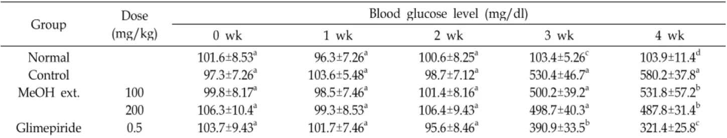
Table 1. Effect of MeOH extract of WSP on the blood glucose levels in STZ-induced diabetic rats
Group Dose
(mg/kg)
Blood glucose level (mg/dl)
0 wk 1 wk 2 wk 3 wk 4 wk
Normal Control MeOH ext.
Glimepiride
100 2000.5
101.6±8.53a 97.3±7.26a 99.8±8.17a 106.3±10.4a 103.7±9.43a
96.3±7.26a 103.6±5.48a 98.5±7.46a 99.3±8.53a 101.7±7.46a
100.6±8.25a 98.7±7.12a 101.4±8.16a 106.4±9.43a 95.6±8.46a
103.4±5.26c 530.4±46.7a 500.2±39.2a 498.7±40.3a 390.9±33.5b
103.9±11.4d 580.2±37.8a 531.8±57.2b 487.8±31.4b 321.4±25.8c Values are the means±SD (n=5). Values within a column with different superscripts are significantly different at <0.05 by the Duncan’s test
lected from the tail vein of animals. The blood glucose level was determined using a one touch glucometer (Roche).
Serum was extracted from the blood collected directly from the abdominal vein after the rats had been subjected to anesthesia. Serum was separated for the estimation of the blood urea nitrogen (BUN), creatinine, lactate dehydrogenase.
BUN was determined by the method of Chaney et al. [9], creatinine content was determined by the method of Slot [40], lactate dehydrogenase activity was assayed by the Cabaud and Wroblewski method [10].
Determination of renal antioxidant enzyme activity of WSP
The level of lipid peroxidation was measured as the amount of malondialdehyde (MDA), a thiobarbituric acid re- active substance (TBARS), using 1’1’3’3’-tetramethoxypropane as the standard [30]. Superoxide Dismustase (SOD) activity was assayed by the method of Marklund and Marklund [28].
This assay procedure involved the inhibition of epinephrine auto-oxidation to adenochrome in alkaline medium (pH 10.2) which is markedly inhibited by the presence of SOD.
Epinephrine was added to the assay mixture, containing tis- sue supernatant with the change in the extinction coefficient observed at 480 nm using spectrophotometer. The catalase activity in tissue was measured at 240 nm spectrophoto- metrically by calculating the degradation of H
2O
2, the sub- strate of enzyme [1]. The glutathion peroxidase (GPx) activ- ity was measured according to the method of Paglia and Valentine [32]. The assay was determined by measuring the decrease in the glutathione (GSH) content after incubation the sample in the presence of H
2O
2and NaN
3. The glutathione S-transferase (GST) was determined according to the method of Habig [17]. This assay was determined by the amount of p-nitrobenzylchloride as the substrate. The absorbance was measured UV-Vis spectrophotometrically at 310 nm.
Statistical analysis
All data were expressed as mean±SD. Total variation present in a set of data was estimated by one-way analysis of variance (ANOVA) followed by Duncan's multiple range tests. p<0.05 was considered significant.
Results Blood glucose level and renal profiles
Table 1 exhibited the effect of WSPMe on the blood glu- cose level. As expected and shown, blood glucose level was increased in STZ-treated rats as compared to nomal group.
The mean blood glucose level in normal group (group I) was stable throughout the experimental period. 2 weeks ad- ministration before STZ-induced (pre experiment), all groups showed no significant difference (p>0.05) in blood glucose level. 7 days after STZ-induced, treatments of WSPMe showed no significant (p>0.05) reduction in blood glucose compared to diabetic control group. Glimepiride showed significant (p
<0.05) reduction in blood glucose level as compared to dia- betic control group. After 14 days treatment (post experi- ment), diabetic animal had significant responses of WSPMe compared to control group. Also glimepiride showed sig- nificant (p<0.05) reduction in blood glucose level compared to control group and WSPMe group. Oral administration of WSPMe dose 200 mg/kg revealed more reduction in blood glucose level compared to WSPMe dose 100 mg/kg, but stat- istically no significant difference (p>0.05).
In diabetic rats, serum levels of all three tested renal
markers: urea, creatinine and lactate dehydrogenase were
significantly increased compared to the normal rats (Table
2). The elevation of these indicated impaired kidney
function. Administration of WSPMe to diabetic rats at 200
mg/kg body weight significantly decreased the levels of
BUN, creatinine and LDH. However it was not noteworthy
Table 2. Effect of MeOH extract of WSP on urea, creatinine and lactate dehydrogenase levels in STZ-induced dia- betic rats
Group Dose
(mg/kg) BUN Creatinine LDH
Normal Control MeOH extract
Glimepiride 100 2000.5
18.4±3.25d 76.9±9.43a 73.2±8.16a 62.4±9.10b 50.7±7.16c
4.97±0.96e 12.60±1.15a 10.30±0.77b 8.42±0.63c 6.17±0.81d
22.5±3.17e 89.4±7.45a 80.5±9.16b 65.2±5.49c 51.7±6.11d Values are the means±SD (n=5). Values within a column with different superscripts are significantly different at <0.05 by the Duncan’s test
BUN: blood urea nitrogen, LDH: lactate dehydrogenase
difference compared to the glimepiride treated groups.
Lipid oxidation activity in kidney
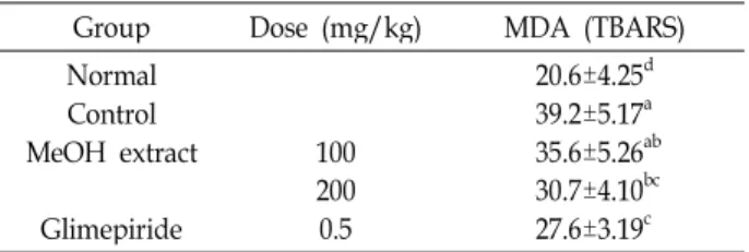
Lipid oxidation has been established as a major mecha- nism of cellular injury in many biological systems of plant and animal origin. Polyunsaturated fatty acid peroxides fur- ther react to form MDA, which has become one of the most widely reported analytes for the purpose of estimating oxi- dative stress effects on lipids [23]. Table 3 showed the level of MDA, a secondary product of lipid peroxidation in the kidney tissue homogenate of control and experimental rats.
STZ-induced diabetic group resulting in a significant (p<
0.05) increased in MDA levels compared to normal group, whereas oral administration of WSPMe at dose 100; 200 mg/kg and glimepiride to diabetic rats exhibited significant (p<0.05) reduction in compared with control group.
Antioxidant enzyme activity in kidney
As shown in Table 4, a significant (p<0.05) decrease in the activities of SOD, CAT and GPx compared to normal
Table 3. Effect of MeOH extract of WSP on lipid peroxide in STZ-induced diabetic rats
Group Dose (mg/kg) MDA (TBARS)
Normal Control MeOH extract
Glimepiride
100200 0.5
20.6±4.25d 39.2±5.17a 35.6±5.26ab 30.7±4.10bc 27.6±3.19c Values are the means±SD (n=5). Values within a column with different superscripts are significantly different at <0.05 by the Duncan's test
MDA: malondialdehyde, TBARS: thiobarbituric acid reactive substance
Table 4. Effect of MeOH extract of WSP on antioxidant enzyme levels in STZ-induced diabetic rats
Group Dose
(mg/kg) SOD Catalase GPx
Normal Control MeOH extract
Glimepiride 100200 0.5
18.7±0.86a 4.3±0.25e 5.7±0.30d 8.5±0.28c 11.4±0.65b
1.98±0.27a 0.92±0.11d 1.06±0.12cd 1.21±0.17c 1.45±0.18b
2.96±0.37a 1.60±0.14c 1.83±0.23c 2.16±0.16b 2.37±0.13b Superoxide Dismustase (SOD, U/mg protein); Glutathione Peroxidase (GPx, NADPH oxidized/min/mg protein); Catalase (CAT, nmol of H2O2 consumed/min/mg protein)
Values are the means±SD (n=5). Values within a column with different superscripts are significantly different at <0.05 by the Duncan’s test
Table 5. Effect of MeOH extract of WSSP on glutathion-S-trans- ferase in STZ-induced diabetic rats
Group Dose (mg/kg) Glutathion-S-Transferase Normal
Control MeOH extract
Glimepiride
100 200 0.5
141.7±11.4a 93.2±11.4d 100.6±13.4cd 110.8±10.6bc 121.2±11.8b Values are the means±SD (n=5). Values within a column with different superscripts are significantly different at <0.05 by the Duncan’s test
group was a notable manifestation of STZ toxicity. The activ- ity of these enzymes was improved significantly (p<0.05) by the administration of WSPMe dose 200 mg/kg when compared with control diabetic group. Also administration of glimepiride, an insulin releaser improved these enzymes activities. While, oral administration of WSPMe dose 100 mg/kg showed no significant difference (p>0.05) reduction in antioxidant enzyme activities compared with control group.
Table 5 showed the concentration of GST in animals treat- ed with STZ which were significantly (p>0.05) decreased in kidney compared to normal group. Whereas Oral admin- istration of WSPMe and glimepiride were significant differ- ence (p>0.05) compared with control group.
Discussion
Diabetes mellitus is a wide-spread disease characterized
by high blood glucose levels which, in turn, induce damages
to cell membranes and generate ROS [41]. Vascular compli-
cations and diabetic nephropathy which are generally asso-
ciated to diabetes are known to be largely related to the oxi- dative stress [16].
Oxidative stress results from an imbalance between the generations of oxygen derived radicals and antioxidant potential.
The pathogenesis of type 2 diabetes involves insulin re- sistance, increased hepatic glucose output, and impaired in- sulin secretion [27]. These features are potential targets for pharmacologic intervention in addition to diet and increased physical activity [14].
The non-insulin dependent diabetic has a less severe form of the disease but nevertheless develops the same complica- tions as the insulin dependent diabetic, that is, neuropathy, retinopathy, kidney disease, and coronary artery disease.
In the present study, as expected, the elevation of blood glucose levels during the experimental period clearly in- dicates the persistent hyperglycemia in the STZ-induced dia- betic rats (Table 1). However, administration with WSPMe markedly reduced the blood glucose concentration in dia- betic rats. The WSPMe can decrease blood glucose levels im- plies WSPMe can block free radical production and prevent the production of ROS during diabetes. This result indicates that WSPMe acts as an antihyperglycemic agent and sig- nificantly reduce the blood glucose level. ROS and advanced glycation end products are known to have a wide range of chemical, cellular and tissue effects implicated in the devel- opment and progression diabetic nephropathy including ac- celerating uremic glomerulopathy with tubulointestinal damage [6].
In experiment, diabetic rats showed symptoms of renal nephropathy. The level of blood urea nitrogen is determined by the amount of protein intake, the production and the ex- cretion of the urea. The most common cause of the elevation of blood urea nitrogen is poor kidney function. Creatinine is one of the main nitrogen compounds in normal urine. The creatinine concentration in blood and body fluids is nor- mally very low, and reaches higher values only if the kid- neys are damaged. The diabetic hyperglycemia induces ele- vation of the plasma levels of urea, creatinine and LDH which are significant markers of renal dysfunction and re- flecting a decline in the glomerular filtration rate. In diabetic rats, serum levels of all three tested renal markers; urea, cre- atinine and lactate dehydrogenase were significantly in- creased when compared to the normal rats (Table 2).
Administration of WSPMe to diabetic rats significantly re- versed these changes to near normal levels. However, no
noteworthy changes were observed in the normal rats treat- ed with WSPMe.
The results of the present study showed significant (p<
0.05) increase in the level of these parameters in the diabetic rats when compared with control rats, while, after the treat- ment with WSPMe, the levels of urea, creatinine and LDH were significantly (p<0.05) decreased. These results are in agreement with other previous studies [2,25,33].
Chronic hyperglycemia was found to produce oxidative stress and increased lipid peroxidation in kidneys, as shown by the increased level of renal MDA, as a lipid peroxidation marker [4]. This increased level of lipid peroxidation could be associated to increase in free radicals generation in dia- betes caused primarily due to high blood glucose levels, which upon autoxidation generates free radicals and sec- ondarily due to the effects of diabetogenic agents.
Elevated levels of lipid peroxidation in tissues and plasma is one of the characteristic features of chronic diabetes [3].
Therefore inhibition of free radical generation and oxidative damage could be considered as an important strategy in the management of diabetes.
In analysis of TBARS, our study clearly showed that MDA
level was decreased in WSPMe treated diabetic rats and may
have role in scavenging hydroxyl and peroxyl radicals gen-
erated by STZ (Table 3). Lipid peroxidation in diabetes melli-
tus can be considered with overproduction of oxidants or
a decrease in antioxidant defenses [46]. Such a decrease of
MDA level in kidney of diabetic rats was also observed by
Sukalski et al. [33]. Three antioxidant enzymes which has
important function are SOD (scavenges superoxide anions),
GPx (removes H
2O
2and lipid peroxides), also CAT that are
considered primary antioxidant enzymes involved in the di-
rect elimination of reactive oxygen species. Associated with
changes in lipid peroxidation, the diabetic kidney showed
decreased activity of the key antioxidant enzymes SOD,
CAT, GPx and GST, which are important in scavenging the
toxic intermediates. According to our results, WSPMe treat-
ment showed significant improved free radical scavenging
enzymes (SOD, CAT, GPx) in the kidney of STZ treated rats
(Table 4). SOD, CAT, and GPx are enzymes that break the
peroxides and play an important role in supplying anti-
oxidant substances to an organism. SOD reduces superoxide
to H
2O
2that can be readily reduced to water principally by
CAT and GPx [35]. The functions of all three enzymes are
interconnected with the lowering of their activities resulting
in the accumulation of lipid peroxides and an associated in-
crease in oxidative stress in diabetic rats [11]. Oral admin- istration of WSPMe improved the activities of these enzymes and thus may help protect the generation of free radicals during diabetes mellitus. The decreased activities of SOD and CAT in tissue are due to excess availability of super- oxide (O
2 -) and hydrogen peroxide (H
2O
2) in the biological systems, which in turn generate hydroxyl and peroxyl radi- cals, resulting in the initiation and propagation of lipid per- oxides [43].
Meanwhile WSPMe treatment has similar effect with gli- mepiride produced no significant (p>0.05) reduction in kid- ney GST compared with control group. Based on the results, protective effect of WSPMe is may be due to the counter- action of free radicals throughout three antioxidant enzymes, increasing antioxidant free radical formation, leads to re- duces LPO and a significant lowering in blood glucose level.
These results suggest that WSPMe administration has pro- tective effect in STZ-induced oxidative stress in rats. However, the precise molecular mechanism by which WSPMe exerts its protective effect against oxidative damage remains to be established.
The present study demonstrated that WSP ameliorates dia- betes-induced oxidative stress by reducing the blood glucose level and prevent diabetes-induced renal damage. Further, Our results provided scientific evidence of the preventive and therapeutic potential of WSP from Indonesia against re- nal damage associated with diabetic oxidative stress.
References
1. Aebi, H. 1974. Catalase in “Method of enzymatic analysis”, In Vergmeyer, H. U. (ed.), Academic press, New York, 2, 673-684.
2. Attele, A. S., Y. P. Zhou, J. T. Xie, J. A. Wu, L. Zhang, L.
Dey, W. Pugh, P. A. Rue, K. S. Polonsky, and C. S. Yuan.
2002. Antidiabetic effects of Panax ginseng berry extract and the identification of an effective component.
Diabetes
51, 1851-1858.3. Baynes, J. W. and S. R. Thorpe. 1997. The role of oxidative stress in diabetic complications.
Curr. Opin. Endocrinol
. 3, 277-284.4. Baynes, J. W. 1991. Role of oxidative stress in the develop- ment of complications in diabetes.
Diabetes
40, 405-412.5. Bernhard, L., W. Werner, P. Rudolf, K. W. Alexandra, and P. Giovanni. 2003. Mode of Action of
Ipomoea Batatas
(Caiapo) in Type 2 Diabetic Patients.Metabolism clinical and experimental.
52, 875-880.6. Bohlender, H. M., S. Franke, G. Steine, and G. Wof. 2005.
Advanced glycation end products and the kidney.
Am. J.
Physiol. Renal Physiol.
289, F645-F659.7. Bonnefont-Rousselot, D., J. P. Nastard, M. C. Jaudon and J. Delattre. 2000. Consequences of the diabetic status on the oxidant/antioxidant balance.
Diabetes Metab.
26, 163-176.8. Cabaud, P. G. and F. Wroblewski. 1958. The determination of serum lactate dehydrogenase.
American Journal Clinical Pathology
30, 234-236.9. Cao, G., E. Sofic, and R. L. Prior. 1996. Antioxidant capacity of tea and common vegetables.
J. Agricul. Food Chem.
44, 3426-3431.10. Chaney, A. L. and E. P. Marbach. 1962. Modified reagents for determination of urea and ammonia.
Clinica Chimica Acta
8, 130-132.11. Chaudhry, J., N. N. Ghoxh, K. Roy, and R. Chandra. 2007.
Antihyperglycemic effect of a new thiazolidine analogue and its role in ameliorating oxidative stress in alloxan-in- duced diabetic rats.
Life Sci
. 80, 1135-1142.12. Craik, F. I. and M. Salthouse. 1992. Handbook of Aging and Cognition, pp. 51-110, Hillsdale, New Jersey.
13. Garber, A. J. 2002. Attenuating CV risk factors in patients with diabetes: clinical evidence to clinical practice.
Diabetes, Obesity and Metabolism
4, S5-S12.14. Goodman, L. S. and A. Gilman. 1985. The Pharmacological Basis of Therapeutics, pp. 1490-1510. 7th ed. Macmillan, New York.
15. Grover, J. K., S. Yadav, and V. Vats. 2002. Medicinal plants of India with anti-diabetic potential.
J. of Ethnopharmacology
81, 1-100.16. Ha, H. and K. H. Kim. 1999. Pathogeniesis of diabetic nephropathy: the role of oxidative stress and proteinkinase C.
Diabetes Res. Clin. Pract.
45, 147-151.17. Habig, W., M. J., Pabst, and W. B. Jakoby. 1974. Glutathione S-transferases the first enzymatic step in mercapturic acid formation.
J. Biol. Chem.
249, 7130-7139.18. Hamden, K., S. Carreau, K. Jamoussi, F. Aya, S. Lasmi, D.
Aloulou, and A. Elfeki. 2009. 1,25-dihydroxyvitamin D3 : therapheutic and preventive effects against oxidative stress and hepatic, pancreatic and renal injury in diabetic rats.
J.
Nutr. Sci. Vitam.
56, 455-461.19. Hiramatsu, K. and S. Aomori. 1988. Increased superoxide production by mononuclear cells of patients with hyper- triglyceridemia and diabetes.
Diabetes
37, 832-837.20. Huang, G. J., H. Y. Chang, H. J. Chen, T. L. Lu, Y. S. Chang, M. J. Sheu, and Y. H. Lin. 2008. Effects of trypsin inhibitor on plasma antioxidant activity and lipid levels in mice from sweet potato roots,
J. Sci. Food Agric.
88, 2556-2562.21. Huang, Y. C., Y. H. Chang, and Y. Y. Shao. 2005. Effects of genotype and treatment on the antioxidant activity of sweet potato in Taiwan.
Food Chemistry
98, 529-538.22. Jennings, P. E., S. Chirico, A. F. Jones, J. Lunec, and A. H.
Barnett. 1987. Vitamin C metabolites and microangiopathy in diabetes mellitus.
Diabetes Res.
6, 151-154.23. Kakkar, R., J. Kalra, S. V. Manth, and K. Parsad. 1995. Lipid peroxidation and activity of antioxidant enzymes in diabetic rats.
Mol. Cell. Biochem.
151, 113-119.24. Kalt, W. 2005. Effects of production and processing factors
on major fruits and vegetable antioxidants.
J. Food Sci.
70, R11-E19.25. Kang, K. S., H. Y. Kim, N. Yamabe, R. Nagai, and T.
Yokozawa. 2006. Protective effect of sun ginseng against di- abetic renal damage.
Biol. Pharm. Bull.
29, 1678-1684.26. Kano, M., T. Takayanagi, K. Harada, K. Makino, and F.
Ishikawa, 2005. Antioxidant activity of anthocyanins from purple sweet potato,
Ipomoea batatas
cultivar Ayamurasaki.Bioscience Biotechnology and Biochemistry
69, 979-988.27. Laakso, M. 2001. Insulin resistance and its impact on the approach to therapy of type 2 diabetes.
International Journal of Clinical Practice Supplemen.
8-12.28. Marklund, S. and G. Marklund. 1974. Involvement of the superoxide anion radical in the autooxidation of pyrogallol and a convenient assay for superoxide dismutase.
Eur. J.
Biochem.
47, 46929. McLennan, S. V., S. Heffnan, L. Wright, C. Rae, E. Fisher, D. K. Yue, and J. R. Turtle. 1991. Changes in hepatic gluta- thione metabolism in diabetes.
Diabetes
40, 344-348.30. Ohkawa, H., N. Ohishi, and K. Yagi. 1979. Assay for lipid peroxides in animal tissues by thiobarbituric acid reaction.
Analytical. Biochemisty
95, 351-358.31. Oki, T., M. Masuda, S. Furuta, Y. Nishiba, N. Terahara, and I. Suda. 2002. Involvement of anthocyanins and other phe- nolic compounds in radical scavenging activity of purple- fleshed sweet potato cultivars.
J. of Food Science
67, 1752-1756.32. Paglia, E. D. and W. N. Valentine. 1967. Studies on the quantitative and qualitative characterization of erythrocyte glutathione peroxidase.
J. Lab. Clin. Med.
70, 158-169.33. Ramkumar, K. M., P. Ponmanickam, S. Velayuthaprabhu, G. Archunan, and P. Rajaguru. 2009. Protective effect of
Gymnema montanum
against renal damage in experimental diabetic rats.Food and Chem. Toxic.
47, 1-6.34. Randle, P. J., P. B. Gailand, C. N. Hales, and E. A.
Neiosholine. 1963. The glucose and fatty acid cycle: its role in insulin sensitivity and metabolic disturbance of diabetes.
The Lancet
1, 785-790.35. Robertson, R. P., J. Harmon, P. P. Tran, Y. Tanaka, and H.
Takahashi. 2003. Glucose toxicity in β-cells: type 2 diabetes, good radicals gone bad, and the glutathione connection.
Diabetes
52, 581-587.36. Rully, M. 1988. Pengaruh infus batang ubi jalar (
Ipomoea ba- tatas
Poir) sebagai antidiabetik pada binatang percobaan tikus. JF FMIPA NHAS 109.37. Ruzaid, A., I. Amin, A. G. Nawalyah, M. Hamid, and H.
A. Faizul. 2005. The effect of Malaysian cocoa extract on glucose levels and lipid profiles in diabetic rats.
J
Ethnopharmacol.
98, 55-60.38. Shuichi, K., A. Hiroyuki, and T. Hirohide. 2001. Isolation of Antidiabetic Components from White-Skinned Sweet Potato (
Ipomoea batatas
L.).Biosci. Biotechnol. Biochem.
65, 109-114.39. Singh, N., V. Kamath, and P. S. Rajini. 2005. Attenuation of hyperglycemia and associated biochemical parameters in STZ-induced diabetic rats by dietary supplementation of potato peel powder.
Clin. Chim. Acta
353, 165-175.40. Slot, C. 1973. Determination of serum creatinine by a direct method.
Clinica Chimica Acta
43, 305-310.41. Stefek, M., N. Tribulova, A. Gajdoski, and A. Gajdosikova.
2002. The pyridoindole antioxidant stobadine attenuates histochemical changes in kidney of STZ-induced diabetic rats.
Acta Histoche.
104, 413-417.42. Szkudelski, T. 2001. The mechanism of alloxan and strepto- zotocin action in β-cells of the rat pancreas.
Physiol. Res.
50, 537-546.43. Tarique, A., M. Sharma, K. K. Pillai, S. E. Haquea, M. M.
Alam and M.S. Zaman. 2007. Protective effect of bezafibrate on streptozotocin-induced oxidative stress and toxicity in rats.
Toxicology
229, 165-172.44. Teow, C. C., V. D. Truong, R. F. McFeeters, R. L. Thompson, K. V. Pecota, and G. C. Yencho. 2007. Antioxidant activities, phenolic and β-carotene contents of sweet potato genotypes with varying flesh colours.
Food Chemistry
103, 829-838.45. Toshiro, M., E. Sumi, K. Mio, F. Keiichi, S. Koichi, T.
Norihiko, and M. Kiyoshi. 2002. Anti-hyperglycemic effect of diacylated anthocyanin derived from
Ipomoea batatas
Cultivar Ayamurasaki can be achieved through the α-gluco- sidase inhibitory action.J. Agric. Food Chem.
5, 7244-7248.46. Ugochukwu, N. H., N. D. Bagayoko, and M. E. Antwi. 2004.
The effects of dietary caloric restriction on antioxidant sta- tus and lipid peroxidation in mild and severe streptozoto- cin-induced diabetic rats.
Clin. Chim. Acta
348, 121-129.47. Wolff, S. P., Z. Y. Jang, and V. J. Hunt. 1991. Protein glyca- tion and oxidative stress in diabetes mellitus and ageing.
Free. Radic. Biol. Med.
10, 339-352.48. Yamagishi, N., K. Nakayama, T. Wakatsuki, and T.
Hatayama. 2001. Characteristic changes of stress protein ex- pression in streptozotocin induced diabetic rats.
Life Sci
. 9, 2603-2609.49. Zhang, Z. F., S. H. Fan, Y. L. Zheng, J. Lu, D. M. Wu, Q.
Shan, and B. Hu. 2009. Purple sweet potato color attenuates oxidative stress and inflammatory response