• 검색 결과가 없습니다.

Xylitol Down-Regulates $1{\alpha},25$-Dihydroxy Vitamin D3-induced Osteoclastogenesis via in Part the Inhibition of RANKL Expression in Osteoblasts

N/A
N/A
Protected

Academic year: 2021

Share "Xylitol Down-Regulates $1{\alpha},25$-Dihydroxy Vitamin D3-induced Osteoclastogenesis via in Part the Inhibition of RANKL Expression in Osteoblasts"

Copied!
8
0
0

로드 중.... (전체 텍스트 보기)

전체 글

(1)

http://dx.doi.org/10.11620/IJOB.2013.38.3.127

*Correspondence to: Syng-Ill Lee, Ph. D. Department of Oral Biology, College of Dentistry, Yonsei University, 134 Shinchon-Dong, Seodaemoon-Gu, Seoul 120-752, Korea, Tel: 82-2-2228-3052, Fax: 82-2-364-1085,

e-mail: lsi@yuhs.ac

This is an Open-Access article distributed under the terms of the Creative Commons Attribution Non-Commercial License(http://creati- vecommons.org/licenses/by-nc/3.0) which permits unrestricted non- commercial use, distribution, and reproduction in any medium, pro- vided the original work is properly cited.

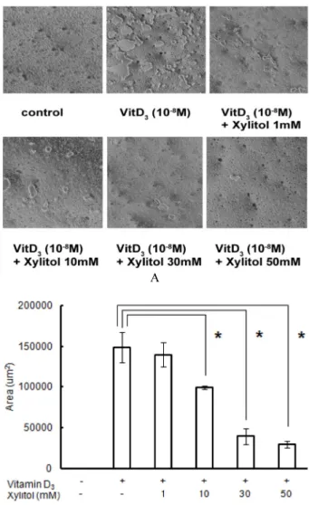
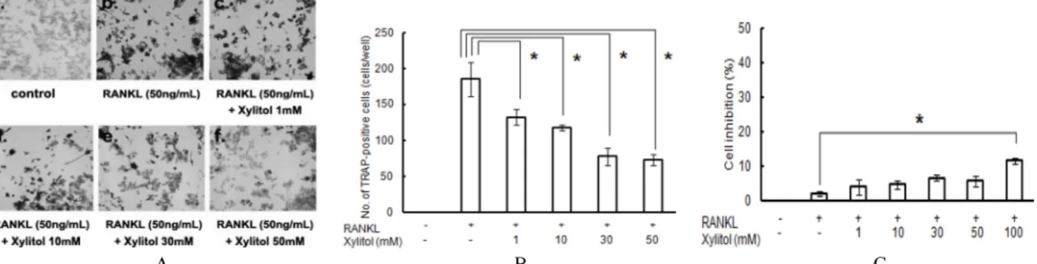
Xylitol Down-Regulates 1 α,25-Dihydroxy Vitamin D3-induced

Osteoclastogenesis via in Part the Inhibition of RANKL Expression in Osteoblasts

Seung-Ho Ohk

1,3,5

, Hyunjoo Jeong

1

, Jong-Pill Kim

2

, Yun-Jung Yoo

1,4

, Jeong-Taeg Seo

1,3,4

, Dong-Min Shin

1,4,♣

, and Syng-Ill Lee

1,3,4,

*

1

Department of Oral Biology,

2

Department of Pedodontics, and

3

Brain Korea 21 Project of Dental Sciences,

4

Oral Science Research Center, Yonsei University, Seoul 120-749,

5

Department of Oral Microbiology, Chonnam National University, Gwangju, 501-190, South Korea (received July 17, 2013 ; revised August 8, 2013 ; accepted August 10, 2013)

Xylitol is a sugar alcohol with a variety of functions including bactericidal and anticariogenic effects. However, the cellular mechanisms underlying the role of xylitol in bone metabolism are not yet clarified. In our present study, we exploited the physiological role of xylitol on osteoclast dif- ferentiation in a co-culture system of osteoblastic and RAW 264.7 cells. Xylitol treatment of these co-cultures reduced the number of tartrate-resistant acid phosphatase (TRAP)- positive multinucleated cells induced by 10 nM 1 α,25(OH)

2

D

3

in a dose ‐dependent manner. A cell viability test revea- led no marked cellular damage by up to 100 mM of xylitol.

Exposure of osteoblastic cells to xylitol decreased RANKL, but not OPG, mRNA expression in the presence of 10

-8

M 1 α,25(OH)

2

D

3

in a dose ‐dependent manner. Furthermore, bone resorption activity, assessed on bone slices in the co- culture system, was found to be dramatically decreased with increasing xylitol concentrations. RANKL and OPG proteins were assayed by ELISA and the soluble RANKL (sRANKL)

concentration was decreased with an increased xylitol con- centration. In contrast, OPG was unaltered by any xylitol con- centration in this assay. These results indicate that xylitol inhibits 1 α,25(OH)

2

D

3

-induced osteoclastogenesis by reducing the sRANKL/OPG expression ratio in osteoblastic cells.

Key words: xylitol, bone resorption, osteoclastogenesis, RANKL

Introduction

It has been reported that xylitol has a variety of function on cells, such as bactericidal, and anticariogenic effects [1].

Xylitol is a five-carbon natural polyhydric alcohol, which is widely distributed in fruits, berries, and plants. The natural dietary carbohydrate xylitol has been used as a source of energy in infusion therapy and found to act curatively in cer- tain clinical situations. Although this sugar alcohol cannot be metabolized, it is taken up by Streptococcus mutans (S.

mutans) and accumulated as a toxic sugar-phosphate in bacterial cells, resulting in growth inhibition. In addition, xylitol has an anticariogenic effects by inhibiting the glu- cosyl transferase (GTF) activity [2] which mediates a sucrose- dependant adherence of mutans Streptococci to the tooth surface. Besides the bactericidal effect of xylitol, a conti-

127

(2)

nuous moderate dietary xylitol supplementation leads to inc- reased bone volume and increased bone mineral content in the long bones of aged rats [3-7]. In spite of the extensive xy- litol research, no experimental evidence for the cellular mec- hanism of xylitol on bone metabolism has been suggested.

In general, bone remodeling is regulated by the activity of bone-forming osteoblasts and bone-resorbing osteoclasts.

Both osteoblasts and osteoclasts are regulated by a variety of hormones and local factors [8-12]. Osteoblasts stem from me- senchymal stem cells, whereas osteoclasts arise by the diffe- rentiation of osteoclast precursors of monocyte/macrophage lineage. Osteoblasts and osteoclasts are required not only for skeletal development, but also for mineral homeostasis and the normal remodeling of bone in adult [13]. An imbalance between bone formation and bone resorption derived from in appropriate RANKL (receptor activator of NF- κB ligand) expression by activated lymphocytes and osteoclasts causes metabolic bone diseases like osteopetrosis and osteoporosis [14,15]. Therefore, osteoblasts and osteoclasts are known to be closely related during the process of remodeling [16-18].

Certain kinds of signaling molecules, such as, RANKL, osteo- protegerin (OPG) and macrophage colony stimulating factor (M-CSF), expressed by osteoblasts, are involved in osteoclas- togenesis. For instance, when osteoblasts/stromal cells are sti- mulated by osteotropic factors such as parathyroid hormone, RANKL is expressed and induces the differentiation of osteo- clast progenitors by binding to the receptor activator of NF- κB (RANK; also known as ODF receptor) [19]. In addition, M- CSF is known to be essential for macrophages to be trans- formed into osteoclasts, while OPG, a decoy receptor of RANKL, is known to participate in the regulation of osteocla- stogenesis [11]. Specifically, OPG, as a member of the tumor necrosis factor receptor (TNFR) family, inhibits the osteo- clastogenesis stimulated by 1 α,25(OH)

2

D

3

, PTH, or IL-11 [17].

Consequently, it is believed that RANKL, M-CSF and OPG, which are expressed by osteoblasts, are associated with osteo- clastogenesis, and that osteoblasts play a major role in the bone remodeling process.

Murine RANKL is a 45 kDa, type II transmembrane glycoprotein with 316 amino acids that exists naturally as non-disulfide-linked homotrimer [17]. The molecule has a cytoplasmic domain with 47 amino acids, a transmembrane segment with 23 amino acids, and a extracellular region with 246 amino acids [20]. Soluble RANKL residue with 177 amino acids is generated by the action of metaloprotease clea-

vage on membrane-bound RANKL. Although both membrane and soluble RANKL are bioactive, the homeostatic form of RANKL might be the membrane-bound form [9], while soluble RANKL might signal underlying pathology [21]. Cells known to express RANKL include odontoblasts and ameloblasts [22,23], osteoblasts, T cell, chrondrocytes, fibroblasts, and skeletal muscle cells [24]. Murine RANKL is active on human cells and shows 85% and 96% amino acid homology to human and rat RANKL, respectively [11]. RANKL binds and signals via a membrane-bound TNF receptor super family member named TRANCE/RANK. RANKL also blinds a na- turally occurring 55 kDa soluble receptor antagonist named osteoprotegerin [17].

With respect to osteoclastogenesis, we have focused on the function of xylitol on the osteoblast and osteoclast. Apart from some knowledge of the general functions of xylitol in a whole body as well as bone density and mass, there is no experimental evidence as to whether xylitol is related to osteo- clastogenesis at the cellular level. Understanding of xylitol on bone metabolism at the molecular level of osteoblast and osteoclast is necessary. Therefore, we hypothesized that xy- litol might concern osteoclastogenesis and bone metabolism with respect to RANKL, OPG, on the osteoblast. Furthermore xylitol might affect osteoclast directly also. To clarify whether xylitol can affect the osteoclastogenesis induced by 1 α,25 (OH)

2

D

3

, we have applied xylitol on an osteoblast/stromal cell co-culture system and RAW 264.7 cells. We have examined the osteoclast differentiation rate and bone resorption activity on the bone slice with xylitol in co-culture system. Also we have compared not only the expression of RANKL and OPG mRNA but also the production of sRANKL and OPG protein.

Materials and Methods

Materials

Routine cell culture media were obtained from GIBCO/

BRL (Grand Island, NY). The Tartrate-Resistant Acid Phosp-

hatase Staining Kit was purchased from the Sigma Chemical

Co., Ltd. (St. Louis, MO). Trizol was purchased from Invi-

trogen Corp. (Carlsbad, CA), and the ICR mice were from

Samtacho Co., Ltd. (Seoul, Korea). Xylitol was purchased from

Borak Corp. (Seoul, Korea). All other chemicals were of the

highest grade commercially available. Recombinant murine

sRANKL was purchased from KOMA Biotech (Seoul, Korea).

(3)

In vitro osteoclast formation assay

The osteoblast formation assay was carried out as prev- iously reported by [25]. Briefly, the osteoblasts were iso- lated from 1 - 2 day-old newborn mice. 30 - 50 calvariae were digested in 10 ml of an enzyme solution containing 0.2% collagenase (Wako, Japan) and 0.1% dispase (GIBCO/

BRL, U.S.A) for 20 minutes at 37

o

C in a shaking water bath.

The supernatant was discarded and 10 ml of the enzyme solution was added. After shaking at 37

o

C for 20 minutes, the supernatant was collected carefully and transferred to a new tube. This digestion of calvariae by collagenase and dispase was repeated three times. The collected supernatant (30 ml) was placed in a centrifuge at 1,500 ×g for 10 minutes, to collect the osteoblastic cells. Cells were resuspended in α-minimum essential medium ( α-MEM) containing 10% fetal bovine serum (FBS) and cultured to confluence in 100 mm culture dishes at a concentration of 1 × 10

5

cells/dish. The cells were then detached from the culture dishes using trypsin-EDTA, sus- pended in α-MEM with 10% FBS and used for the co- culture as osteoblastic cells.

Femoral and tibial bone marrow cells were collected from 4-week-old mice. The tibiae and femora were removed and dissected free of adhering tissues. The bone ends were re- moved and the marrow cavities were flushed by slowly in- jecting media at one end using a 25-gauge needle. The cal- variae and bone marrow cells collected were washed and used in the co-culture. Mouse calvarial cells (1 × 10

4

cells/well) were co-cultured with bone marrow cells (1 × 10

5

cells/well) in α-MEM containing 10% FBS in 48-well plates (Corning Inc., Corning, NY). The culture volume was made up to 400 μ l per well with α-MEM supplemented with 10% FBS, in the presence of 1 α,25(OH)

2

D

3

(10

-8

M), without or with xylitol (1, 10, 30, 50 or 100 mM). All cultures were maintained at 37

o

C in a humidified atmosphere containing 5% CO

2

in at- mosphere. After incubation for 4 days, the cells were sub- jected to tartrate-resistant acid phosphatase (TRAP, an osteo- clast marker enzyme) staining. In vitro formation assay of osteoclast was repeated four times.

Viability test

The MTT (3-4,5-dimethlthiazol-2-yl-)-2,5-diphenyltetrazolium bromide) test is based on the principle that tetrazolium salts are reduced by reducing mitochondrial enzymes (succinate, dehydrogenase), which allows the toxicity of viable cells and the level of cellular differentiation to be measured. MTT was

dissolved in phosphate-buffered saline (PBS) at 5 mg/ml and filtered to remove any insoluble residue. MTT solution was added directly to the assay plates. The cells were sub- sequently incubated for an additional 4 hours at 37

o

C. The purple formazan crystals that formed were dissolved in DMSO, and the plates were read on a spectrophotometer at 570 nm.

Bone resorption activity assay (Pit formation assay)

Osteoblastic cells obtained from the calvariae of newborn ICR mouse and bone marrow cells obtained from the tibiae and femora of male ICR mouse were co-cultured in α-MEM in calcium phosphate apatite-coated 24-well plate, (OAAS plate, Oscotec Inc., Korea) at 2 × 10

5

cells/0.8 ml/well and 2 × 10

6

cells/0.8 ml/well, respectively. The cells were cultured for 4 days at 37°C in a humidified 5% CO

2

atmosphere. Then the cells were treated with 10 nM 1 α,25(OH)

2

D

3

and xylitol with different concentrations. Cultures were maintained for 4 days. The medium in each well was replaced with the respec- tive fresh medium with 1 α,25(OH)

2

D

3

(10

-8

M) and xylitol. The experiments were performed four times. At the end of cul- ture, attached cells were removed from the plate by abrasion with 4% sodium hypochloride solution (Sigma, St. Louis, MO).

Images of pit were acquired with a digital camera attached to a microscope at x100 magnification, and total areas of resorption pits were analyzed by the Meta Morph program (Molecular Devices, LLC., CA).

Reverse Transcriptase-PCR

The expressions of RANKL, OPG, and β-actin were eva- luated by RT-PCR using total RNA isolated from murine osteoblastic cells. Total RNA was isolated using Trizol reagent.

The primers used were : for RANKL (750 bp), 5'-ATCAG- AAGACAGCACTCACT-3' (forward), 5'-ATCTAGGACATCCA- TGCTAATGTTC-3' (reverse); for OPG (636 bp), 5'-TGAG TGTGAGGAAGGGCGTTA C-3' (forward), 5'-TTCCTCG- TTCTCTCAATCTC-3' (reverse) and for β-actin (366 bp), 5'-GGACTCCTATGGTGGGTGACGAGG-3' (forward), and 5'-GGGAGAGCATAGCCCTCGTAGAT-3' (reverse).

Relative RT-PCR was performed to measure gene exp-

ression of RANKL, OPG, and β-actin mRNAs. Polymerase

chain reactions were performed on a T gradient 96 PCR

machine (Biometra Co., Gottingen, Germany) using 1~2 ng

of cDNA, 5 pmoles of each oligonucleotide primer, 200 μM

of each dNTP, 1 unit of Taq Polymerase (Applierd Biosy-

stems, CA, USA) and 10 x Taq polymerase buffer in a 50 μl vo-

(4)

lume. The PCR program initially started with a 95

o

C dena- turation for 5 min, followed by 25 to 35 cycles of 95

o

C/1 min, T

a

/1 min, 72

o

C/1 min (T

a

, annealing temperature; 45.3

o

C for RANKL, 47.9

o

C for OPG, and 58

o

C for β-actin). Linear amplification range for each gene was tested on the adjusted cDNA. The less expressed transcripts of RANKL and OPG required 35 cycles of PCR for detection. For β-actin, 25 cycles of PCR was performed, respectively. Densitometry values were measured at each cycle sampling using the TINA software (University of Manchester, Manchester. U.K.).

RT-PCR values are presented as a ratio of the specified gene's signal in the selected linear amplification cycle divided by the β- actin positive control signal.

ELISA

Quantikine

M murine Mouse RANK Ligand kit (R & D systems Inc., Minneapolis, IN) was used to analyze RANKL protein. Briefly, mRANKL standard was diluted in Calibrator Diluent RD6-12 solution to make final concentration of 0, 31.2, 62.5, 125, 250, 500, 1000, and 2000 pg/ml. Assay Diluent RD1W and standards (50 μl each) were added to each well and incubated for 2 hours at room temperature.

Each well was aspirated and washed, repeating the process four times for a total of five washes. mRANKL conjugate (100 μl) was added to each well and incubated for 2 hours at room temperature. Washing was repeated as described above.

Substrate solution (100 μl) was added to each well and incubated for 30 minutes at room temperature in dark room.

Stop Solution (100 μl) was added to each well and mixed by gentle tapping. Then the enzyme reaction yields a blue product that turns yellow. The intensity of the color of each well was determined within 30 minutes, using a microplate reader at 450 nm.

Data analysis and statistics

The results are expressed as the mean ± S.E.M. The statis- tical significances of differences between the groups were deter- mined using the one-way ANOVA test. In statistical tests, the p value < 0.05 was considered to be significant.

Results

Xylitol inhibits 1 α,25(OH)

2

D

3

-induced osteoclast formation Osteoclastogenesis was induced by 1 α,25(OH)

2

D

3

in

osteoblastic cells/bone marrow co-culture. To clarify the role of xylitol on bone metabolism, 1, 10, 30, 50, or 100 mM of xylitol were added to co-cultures and incubated at 37

o

C for 4 days. When 10 nM of 1 α,25(OH)

2

D

3

was added to the co-culture TRAP positive multinucleated cells were formed, whereas no TRAP positive cells were detected in media only. In the presence of xylitol, 1 α,25(OH)

2

D

3

-indu- ced osteoclast differentiation was reduced (Fig. 1A). The addition of 1, 10, 30, 50 or 100 mM of xylitol reduced the number of TRAP positive multinucleated cells up to about 35% in 50 mM of xylitol (Fig. 1B). However, it might be possible that xylitol could cause cell damage directly without interrupting the normal maturation of osteoclasts. To con- firm possibility, we have carried out a viability test. As shown in Fig. 1C, xylitol did not show any remarkable toxic effect when treated with xylitol at up to 50 mM. These results suggest that the effect of xylitol on bone metabolism was not caused by its direct toxic effect upon the cells.

Bone resorption activity assay (pit formation assay)

We have measured resorbed bone lacuna and sum up on each bone slice. The addition of 10 mM of 1 α,25(OH)

2

D

3

effectively caused the formation of lacuna on bone slices whereas no resorption lacuna has been observed without 1 α,25(OH)

2

D

3

-induction. However, the resorbed area was gradually decreased as the concentration of xylitol was increased up to 50 mM (Fig. 2A). At 50 mM of xylitol about 80% of bone resorption area has been decreased. The ave- rage areas of resorption pit were measured and depicted in Fig. 2B. This indicated that xylitol might be effective on osteo- clast activation and function.

Xylitol caused changes in mRNA expression of RANKL As shown in Fig. 3, the expressions of RANKL and OPG mRNA in osteoblasts were monitored by RT-PCR in the presence or absence of xylitol. As the xylitol concentration in the co-culture medium was increased, the 1 α,25(OH)

2

D

3

- induced expression of RANKL mRNA was decreased (Fig.

3A). RANKL mRNA expression in osteoblasts was inversely

proportional to xylitol concentration. On the other hand, the

expression of OPG mRNA was not changed regardless of

xylitol concentration. These findings indicate that xylitol inhibits

osteoclast differentiation by down-regulating the expression

of RANKL. The ratio of RANKL to OPG mRNA in osteo-

blast is illustrated in Fig. 3B. As the xylitol concentration was

(5)

A

B

C

Fig. 1. Inhibition of osteoclast differentiation by xylitol. (A), In the presence of xylitol, 1 α,25(OH)

2

D

3

-induced osteoclast differen- tiation was reduced (×200). (B), TRAP-positive multinucleated cells containing three or more nuclei were counted as osteo- clasts. (C), MTT test. The statistical significance of differences bet- ween the groups was determined using the one-way ANOVA test. (*), In all statistical tests, a p value < 0.05 was considered to be statistically significant. Each data was shown in mean ± SEM of four cultures.

A

B

Fig. 2. Effects of xylitol treatment on resorption pit formation.

(A), Resorbed lacuna on the OAAS plates were photographed the microscope (x100). (B), Total resorption area per well measured by image analyzer and graphed. (*), A p value <

0.05 was considered to be statistically significant.

increased, the ratio of RANKL to OPG mRNA decreased, which means RANKL and OPG, which are closely linked to osteoclastogenesis.

In addition, RANKL and OPG proteins were also analy- zed with ELISA using anti-RANKL antibody (Fig. 4A).

RANKL protein was decreased with the increase of xylitol

concentration. However, the addition of xylitol did not

change the amount of OPG protein which is consistent with

OPG mRNA data (Fig. 4B). Consequently, xylitol inhibited

1 α,25(OH)

2

D

3

-induced RANKL mRNA and protein, and led

to alter osteoclastogenesis. In addition, such changes of

signaling molecules were dependent on the xylitol

concentration.

(6)

A B C

Fig. 5. Inhibition of sRANKL-induced RAW 264.7 cell differentiation by xylitol. (A), RANKL-induced osteoclast differentiation was reduced in the presence of xylitol (×100). (B), TRAP-positive multinucleated cells containing three or more nuclei were counted. (C), MTT test. (*), In all statistical tests, a p value < 0.05 was considered to be statistically significant. Each data was shown in mean ± SEM of four cultures.

A

Fig. 3. Effects of xylitol on mRNA expression of RANKL and B

OPG in osteoblastic cells. (A), Various concentrations of xylitol were added to the mouse calvarial osteoblast culture with 10 nM of 1 α,25(OH)

2

D

3

. After incubation for 4 days, total RNA was then extracted from osteoblasts, and the expressions of RANKL and OPG mRNAs were analyzed by RT-PCR products. (B), The expression of RANKL mRNA compared with OPG mRNA. The results were expressed as the means ± SEM of four experiments. (*), A p value < 0.05 was considered to be statistically significant.

Xylitol inhibits RANKL-induced osteoclastogenesis.

To clarify the role of xylitol on bone metabolism, 1, 10, 30, 50, or 100 mM of xylitol were added to cultures and incu-

bated at 37

o

C for 6 days to investigate osteoclast differen- tiation. Osteoclastogenesis was induced by RANKL in RAW

A

Fig. 4. Expression of RANKL and OPG protein in mouse cal- B

varial osteoblastic cells. (A), Protein analysis using ELISA showed

that xylitol inhibited the expression of sRANKL. (B), OPG level

were slightly increased in osteoblasts stimulated by xylitol, but

it was not statistically significant. All data were expressed as

the means ± SEM of four experiments.

(7)

264.7 cell culture. When 50 ng/ml of RANKL was added to the RAW 264.7 cell culture, TRAP positive multinucleated cells were formed whereas no TRAP positive cells were detected in media only. In the presence of xylitol, RANKL- induced osteoclast differentiation was reduced (Fig. 5A).

The addition of 1, 10, 30, 50 or 100 mM of xylitol reduced the number of TRAP positive multinucleated cells (Fig. 5B).

However, it might be possible that xylitol causes cell damage directly without interrupting the normal maturation of osteo- clasts. To confirm this possibility, we have carried out a via- bility test. As shown in Fig. 5C, xylitol did not show any toxic effect at up to 100 mM.

Discussion

In this study, the effects of xylitol on osteoclastogenesis in osteoblast-osteoclast co-culture system and differentiation of RAW264.7 into osteoclast-like cells were investigated. As mentioned earlier, we found xylitol affects the bone meta- bolism, leading to the changes in 1 α,25(OH)

2

D

3

-induced osteo- clastogenesis. Interestingly, xylitol inhibited the 1 α,25(OH)

2

D

3

- induced osteoclastogenesis (by up to 65 % of the control) in co-culture system (Fig. 1). Although xylitol inhibited 1 α,25 (OH)

2

D

3

-induced osteoclastogenesis, it could be argued that such an inhibition of osteoclastogenesis might not due to the physiological intervention of xylitol in the normal process of osteoclastogenesis, but the cell damage non-physiologi- cally. To rule out the possibility that xylitol might cause non- physiological cell damage we have performed the MTT test.

The test showed that xylitol did not exert any harmful effect upon the cells in this co-culture system, which suggests that xylitol inhibits the formation of TRAP positive cells, without a toxic effect upon the cells.

Here, we raised the question about how xylitol triggers the down-regulation of 1 α,25(OH)

2

D

3

-induced osteoclasto- genesis. Since osteoclast differentiation is mediated by critical signal molecules, such as RANKL, OPG and M-CSF [8-11], we used an osteoblast/stromal cell co-culture system to evaluate whether xylitol alter the 1 α,25(OH)

2

D

3

-induced osteoclast differentiation in terms of the expression profiles of RANKL and OPG mRNA. The expression of RANKL in osteoblastic cells by the treatment of 1 α,25(OH)

2

D

3

was down-regulated upon increasing the xylitol concentration, and the expression of OPG mRNA was not changed signi-

ficantly (Fig. 3). In addition, the expression of sRANKL was decreased with xylitol concentration in the process of 1 α,25(OH)

2

D

3

-induced osteoclastogenesis, being in consis- tency with the decrease in RANKL mRNA expression (Fig.

4A). On the other hand, OPG protein was slightly increased with xylitol treatment but it was not statistically significant (Fig. 4B). From the findings described above, such an inhi- bitory mechanism of osteoclastogenesis by xylitol might be associated with modulating RANKL, not OPG expression in osteoblasts. Previous study showed that Bumethnide, NaKCl cotransmitter inhibitor, reduced expression of RANKL via cell volume shrinkage of osteoblast due to the hyperos- molarity [26]. Molarity of xylitol under 50 mM used in this study may not be considered enough to explain effect of xylitol. Since, it might be insufficient to cause the hyperos- motic shrinkage. Then, probability which we can speculate is direct effect on inner cell structure or physiologic process, which is possible through membrane transportation as in S.

mutans. If osteoblast and osteoclast have transporter like phosphorylation transferase system of fructose (PTS-Fru) or similar one, absorbed xylitol-phosphate cannot be metaboli- zed and it may have toxic effect on bone cells like in S.

mutans. However, understanding of the exact nature of down- regulation of osteoclastogenesis by xylitol requires further studies at the level of osteoblast and osteoclast.

In summary, we have provided the first evidence that xylitol inhibit not only RANKL protein synthesis in osteoblastic cells but also osteoclast function (bone resorption activity) in 1 α,25(OH)

2

D

3

-induced osteoclastogenesis. Xylitol down- regulated osteoclastogenesis in 1 α,25(OH)

2

D

3

-induced osteoc- lastogensis in co-culture system via reduction of RANKL mRNA expression and sRANKL synthesis.

Acknowledgement

This study was supported by a faculty research grant of Yonsei University College of Dentistry for 2011(6-2011-0029).

References

1. Kitchens DH. Xylitol in the prevention of oral diseases.

Spec Care Dentist. 2005;25(3):140-4.

2. Wunder D, Bowen WH. Action of agents on glucosy-

ltransferases from Streptococcus mutans in solution and

adsorbed to experimental pellicle. Arch Oral Biol. 1999;

(8)

44(3):203-14.

3. Svanberg M, Hietala EL, Knuuttila M. The effect of die- tary xylitol on dentin formation in ovariectomized rats.

Acta Odontol Scand. 1994;52(2):82-5.

4. Svanberg M, Knuuttila M. The effect of dietary xylitol on recalcifying and newly formed cortical long bone in rats.

Calcif Tissue Int. 1993;53(2):135-8.

5. Svanberg M, Knuuttila M. Dietary xylitol prevents ovariec- tomy induced changes of bone inorganic fraction in rats.

Bone Miner. 1994;26(1):81-8.

6. Svanberg M, Knuuttila M. Dietary xylitol retards bone re- sorption in rats. Miner Electrolyte Metab. 1994;20(3):

153-7.

7. Svanberg M, Mattila P, Knuuttila M. Dietary xylitol retards the ovariectomy-induced increase of bone turnover in rats.

Calcif Tissue Int. 1997;60(5):462-6.

8. Burgess TL, Qian Y, Kaufman S, Ring BD, Van G, Cap- parelli C, et al. The ligand for osteoprotegerin (OPGL) di- rectly activates mature osteoclasts. J Cell Biol. 1999;

145(3):527-38.

9. Fuller K, Wong B, Fox S, Choi Y, Chambers TJ. TRANCE is necessary and sufficient for osteoblast-mediated acti- vation of bone resorption in osteoclasts. J Exp Med. 1998;

188(5):997-1001.

10. Kong YY, Yoshida H, Sarosi I, Tan HL, Timms E, Cap- parelli C, et al. OPGL is a key regulator of osteoclas- togenesis, lymphocyte development and lymph-node orga- nogenesis. Nature. 1999;397(6717):315-23.

11. Lacey DL, Timms E, Tan HL, Kelley MJ, Dunstan CR, Burgess T, et al. Osteoprotegerin ligand is a cytokine that regulates osteoclast differentiation and activation. Cell. 1998;

93(2):165-76.

12. Jeong HJ, Tian Y, Kim BH, Nam MY, Lee HA, Yoo YJ, et al. Hypertonicity Down-regulates the 1 α,25(OH)

2

Vitamin D

3

-induced Osteoclastogenesis Via the Modulation of RANKL Expression in Osteoblast. Int J Oral Biol. 2005;30(1):

23-30.

13. Eriksen EF. Cellular mechanisms of bone remodeling. Rev Endocr Metab Disord. 2010;11(4):219-27.

14. Aubin JE. Advances in the osteoblast lineage. Biochem Cell Biol. 1998;76(6):899-910.

15. Kim MJ, Jun YJ, Yu HI, Yang SY, Oh YM, Kim SH, et al.

Altered Expression of RANKL/OPG after Alendronate Ad- ministration in the Developing Teeth of Postnatal Rats. Int J Oral Biol. 2011;36(1):37-42.

16. Suda T, Udagawa N, Nakamura I, Miyaura C, Takahashi N.

Modulation of osteoclast differentiation by local factors.

Bone. 1995;17(2 Suppl):87S-91S.

17. Takahashi N, Udagawa N, Suda T. A new member of tumor necrosis factor ligand family, ODF/OPGL/TRANCE/

RANKL, regulates osteoclast differentiation and function.

Biochem Biophys Res Commun. 1999;256(3):449-55.

18. Tsukii K, Shima N, Mochizuki S, Yamaguchi K, Kinosaki M, Yano K, et al. Osteoclast differentiation factor mediates an essential signal for bone resorption induced by 1 alpha, 25-dihydroxyvitamin D3, prostaglandin E2, or parathyroid hormone in the microenvironment of bone. Biochem Biop- hys Res Commun. 1998;246(2):337-41.

19. Jimi E, Akiyama S, Tsurukai T, Okahashi N, Kobayashi K, Udagawa N, et al. Osteoclast differentiation factor acts as a multifunctional regulator in murine osteoclast differentia- tion and function. J Immunol. 1999;163(1):434-42.

20. Wong BR, Rho J, Arron J, Robinson E, Orlinick J, Chao M, et al. TRANCE is a novel ligand of the tumor necrosis factor receptor family that activates c-Jun N-terminal ki- nase in T cells. J Biol Chem. 1997;272(40):25190-4.

21. Udagawa N, Takahashi N, Yasuda H, Mizuno A, Itoh K, Ueno Y, et al. Osteoprotegerin produced by osteoblasts is an important regulator in osteoclast development and function. Endocrinology. 2000;141(9):3478-84.

22. Rani CS, MacDougall M. Dental cells express factors that regulate bone resorption. Mol Cell Biol Res Commun.

2000;3(3):145-52.

23. Wise GE, Que BG, Huang H, Lumpkin SJ. Enhancement of gene expression in rat dental follicle cells by parathyroid hormone-related protein. Arch Oral Biol. 2000;45(10):903-9.

24. Kartsogiannis V, Zhou H, Horwood NJ, Thomas RJ, Hards DK, Quinn JM, et al. Localization of RANKL (receptor acti- vator of NF kappa B ligand) mRNA and protein in skeletal and extraskeletal tissues. Bone. 1999;25(5):525-34.

25. Choi BK, Ohk SH, Lee HJ, Kang JH, Jeong GJ, Yoo YJ.

Effects of whole cell sonicates of Treponema lecithinoly- ticum on osteoclast differentiation. J Periodontol. 2001;

72(9):1172-7.

26. Lee HA, Jeong H, Kim EY, Nam MY, Yoo YJ, Seo JT, et al.

Bumetanide, the specific inhibitor of Na+-K+-2Cl- cotran- sport, inhibits 1alpha,25-dihydroxyvitamin D3-induced osteo- clastogenesis in a mouse co-culture system. Exp Physiol.

2003;88(5):569-74.

수치

Fig. 1. Inhibition of osteoclast differentiation by xylitol. (A), In the  presence of xylitol, 1 α,25(OH) 2 D 3 -induced osteoclast  differen-tiation was reduced (×200)
Fig. 4. Expression of RANKL and OPG protein in mouse cal- B

참조

관련 문서

Recognizing that a S&amp;T Co–operation Fund of 10 million USD, contributed for the period 2011–15, has been utilized to implement joint research.. projects in the

- CERAGEM center owners, staffs and customers from Bihar state participated by donation to support daily necessities &amp; living expenses

Rule 6(1)에 따라 원칙적으로 매입 재료 또는 서비스가 면세 재화의 제조 또는 면세 서비스의 공급에 사용될 경우 Cenvat Credit를 이용할 수 없다. 따라서

[r]

11-1 not only bibimbap, but also pizza 11-2 not only ate Korean food, but also met Korean people 11-3 (1) in not only going to the photo museum, but also taking photos

Ross: As my lawfully wedded wife, in sickness and in health, until

□ Heat is defined as transferred energy dew to temperature differences through boundary.. - Conduction by molecular

다양한 번역 작품과 번역에 관한 책을 읽는 것은 단순히 다른 시대와 언어, 문화의 교류를 넘어 지구촌이 서로 이해하고 하나가