내분비계 장애물질이 어류의 HPG, HPT, HPA 축에 미치는 연계영향
장솔*·지경희*, **†
*용인대학교 일반대학원 환경보건학과
**용인대학교 환경과학대학 산업환경보건학과
A Review on the Effects of Endocrine Disruptors on the Interaction between HPG, HPT, and HPA Axes in Fish
Sol Jang* and Kyunghee Ji*,**†
*Department of Environmental Health, Graduate School of Yongin University
**Department of Occupational and Environmental Health, College of Environmental Sciences, Yongin University
ABSTRACT
Objectives: The objective of this review was to summarize the primary role of three representative endocrine axes in aquatic vertebrates and discuss the effects on endocrine systems and their interactions in teleost fish after exposure to environmental contaminants.
Methods: We summarized individual traits and mechanisms for hormonal and transcriptional interactions between the hypothalamic-pituitary-gonad (HPG), hypothalamic-pituitary-thyroid (HPT), and hypothalamic- pituitary-adrenal (HPA) axes in fish. We also provided a brief discussion on the effects of nonylphenol-induced toxicity on endocrine systems and their interactions in fish as a demonstration of holistic explanation.
Results: Currently-available data showed that thyroid dysfunction is associated with reproductive toxicity due to changes in steroidogenic gene expressions and sex hormone levels as well as gonad glands in fish. As an example, we demonstrated that exposure to nonylphenol could induce estrogenicity in male fish by decreasing thyroid hormones, which contributes to increased aromatase expression. Although the mechanisms are complicated and involved in multiple ways, a number of studies have shown that sex steroids influence the HPT axis or the HPA axis in fish, indicating bi-directional crosstalk. Critically missing is information on the primary target or toxicity mechanisms of environmental contaminants among the three endocrine axes, so further studies are needed to explore those possibilities.
Conclusions: This review highlights the interactions between the HPG, HPT, and HPA axes in fish in order to better understand how these endocrine systems could interact with each other in situations of exposure to endocrine disrupting chemicals.
Key words: Interaction, endocrine disruption, HPG axis, HPT axis, HPA axis
I. 서 론
수서척추동물인 경골어류(teleost fish)는 육상척추
동물과 마찬가지로 생식선, 갑상선, 부신 내분비계가 각각 시상하부-뇌하수체-생식선 축(hypothalamus- pituitary-gonad axis; HPG axis), 시상하부-뇌하수체
†
Corresponding author: Department of Occupational and Environmental Health, College of Environmental Sciences, Yongin University, Yongin, Gyeonggi, 449-714, Republic of Korea, Tel: +82-31-8020-2747, Fax: +82-31-8020-2886, E- mail: kyungheeji@yongin.ac.kr
Received: 4 May 2015, Revised: 24 June 2015, Accepted: 26 June 2015
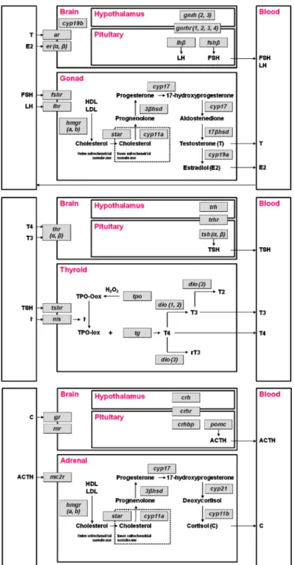
초빙논문 Invited reviewFig. 1. The pathways of hypothalamic-pituitary-gonad (HPG), hypothalamic-pituitary-thyroid (HPT), and hypothalamic-
pituitary-adrenal (HPA) axes in fish. Abbreviations of genes and hormones are shown as italics and bold character,
respectively. Gene acronyms are defined in Table 1.
-갑상선 축(hypothalamus-pituitary-thyroid axis; HPT axis), 시상하부-뇌하수체-부신 축(hypothalamus- pituitary-adrenal axis; HPA axis)에 의해 호르몬 분비 가 조절된다.1) 이러한 내분비계에서 합성·분비되는 호르몬은 어류의 생식, 성장, 발달, 대사 등에 중요한 역할을 한다. 각각의 내분비계 축은 서로 상호작용하 고 있어 하나의 내분비계 축이 영향을 받으면 다른 내분비계 축도 함께 영향을 받을 수 있다.1-3)척추동 물은 서식지(육상 또는 물)에 관계없이 비슷한 내분 비계 축을 공유하고 있으며,4) 환경오염물질로 인한 수 서척추동물의 영향을 다각적으로 연구하는 것은 전일 적(holistic)인 관점에서 내분비계 영향을 이해하는 데 도움을 줄 수 있다. 본 연구에서는 노닐페놀을 포함 한 물환경 중 내분비계 장애물질에 노출되었을 때 경 골어류의 생식선, 갑상선, 부신 내분비계 축의 다양한 호르몬, 유전자 발현에 미치는 영향과 축의 상호작용 에 초점을 맞추어 논문들을 종합적으로 정리하고자 하며, 앞으로의 연구방향에 대해 제안하고자 한다.
II. 내분비계의 역할
수서척추동물의 HPG, HPT, HPA 축의 중요한 호 르몬과 유전자 발현 경로를 Fig. 1에 도식화하였으 며, 유전자의 약어를 Table 1에 제시하였다. 물고기 도 다른 육상척추동물과 같이 내분비계 축의 중요한 호르몬 수준이 증가하면 음성 되먹임(negative feedback) 과정을 통해 시상하부와 뇌하수체에서 발 현되는 자극호르몬 수준을 감소시켜 항상성 (homeostasis)을 유지한다.
1. HPG 축의 역할
수서척추동물의 생식세포 발달과 번식은 HPG 축 의 다양한 스테로이드 호르몬과 비텔로제닌 합성을 위한 간의 상호작용에 의해 조절된다. HPG 축의 정 점에 있는 시상하부에서는 성선자극호르몬방출호르 몬(gonadotropin releasing hormone; GnRH)이 합성·
분비되고 있으며, 이 신경호르몬은 10개의 아미노산 이 연결된 중합체(decapeptide)로 구성되어 있다.
GnRH는 모든 척추동물에서 발현되고 있으며, 생물 군(taxa)에 따라 여러 가지 형태를 지니고 있다.5)예 를 들어, 대부분의 포유류는 2가지 형태의 GnRH를 보유하고 있으나, 경골어류에서는 뇌의 서로 다른 부
위에서 2개 또는 3개의 GnRH가 개별적인 기능을 담당하고 있다.6) GnRH3 ({Trp7Leu8}-GnRH)는 어 류에서만 발견되며, GnRH2 ({His5Trp7Tyr8}-GnRH) 는 사람을 포함한 모든 척추동물에서 발견된다.4)
Table 1. Gene acronyms of HPG, HPG, and HPA axes in
fish
Abbreviation Gene name
gnrh Gonadotropin-releasing hormone gnrhr Gonadotropin-releasing hormone receptor fsh β Follicle stimulating hormone β
lh β Luteinizing hormone β cyp19b Cytochrome P450 19B er Estrogen receptor ar Androgen receptor
fshr Follicle stimulating hormone receptor lhr Luteinizing hormone receptor hmgr Hydroxymethylglutaryl CoA reductase star Steroidogenic acute regulatory protein cyp11a Cytochrome P450 side-chain cleavage 3βhsd 3β-hydroxysteroid dehydrogenase cyp17 Cytochrome P450 17
17 βhsd 17 β-hydroxysteroid dehydrogenase cyp19a Cytochrome P450 19A
trh Thyrotropin-releasing hormone trhr Thyrotropin-releasing hormone receptor tsh Thyroid stimulating hormone
thr Thyroid hormone receptor
tshr Thyroid stimulating hormone receptor nis Sodium iodide symporter
tpo Thyroperoxidase tg Thyroglobulin
dio Deiodinase
crh Corticotropin-releasing hormone crhr Corticotropin-releasing hormone receptor crhbp Corticotropin-releasing hormone binding
protein
pomc Proopiomelanocortin
gr Glucocorticoid receptor
mr Mineralocorticoid receptor
mc2r Melanocortin 2 receptor
cyp21 Cytochrome P450 21
cyp11b Cytochrome P450 11B
GnRH는 수용체(GnRH receptor; GnRHR)에 결합 하여 뇌하수체에서 성선자극호르몬(gonadotropin hormone, GH)을 분비할 수 있도록 한다.
GH인 난포자극호르몬(follicle stimulating hormone;
FSH)과 황체형성호르몬(luteinizing hormone; LH)은 각각 난포자극호르몬 수용체(FSH receptor; FSHR) 와 황체형성호르몬 수용체(LH receptor; LHR)에 결 합하여 스테로이드 호르몬의 생합성(steroidogenesis) 과 생식세포 형성(gametogenesis)에 영향을 준다. 수 컷 어류에서 FSH는 주로 정자가 생성되는 초기단계 에 정원세포(spermatogonia), 정모세포(spermatocyte), 정자세포(spermatid)에 이르는 생식세포가 성숙·분 화할 수 있도록 조절하며, LH는 레이디히 세포(Leydig cell)에서 테스토스테론(testosterone; T)을 생성하고 운동성이 있는 성숙된 정자(spermatozoa)를 방출하 는 데 중요한 역할을 한다.7) 암컷 어류에서 FSH는 주로 비텔로제닌(vitellogenin; VTG) 합성을 조절하 며, LH는 난자의 성숙과정을 조절한다.8)
성 스테로이드 호르몬인 T와 에스트로겐(17β- estradiol; E2)은 GH의 자극을 받아 생식기에서 분 비되며, 스테로이드 호르몬 생합성과 연관된 여러 효 소와 유전자들의 발현에 의해 합성이 조절된다. 스테 로이드급성조절단백질(Steroidogenic acute regulatory protein; StAR)은 신장의 앞쪽과 생식기에서 검출되 고 있으며,9)막 외부의 콜레스테롤을 내부로 이송하 는 역할을 한다. Cytochrome P450 (CYP) 11A 효 소는 스테로이드 호르몬 생합성 시 속도결정단계 (rate-limiting step)에서 촉매역할을 하며, 3β- hydroxysteroid dehydrogenase (3βHSD)는 성 스테 로이드 호르몬인 프로게스테론(progesterone; P) 형 성에 필수적인 효소이다.10) CYP17은 안드로겐 생합 성을 촉진시키며, 아로마타제(aromatase, CYP19)는 T 호르몬을 E2 호르몬으로 변환시켜주는 중요한 효 소이다.11) E2 호르몬은 에스트로겐 수용체(estrogen receptor; ER)와 결합하여 암컷 어류의 간에서 난황 단백 전구체인 VTG를 형성할 수 있도록 한다.12)
2. HPT 축의 역할
갑상선은 물고기의 발달, 성장, 대사에서 중요한 역할을 담당한다. HPT 축의 시상하부에서는 갑상선 자극호르몬방출호르몬(thyrotropin-releasing hormone;
TRH)을 방출하고, 이 호르몬은 수용체(TRH receptor;
TRHR)에 결합하여 갑상선자극호르몬(thyroid- stimulating hormone; TSH)을 뇌하수체에서 분비하 도록 한다. TSH는 갑상선호르몬(thyroid hormone;
TH) 합성과 분비를 조절하는 역할을 하며, 2개(TSHα, TSHβ)의 아단위(subunits)를 가지고 있다. 물고기에 서 TH는 갑상선호르몬수용체(TH receptor; THR)에 부착되어 호르몬 활동이 조절된다.13) 어류에서는 2 가지 종류의 THR (THRα, THRβ)이 존재한다고 알 려졌으며, 배아(embryo)-치어(larvae)-유어(juvenile fish)에 이르는 초기 성장 단계의 서로 다른 시기에 발현되어 발달과 성장을 촉진시킨다.14) THRα는 TH 의 목표조직에서 더 많이 발현되며, THRβ는 주로 시상하부와 뇌하수체에서 발현되어 HPT축을 조절하 는 역할을 한다.15)
수서척추동물에서 탈요오드화 과정(deiodination)은 TH의 활성을 조절하는 데 중요한 역할을 한다. 어 류에 존재하는 탈요오드화 효소(deiodinase; DIO)는 티록신(thyroxine, T4)을 생체 내에서 활성을 가지는 트리요오드티로닌(triiodothyronine; T3)으로 변환시 켜 주거나, 불활성을 띠는 대사체인 역트리요오드티 로닌(reverse T3; rT3) 또는 디요오드티로닌(3,3’- diiodothyronine; T2)으로 바꾸어주는 역할을 한다.16) 현재까지 물고기에서는 3가지 종류의 DIO 효소(type I, type II, type III)가 발견되었으며, DIO2는 T4 호 르몬을 T3 호르몬으로 변환시키는 역할을 한다.
갑상선글로불린(thyroglobulin; TG)은 T4 호르몬과 T3 호르몬을 합성하는 단백질이며,17)갑상선 과산화 효소(thyroid peroxidase; TPO)는 TG의 타이로신 잔 기(tyrosyl residues)를 요오드화하여 T4 호르몬을 형 성하도록 촉매작용을 한다.18) 나트륨-요오드 공동운 반체(sodium iodide symporter; NIS)는 갑상선 소포 세포(follicular cells) 안으로 요오드를 능동수송 시 켜주는 혈장 막 단백질이다.19)
3. HPA 축의 역할
물고기의 HPA 축은 스트레스에 대한 반응과 대사, 면역, 생식 시스템 등의 많은 신체 과정을 조절하는 역할을 한다. HPA 축의 시상하부에서는 부신피질자 극호르몬방출호르몬(corticotropin-releasing hormone;
CRH)이 방출되어 뇌하수체의 부신피질자극호르몬 (adrenocorticotropic hormone; ACTH)을 분비하도록 한다. 프로오피오멜라노코르틴(proopiomelanocortin;
POMC) 단백질은 ACTH 호르몬이 합성되는 데 관 여하며,20) ACTH는 멜라노코르틴 수용체(melanocortin 2 receptor; MC2R)와 결합하여 코티졸(cortisol; C) 을 합성하고 방출할 수 있도록 한다.
물고기의 부신/신간 조직(adrenal/interrenal tissue) 에서는 스테로이드 호르몬 생합성과 연관된 효소들 (StAR, CYP11A, CYP17, CYP19)이 일부 발현된다.
CYP11B는 11-데옥시코티솔(11-deoxycortisol)을 C 호르몬으로 변환하여 주는 효소로, C 호르몬 생합성 단계에서 마지막으로 작용하여 생산량을 조절하는 역할을 한다.21) 11β수산화스테로이드탈수소효소2(11β- hydroxysteroid dehydrogenase-2; 11βHSD2)는 C 호 르몬을 비활성 대사체인 코티손(cortisone)으로 산화 시키는 효소로, 물고기의 배아발달 시기에는 과도한 C 호르몬의 분비로 인한 성장 억제 및 세포 자멸사 유발(pro-apoptotic) 영향을 방지해 준다.21,22)
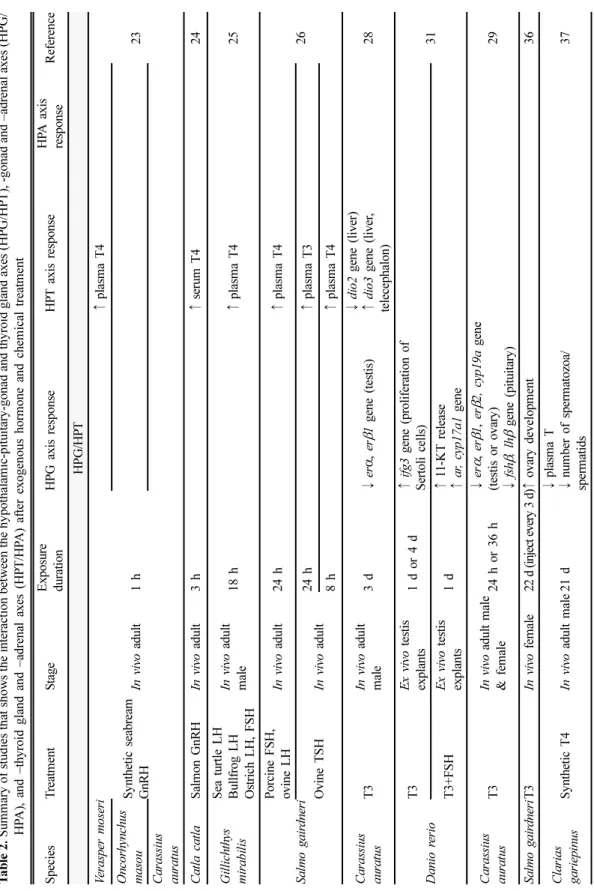
III. HPG, HPT, HPA 축의 상호작용 예전에는 생식기, 갑상선, 부신이 각각 고유의 분 리된 역할을 담당하고 있다고 생각하였으나, 최근 여 러 연구들에서 내분비계 축이 서로 상호작용하고 있 음이 제안되고 있다. 아래에는 경골어류의 HPG 축 과 HPT 축의 상호작용, HPG 축과 HPA 축의 상호 작용, HPT 축과 HPA 축의 상호작용을 설명하는 연 구들을 각각 정리하였다(Table 2, Fig. 2).
1. HPG, HPT axes
물고기의 HPG 축에 관련된 호르몬은 TH의 활성 에 영향을 미칠 수 있다. 예를 들어, 경골어류(노랑 가자미(barfin founder), 산천어(masu salmon), 금붕 어, 잉어(carps))에 외인성 GnRH를 노출하였을 때 혈중 T4 호르몬 수준이 증가되었다.23,24) GnRH는 직 접반응으로24)또는 뇌하수체의 TSH, GH 영향에 대 한 간접반응으로 혈중 T4 호르몬 수준에 영향을 미 칠 수 있다.25) 외인성 FSH, LH도 물고기의 혈장 T4 호르몬 수준을 증가시킬 수 있다.25,26) 이러한 연구 결과는 수서척추동물의 시상하부와 뇌하수체에서 분 비되는 생식 내분비계 호르몬이 TH 활성을 증가시 킴을 뒷받침한다.
물고기의 갑상선 기능이 항진(hyperthyroidism)되 거나 저하(hypothyroidism)되었을 경우 HPG 축의
유전자 발현과 호르몬 활성을 변화시켜 궁극적으로 번식에 영향을 미칠 수 있다.27) 예를 들어, 외인성 T3 호르몬에 노출된 금붕어(goldfish)에서는 뇌하수 체의 fshβ, lhβ 유전자와 생식기의 cyp19a, erα, erβ1, erβ2 유전자 발현이 감소되어 E2 호르몬의 합성과 분비가 저해되었다.28,29)외인성 T4 호르몬에 노출된 물고기(Clarias gariepinus)는 아로마타제 활성이 억 제되어 혈청 E2 호르몬의 농도가 감소되었다.30) T3 호르몬과 TSH가 함께 투여된 제브라피쉬 정소에서 는 생물학적 활성이 강한 안드로겐인 11-케토테스테 론(11-ketotestosterone; 11-KT) 호르몬 방출이 증가 되었고, androgen receptor (ar), cyp17a1 유전자 발 현도 증가되었다.31)일반적으로 외인성 T3, T4 호르 몬을 투여하였을 때에는 TH의 증가와 함께 여성 호 르몬(E2)의 감소와 남성 호르몬(11-KT)의 증가가 관 찰되었다.
이와 반대로 TH가 저하될 경우에는 여성 호르몬 의 증가와 남성 호르몬의 감소패턴이 관찰되었다. 예 를 들어, 갑상선 활성을 억제하는 티오요소(thiourea) 에 노출된 수컷 공기호흡메기(air-breathing catfish) 는 혈청 11-KT 호르몬과 T 호르몬의 농도가 줄어 들었으며,32) 남성 호르몬 생산에 관여하는 11βhsd 유 전자 발현도 감소하였다.33) 6-propyl-2-thiouracil은 TPO 활동을 방해하여 갑상선 활성을 억제하는 물질 로, 이 물질에 노출된 암컷 제브라피쉬에서는 혈장 T4, T3 호르몬의 감소와 함께 FSH, LH의 증가가 관찰되었다.1) TH의 음성 되먹임 작용은 뇌에서 분 비되는 GnRH, FSH, LH와 연관되어 있어 TH의 분 비가 줄어들 경우 번식에도 영향을 미칠 수 있다.34)
TH는 성분화(gonadal differentiation) 뿐만 아니 라 생식세포 성숙(gonadal maturation)에도 영향을 줄 수 있다. T3 호르몬을 수컷 fathead minnow (Pimephales promelas)에 10일간 투여하였을 때, 성 숙된 정자 생산이 증가되었다.35)또한 암컷 rainbow trout에서는 T3가 높아졌을 때 난소 성장이 증진되 었다.36) 일부 연구에서는 외인성 T4를 투여하였을 때 성숙된 정자와 정자세포의 수가 감소되었다고 보 고하였으나,37) 대부분의 경우에는 수컷화가 유발되 었다. 예를 들어, 갑상선 기능 저하를 유발하는 퍼 클로레이트(perchlorate)와 메티마졸(methimazole)에 노출된 제브라피쉬 치어는 정자 생산이 억제되었으 나, T4 호르몬과 함께 노출되었을 때에는 방향성이
J Environ Health Sci 2015; 41(3): 147-162 http://www.kseh.org/
Ta bl e 2 . Sum m ar y of st udi es t h at s hows t h e i n te ra ct ion be twe en t h e hy p o th al am ic -p it u it ar y -g o n ad an d thyro id gl and axe s (HPG/ H P T ), - gon ad an d – adr en al a x es (HPG/ HP A ), a nd –t hyro id gl and and –a dre n al a x es ( H P T /H P A ) aft er e xoge nous ho rm one and ch em ic al t re at m ent S p ecies T re at m en t S tag e E xpos ure du ra ti on H P G axis respo n se HP T axis re spons e HP A ax is resp onse Re fe re nc e HPG /HP T V erasper m o seri S y nthetic s eabream Gn RH In vivo adult 1 h
↑ pl as m a T4 23 O n co rh ynchu s ma so u C a ra ssius au ra tu s C a tla c a tl a S alm on G n RH In vivo adult 3 h ↑ seru m T 4 24 G ill ich th ys m irab ilis
S ea turtle L H B u llf ro g L H O stri ch L H , FS H
In vivo adult ma le 18 h ↑ pl as m a T4 2 5 Sa lm o g a ir d n er i Po rc in e FS H, ov ine L H In vivo adult 2 4 h ↑ pl as m a T4 26 O v ine TS H In vivo adult 24 h ↑ pl as m a T3 8 h ↑ pl as m a T4 C a ra ssius au ra tu s T3 In vivo adult ma le 3 d ↓ er α , er β 1 g ene (testis) ↓ di o2 g ene (liver) ↑ di o3 g ene (liver , te lece phalon ) 28 Da ni o r er io
T3 Ex vi vo testis explan ts 1 d or 4 d ↑ if g 3 g ene (p ro li fe ration of S er to li c el ls ) 31 T3 +F SH Ex vi vo testis explan ts 1 d ↑ 11 -K T r el ea se ↑ ar , cyp1 7a1 gen e C a ra ssius au ra tu s T3 In vivo adu lt m ale & f em ale 24 h o r 36 h ↓ er α , er β 1 , er β 2, cyp 19a gen e (testis or o v ary ) ↓ fsh β , l hβ gen e (pituitary ) 29 Sa lm o g a ir d n er iT3 In vivo f em al e 22 d (inje ct ev ery 3 d) ↑ ova ry dev elopm en t 3 6 Cl a ri a s ga ri ep in us S y nthetic T 4 In vivo adult m ale 21 d ↓ plasm a T ↓ num ber of sperm atozo a/ spe rm at ids 37
Ta bl e 2 . Cont inue d S p ecies T re at m en t S ta ge E xpo sure du rati o n H P G axis respo n se HP T axis re spons e HP A axis re spon se Re fe re nc e Cl a ri a s ga ri ep in us T4 In vivo juven ile, imma tu re , ma tu re fe m al e 21 d ↑ rapid oocy te grow th ↓ arom atase i m m u norea ct iv it y ↓ serum E2 30 P ime phales pr o m el as
T3
In vivo adu lt ma le 10 d ↑ ma tu re s p er ma to zo a ↑ pl as m a T3 ↑ thr α , thr β ge n e ( b ra in , l ive r) 35 In vivo adu lt fe m al e 10 d ↓ cortical alveo la ↑ pl as m a T3 ↑ thr α , thr β ge n e ( b ra in , l ive r) M eth im az o le (go itrog en)
In vivo adu lt ma le 10 d ↑ ma tu re s p er ma to zo a ↓ pl as m a T4 ↑ tsh β gene ( p it u itary ) In vivo adu lt fe m al e 10 d ↑ cortical alveo la ↓ pl as m a T4 ↑ tsh β gene ( p it u itary ) Da ni o r er io
T4 In vivo larvae 30 d altered s ex ratios (m asc u li n ize) 38 P erchlorate, M eth im az o le (go itrog en) In vivo larvae 30 d altered s ex ratios (f em inize) Da ni o r er io P erchlorate+ T4 Larv ae 30 d altered s ex ratios (m asc u li n ize) ad vancin g the onset o f spe rm at o gene si s 39 Cl a ri a s ga ri ep in us T h iourea (go itrog en) In vivo ad ul t 2 1 d ↓ 11 β h , 11 β hs d g ene (testis) ↑ cyp1 9a1 g ene ( o va ry ) 33 Cl a ri a s ga ri ep in us T h iourea (go itrog en) In vivo adu lt ma le 21 d ↓ serum 1 1 -KT , T ↓ im m uno reacti v it y of G n RH (brain) a nd L H (pituitary ) 32 Da ni o r er io P ro p y lthiou racil (anti-thy roid agen t) in vivo adult fe m al e 48 h
↑ plasm a FS H, L H , T ↑ lhr , star , 3β hsd , 17 β hsd gene (go n ad) - ar , cyp1 9a2 , er α , er β gene (brain)
↓ p la sm a T 4 , T3 ↑ tg gen e ( th y roid)
no s ig n if ic an t ef fect on C 1
J Environ Health Sci 2015; 41(3): 147-162 http://www.kseh.org/
Ta bl e 2 . Cont inue d S p ecies T re at m en t S ta ge E xpo sure du ra ti on HP G ax is r es p on se HP T a x is r es p o n se HP A a x is resp onse Re fe re n ce HPG /HP A Sa lm o s a la r E2 In vivo juven ile 21 d ↑ p la sm a VTG ( ) ↓ plasm a T3 n o sign ifican t ef fect o n T 4 ↑ plasm a C 4 1 O d o n testhes b onar ie n sis C o rtisol In vitr o testicular explan ts up to 6 w ph ↑ m asculinization ↑ 11 -K T (testis ex plants) ↑ 11 β hs d 2 gene 43 O d o n testhes b onar ie n sis C o rtisol In vivo larvae up to 7 w ph ↑ m asculinization ↑ 11 -K T , T (w hole bod y ) ↓ cyp1 9a1 gen e 42 P a ralichth ys ol iv ac eu s C o rtisol In vivo juven ile 70 d ↑ m asculinization ↓ cyp1 9a1 gen e 46 P a ralichth ys ol iv ac eu s C o rtisol In vitr o go nada l or g an cu lt u re 14 d ↑ m asculinization ↑ cyp2 6b1 gen e 45 HP T/ HP A C yprin us ca rpio T4 In vivo ad ul t 1 4 d ↓ plasm a C ↑ cr hbp gen e (hyp o tha la m u s) 49 Da ni o r er io 6-P ropy l- 2 -th io uracil (anti-thy roid agen t) In vivo em bry o 5 dpf ↑ nis , tg gene (t h y roid) ↑ hm g ra , cy p1 1 b ge n e (interrenal) 48 Me ty ra pon e (cy p 11b enzy m e in hi bi to r) In vivo em bry o 5 dpf ↑ tsh β , dio1 , dio2 gene (thy roid)
↑ po m c, mc 2r , hm gr a , hm g rb , sta r, cyp1 1 a , 3β hs d , cyp 17 g ene (interrena l) Gen e ac ro n y m s ar e sh ow n in T ab le 1 . Oth er A b b rev iat ion : C – co rtiso l, d p f – da y po st f erti liza tio n , ig f3 – insu lin -lik e gr o w th f act o r 3, T – t es tos te ron e, T3 – t rii od ot hy roni ne , T4 – th y ro x ine , E2 - 1 7β -e st ra dio l, FSH – f o lli cle stim ula tin g ho rm o n e, L H – lu tein iz in g h o rm o n e, wp h – w eek s p o st h atc hin g Ef fec t ( ↑ sign ific an t in cr ea se, ↓ sig n if ican t d ecr ease ) a n d t ar g et a re in d ica ted .
바뀌어 수컷화가 진행되었다.38) 또한 외인성 T4 호 르몬과 퍼클로레이트에 함께 노출된 제브라피쉬에서 는 정자세포 발생과정이 촉진되어 수컷화가 진행되 었다.39)즉, TH 분비가 많아지면 물고기의 수컷화를 유발할 수 있으며, 정자생성 초기 단계와 비텔로제 닌 소포세포 성장에 영향을 줄 수 있다.2)
2. HPG, HPA axes
수서 척추동물에서는 뇌(brain)와 부신 조직(adrenal/
interregnal tissue)에서 부분적으로 스테로이드 호르 몬 생합성과 연관된 경로(star, cyp11a, cyp17 and
cyp19)를 공유하기 때문에, HPG 축과 HPA 축의 호 르몬들이 상호작용할 수 있다. 성호르몬인 E2 호르 몬 농도가 증가하면 HPA 축의 CRH가 증가하고, 이 로 인하여 부신피질자극호르몬(adrenocorticotropic hormone; ACTH)과 글루코코르티코이드 호르몬 (gluticorticoid hormone)의 농도가 변화하게 된다.1,40) 한 연구에서는 성호르몬인 E2에 노출된 연어(Atlantic salmon)에게서 C 호르몬 농도의 증가가 관찰되었다.41)
또한 스트레스가 유발되면 HPA 축을 통해 성호르 몬의 합성과 방출뿐만 아니라 성분화에 영향을 미칠 수 있다. 최근의 연구들에서는 따뜻한 수온에서 물고
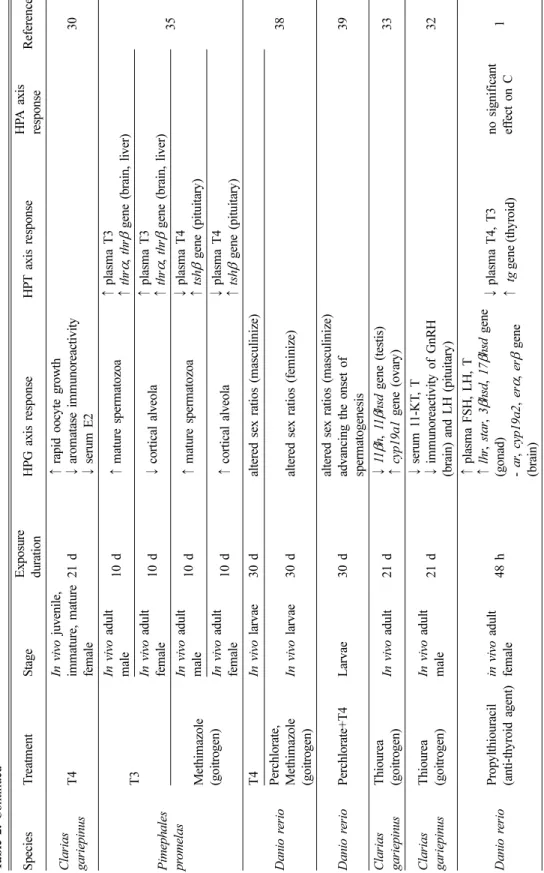
Fig. 2. Schematic representation of hypothalamic-pituitary-gonad (red), -thyroid (blue), and –adrenal (green) axes interaction. Dashed arrows represent the points of interaction between the different axes highlighted in this review.
Shadowed areas show the bi-directional interaction of nonylphenol effects.
① GnRH could increase plasma T4 levels in fish.
23,24)② E2 contributes to the depression in plasma T3 by modifying several aspects of the peripheral metabolism of TH and inhibiting dio2 converting T4 to T3. T and other androgens tend to enhance T3 formation and thyroidal status.
2)③ Action of TH diminish the reproductive axis by 1) inhibiting pituitary LH expression and steroidogenesis,
12)2) reducing gonadal aromatase expression which contributes to reduced estrogen synthesis,
12,30,27)and 3) diminishing estrogen responsiveness by reducing the ER subtypes expression.
27,28)④ Cortisol could increase the androgen-related machinery and subsequent masculinization in fish.
42,43,45,46)⑤ Corticotropin-releasing hormone (CRH) could induce the thyroid stimulating hormone (TSH) in pituitary in
fish.
47)J Environ Health Sci 2015; 41(3): 147-162 http://www.kseh.org/
기의 C 호르몬 농도가 높아지면 pejerrey,42,43) medaka,44) Japanese flounder45,46)의 수컷화(masculinization)를 유발한다고 보고하였다. 성어에서는 HPG 축과 HPA 축에서 글루코코르티코이드 호르몬의 합성과 대사를 관장하는 cyp11β와 11-oxigenates androgens의 합성 을 관장하는 11βhsd2를 공유하고 있다. 글루코코르 티코이드 호르몬(스트레스)과 안드로겐 호르몬(수컷 화)은 성분화 시기에 상호작용할 수 있으며, C 호르 몬은 11-KT 호르몬 생산을 증진시켜 정자세포 형성 을 증진시킬 수 있다.43)
3. HPT, HPA axes
수서척추동물의 CRH는 갑상선, 부신 내분비계 축 을 모두 조절할 수 있는 신경조절제(neuroregulator) 이며, HPT 축과 HPA 축을 연결시키는 중요한 연결 고리로 작용한다. 즉, 시상하부에서 분비되는 CRH 는 HPA 축을 조절하는 동시에 TSH를 자극하기도 한다(Fig. 2).47) 예를 들어, CYP11B 효소 억제제인 metyrapone에 노출되었을 때에는 갑상선 조직의 tshβ, dio1, dio2 유전자 발현이 증가되었는데,48) C 호르몬 농도 저하에 대한 보상작용으로 CRH가 증가되고 이 에 따라 갑상선 내분비계의 유전자 발현도 증가되었 다고 설명하고 있다. CRH가 갑상선 축의 여러 호 르몬을 조절, 억제, 자극하는 것은 물고기의 생애주 기에 따라 다를 수 있다.
TH 역시 HPA 축의 호르몬 분비에 영향을 줄 수 있다. 예를 들어, 갑상선 기능 항진증(hyperthyroidism) 이 유발된 잉어(Cyprinus carpio)에서는 혈장 C 호 르몬 수준이 감소하였다.49) 대표적인 항갑상선 약물 인 6-propyl-2-thiouracil에 노출된 제브라피쉬 배아/
치어는 부신 조직의 hmgra, cyp11b 유전자 발현이 증가하였다.48)이 연구에서는 6-propyl-2-thiouracil 노 출이 제브라피쉬의 TH 농도를 감소시키고, 음성 되 먹임 작용으로 CRH의 민감도를 증가시켜 hmgra, cyp11b 유전자 발현이 증가되었다고 설명하고 있다.
IV. 노닐페놀 노출로 인한 경골어류 내분비계의 영향
노닐페놀은 계면활성제, 에폭시 수지, 페인트 첨가 제 등으로 널리 사용되고 있으며, 물환경 중으로 유 입되어 오염이 되면 환경과 생태계에 축적되어 인간
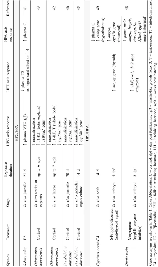
과 환경에 영향을 줄 수 있는 대표적인 내분비계 장 애물질이다. 2013년 9월부터는 어린이용품의 관리를 강화하기 위해 함량기준이 설정되었으며, 최근 「유 해화학물질 관리법」의 취급제한 물질로 지정되었 다. 본 연구에서는 상대적으로 연구자료가 풍부하고 대표적인 내분비계 장애물질로 알려진 노닐페놀의 독성을 정리하였고, 물고기의 번식, 갑상선, 부신 내 분비계에 미치는 영향을 전일적으로 설명하여 추후 연구들의 방향에 대해 제시하고자 하였다(Table 3).
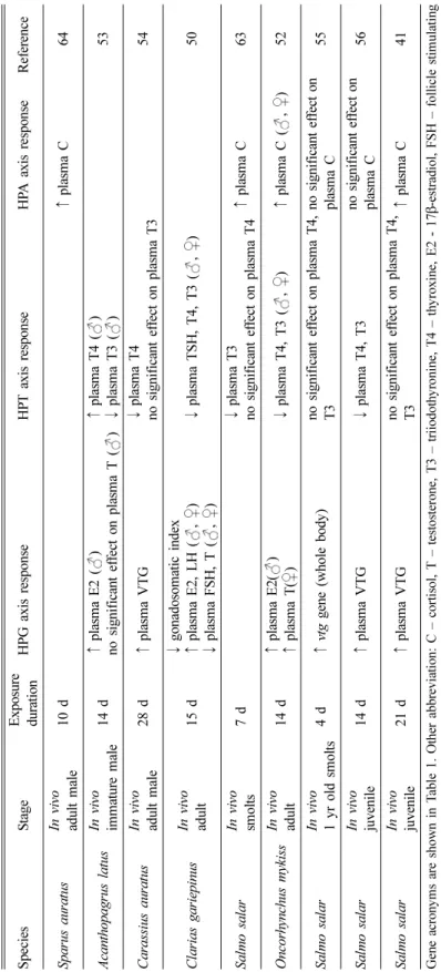
노닐페놀의 생식선 내분비계 독성영향은 성별로 다 르게 나타났으며, 주로 수컷 물고기의 암컷화가 관찰 되었다. 노닐페놀에 노출된 수컷 물고기에서 여성호 르몬인 E250-53)와 비텔로제닌이 증가하였고,41,51,54-62)스 테로이드 호르몬 생합성과 연관된 경로의 여러 유전 자들(cyp17, cyp19a, cyp19b 등)의 발현이 변화하였다.51) 이러한 영향은 조직학적인 수준에서 생식소중량지수 (gonadosomatic index)의 감소와도 연관이 있다.50,57)
노닐페놀은 갑상선 내분비계의 영향에 대한 간접 적인 반응으로 생식선에 영향을 미칠 수 있다. 즉, 수컷 물고기의 혈장 T4와 T3가 감소함50,52,53,56,63)으 로 인해 T에서 E2로 전환시켜주는 cyp19 효소가 증 가51)하여 궁극적으로 E2가 증가될 수 있다(Fig. 2의
②③). 예를 들어, 노닐페놀에 14일간 노출된 수컷 무지개 송어(Oncorhynchus mykiss)에서는 혈장 T4 와 T3가 감소하였고, 이러한 결과가 생식선 내분비 계의 혈장 E2를 증가시키고 T를 감소시키는 데 영 향을 미칠 수 있다.52)
수서척추동물에서 CRH는 다른 내분비계 축과 연 결하는 중요한 고리로 작용한다. 노닐페놀에 노출된 수컷 물고기에서는 C 호르몬이 증가41,52,64,65)하였고, 혈장 TSH, T4, T3가 감소되었다.50) C 호르몬이 증 가되었을 때 음성되먹임 작용으로 시상하부의 CRH 가 감소될 수 있으며, 이는 TSH와 TH의 감소로 이 어질 수 있다(Fig. 2의 ⑤). 또한 혈장 C 호르몬이 증가할 경우 HPG 축과 연결된 비텔로제닌도 증가 할 수 있다.41) 그러나 노닐페놀이 HPG, HPT, HPA 축의 어느 축에 먼저 영향을 주는지 선후관계가 밝 혀지지는 않았다.
V. 결 론
본 연구에서는 물환경 중 내분비계 장애물질이
Ta bl e 3 . Sum m ar y of s tu d ie s th at s hows th e i n te ra ct io n be tw ee n th e h y pot ha la m ic- pi tu it ar y -gona d ( H PG ), - thy ro id ( H P T ), a nd – adr en al ( H PA ) axe s af te r nonylp h eno l expo sur e S p ecies S tag e Ex posu re duration HP G axis re spons e H P T axis re spons e H PA axis resp onse R ef er en ce A can th opa grus l at us In vivo imma tu re ma le 14 d ↑ pl as m a VT G ( ) 58 C ara ssius a uratus In vivo adult m ale 28 d ↑ p las m a E 2 , T ( ) ↑ er α , er β , vtg gene ( liver) ↓ cyp 17 gene ( testis) ↑ cyp 19a , cyp1 9b g ene ( brain)
51 C yprin us ca rpio In vivo adult m ale 7 d ↑ pl as m a VT G ( ) ↓ g onad o som atic in dex ( ) 57 Da ni o r er io In vivo adult m ale 21 d ↑ vtg1 , vtg2 gene ( liver) ↑ er α , er β g ene ( liver) 59 G ob io cypris r arus In vivo adult 21 d ↑ pl as m a VT G ( ) 62 O nco rh ynchu s kisutch In vivo ju venile 125 d n o si gn if ic an t e ffe ct on pl as m a T4 , T3 66 O nco rh ynchu s m ykiss In vivo ju venile 14 d no signif icant ef fe ct on plasm a C 67 O ryzias l ati pe s In vivo adult 21 d ↑ pl as m a VT G ( ) 60 O ryzias l ati pe s In vivo em b ryo 4 h p f ↓ gn rh 1, gn rh 2, gn rh 3 ge n e (w ho le bo d y ) ↑ er α g ene (w h o le bo dy ) 68 Se bastes schlegeli In vivo adult m ale 14 d ↑ pl as m a VT G ( ) n o s ign if ic an t e ffe ct on pl as m a E2 , T 61 C larias gariepin us In vivo adult 7 d ↑ serum C 6 5
J Environ Health Sci 2015; 41(3): 147-162 http://www.kseh.org/
Ta bl e 3 . Cont inue d S p ecies S tag e Ex posu re du ra tion HP G ax is r es p on se HP T a x is r es p o n se HP A a x is r es p o n se Re fe re n ce Sparu s aur atus In vivo adult m ale 10 d ↑ plasm a C 6 4 A can th opa grus l at us In vivo imma tu re ma le 14 d ↑ plasm a E 2 ( ) no signif icant ef fect on plasm a T ( ) ↑ plasm a T 4 ( ) ↓ plasm a T 3 ( ) 53 C ara ssius a uratus In vivo adult m ale 28 d ↑ plasm a V T G ↓ plasm a T 4 no signif icant ef fec t on plasm a T 3 54 C larias gariepin us In vivo adult 15 d ↓ gon adoso m ati c index ↑ p la sm a E2 , LH ( , ) ↓ plasm a F S H , T ( , ) ↓ p la sm a TS H, T4 , T3 ( , ) 5 0 Sa lm o s al ar In vivo sm olt s 7 d ↓ plasm a T 3 no signif icant ef fec t on plasm a T 4 ↑ plasm a C 6 3 O nco rh ynchu s m ykiss In vivo adult 14 d ↑ plasm a E 2 ( ) ↑ plasm a T ( ) ↓ p la sm a T4 , T3 ( , ) ↑ plasm a C ( , ) 5 2 Sa lm o s al ar In vivo 1 yr ol d s m ol ts 4 d ↑ vtg g ene (w ho le bod y ) no signif icant ef fec t on plasm a T 4 , T3 no signif icant ef fect on plasm a C 55 Sa lm o s al ar In vivo ju venile 14 d ↑ plasm a V T G ↓ p la sm a T4 , T3 no signif icant ef fect on plasm a C 56 Sa lm o s al ar In vivo ju venile 21 d ↑ plasm a V T G no signif icant ef fec t on plasm a T 4 , T3 ↑ plasm a C 4 1 G ene ac ron y m s ar e s h o w n i n T abl e 1 . O th er a b bre v ia ti o n : C – c o rt is ol , T – t es to st er one , T3 – t ri iodo th y ro n in e, T4 – t h y roxi ne , E2 - 17 β -e str ad io l, FSH – foll icle stim ul atin g h o rm o n e, L H – lu tei n iz in g ho rm o n e, VT G – v itel log en in , d p f – d ay p ost f ert iliz atio n . Ef fec t ( ↑ sig n ific an t in cr ea se, ↓ sig n ifica n t de cr ea se ) an d t ar g et ar e in d ica ted .
HPG, HPT, HPA 축의 다양한 유전자와 호르몬에 미 치는 영향을 조사하였으며, 이러한 결과를 종합적으 로 연계하여 해석하고자 하였다. 또한 비교적 자료 가 풍부하고 대표적인 물환경 내분비계 장애물질인 노닐페놀의 주요 내분비계 독성 영향들을 정리하여 생식선, 갑상선, 부신 내분비계 축의 다양한 분자생 물학적 지표들이 어떠한 상호작용으로 영향을 미치 는지 전일적으로 해석하고자 하였다. 현재까지의 연 구들은 내분비계 장애물질이 주요 3가지 내분비계 축의 어느 조직에 먼저 독성을 일으키는지 또는 독 성기전들이 어떻게 연관되어 해석되는지의 정보가 부족하다. 앞으로의 연구에서 환경오염물질이 물고 기의 HPG, HPT, HPA 축의 다양한 호르몬과 유전 자에 미치는 영향을 함께 관찰한다면 통합적으로 이 해하는 데 도움이 될 것이다.
Acknowledgement
This study was supported by National Research Foundation of Korea (NRF; Project no.
2013R1A1A1061684).
References