Congenital and perinatal cytomegalovirus infection
Chun Soo Kim, M.D., Ph.D.
Departments of Pediatrics, Keimyung University School of Medicine, Daegu, Korea
= Abstract =
Cytomegalovirus (CMV) is currently the most common agent of congenital infection and the leading infectious cause of brain damage and hearing loss in children. Symptomatic congenital CMV infections usually result from maternal primary infection during early pregnancy. One half of symptomatic infants have cytomegalic inclusion disease (CID), which is characterized by involvement of multiple organs, in particular, the reticuloendothelial and central nervous system (CNS).
Moreover, such involvement may or may not include ocular and auditory damage. Approximately 90% of infants with congenital infection are asymptomatic at birth. Preterm infants with perinatal CMV infection can have symptomatic diseases such as pneumonia, hepatitis, and thrombocytopenia. Microcephaly and abnormal neuroradiologic imaging are associated with a poor prognosis. Hearing loss may occur in both symptomatic and asymptomatic infants with congenital infection and may progress through childhood. Congenital infection is defined by the isolation of CMV from infants within the first 3 weeks of life. Ganciclovir therapy can be considered for infants with symptomatic congenital CMV infection involving the CNS. Pregnant women of seronegative state should be counseled on the importance of good hand washing and other control measures to prevent CMV infection. Heat treatment of infected breast milk at 72℃ for 5 seconds can eliminate CMV completely. (Korean J Pediatr 2010;53:14-20)
Key Words : Cytomegalovirus, Congenital, Perinatal, Hearing loss
1)
jtj
Introduction
Human cytomegalovirus (CMV) is a member of the Her- pesviridae family of DNA viruses and ubiquitous in the ge - neral population. Most CMV infections are inapparent, but the virus can cause a wide range of diseases in neonates and immunocompromised patients. CMV is the most com - mon cause of congenital infection worldwide, affecting 0.2
% to 2.4% of all live births
1-4). CMV is now the most com - mon viral cause of mental retardation and hearing deficit of children in developed countries
5-7).
CMV causes deformation of preformed tissues rather than malformation of developing organs
8), so CMV can affect fe- tuses beyond the first trimester, although earlier infection is usually more severe
8-10). Primary maternal infection is most likely to cause severe symptomatic congenital infec - tion
11), but recurrent infection may also rarely do so. Peri -
Received : 2 December 2009, Accepted : 6 December 2009 Address for correspondence : Chun Soo Kim, M.D., Ph.D.
Departments of Pediatrics, Keimyung University School of Medicine 194 Dongsan-dong, Jung-gu, Daegu, 700-712, Korea
Tel : +82.53-250-7526, Fax : +82.53-250-7783 E-mail : [email protected]
natal CMV infection is usually not associated with clinical illness in term babies, but may also cause serious problems in preterm infants
12-15).
Transmission
Transmission of CMV occurs from person to person through body fluids, and requires close contact with con- taminated secretions because the virus in not very conta- gious. CMV can be found in blood, urine, semen, cervical secretions, saliva, breast milk and transplanted organs, all these sites intermittently excrete viruses
1, 12). Viral excre- tion is particularly prolonged after primary infection over years but also occurs with reactivation of infection
1, 5).
Transmission of CMV to fetuses and newborn infants
occurs in one of following modes
12, 15-18): (1) in utero by
transplacental passage from hematogenous spread to the
fetus during maternal viremia; (2) at birth by intrapartum
passage after exposure to infected cervical and vaginal
secretions; (3) postnatally by ingestion of CMV-positive
breast milk or infected blood transfusion.
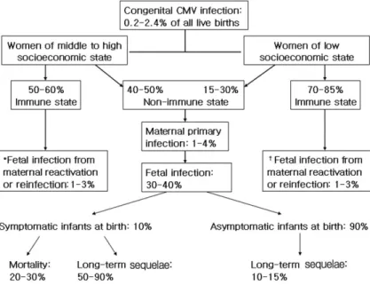
Fig. 1. A schema of congenital cytomegalovirus infection and its outcome. *
†The rate of symptomatic infection at birth is less than 1%. The suggested incidence rate reflects data reported in the pediatric literature
1-6, 8, 9).
Epidemiology
CMV infection is endemic and lacking in seasonal varia- tion. Congenital CMV infection can result from maternal primary or reactivated infection during pregnancy
1, 5). And also maternal reinfection of different strains of CMV can rarely lead to congenital symptomatic infection
19, 20). The prevalence of congenital CMV infection varies between 0.2-2.4% in different countries
1-4). In developed countries, around 50-60% of pregnant, middle to high class women have antibodies to CMV, compared with around 70-85% of those from lower socioeconomic groups
3, 4). Overall, 1-4%
of susceptible women are affected with primary CMV infec- tion during pregnancy, and around 30-40% of the fetus are transplacentally infected, and also about 10% infants mani- fest clinical signs and symptoms at birth
1-5). Approximately 1-3% of infants born of women with preexisting antibody to CMV are infected in utero, but they have rarely sympto- matic illnesses at birth (less than 1%)
5, 19). It has been known that the risk of symptomatic infections at birth or the baby who will develop subsequent sequelae is higher if maternal infection occurs during the first half of pregnancy, and also most of severe congenital infection is caused by primary rather than recurrent infection of pregnant women
5, 10, 11)
. A suggested schema of congenital CMV infection and its outcome may be a possible scenario in the commu-
nity (Fig. 1).
Perinatal CMV infection can result from exposure to infected secretions of maternal genital tract at delivery or breast milk that contains the virus. Around 2-28% of sero- positive mothers shed CMV in cervical and vaginal secre- tions during delivery, and approximately 50% of exposed infants are infected
15). Around 9-88% of seropositive women shed CMV into their milk, and approximately 50- 60% of infants fed breast milk that contains the virus become infected
15, 21).
Transfusion-acquired perinatal CMV infection occurs in infants of seronegative mothers who are exposed to blood products that contain the virus. The incidence of infection is about 10-30% and occurs usually preterm infants with a birth weight of less than 1,500 g
1, 22). The risk of infec- tion is related the volume of transfused blood or number of donors, and the titer of CMV in donor blood
15).
Clinical manifestation
1. Congenital infection
Approximately 10% of infants with congenital CMV
infection have signs and symptoms at birth, and only one
half of these babies have the fulminant illness; cytomegalic
inclusion disease (CID)
13, 15). Another 5% of these infants
show mild and atypical involvement, and 90% are born with
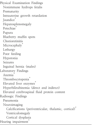
Table 1. Clinical Manifestation of Congenital Cytomegalovirus Infection
Physical Examination Findings Nonimmune hydrops fetalis Prematurity
Intrauterine growth retardation Jaundice
*Hepatosplenomegaly Petechiae
*Pupura
Blueberry muffin spots Chorioretinitis Microcephaly
*Lethargy Poor feeding Hypotonia Seizures
Inguinal hernia (males) Laboratory Findings Anemia
*Thrombocytopenia
*Elevated liver enzymes
*Hyperbilirubinemia (direct and indirect) Elevated cerebrospinal fluid protein content Radioogic Findings
Pneumonia Neuroimaging
Calcifications (periventricular, thalamic, cortical)
*Ventriculomegaly
Cortical dysplasia Hearing impairment
**
Prominent features
Data from Stehel EK, et al.
15)subclinical but chronic infection
2, 15). The symptomatic in- fection of infants usually results from primary infection of the mother
5, 9). And it appear that the risk is greater, the earlier in gestation the infection occurs
5, 11).
Infants with CID typically have a purpuric rash due to thrombocytopenia (usually with petechiae), intrauterine growth retardation (IUGR), hepatosplenomegaly, jaundice, hearing loss, microcephaly, chorioretinitis and intracerebral calcification. More commonly, infected infants may exhibit a wide spectrum of disease with only one or a few of these manifestations
15)(Table 1). Premature infants or babies who are small for gestational age with hepatosplenomegaly and abnormal liver function are most common findings of symptomatic congenital CMV infection
4). Hyperbilirubinemia may be transient with a rise in the direct component. Throm- bocytopenia is developed one third of infants with congeni- tal infection, and may persist for weeks
5). There may also be a Coombs-negative hemolytic anemia
5, 11). Pneumonia
may develop as a late findings of congenital CMV infection, usually occurring at 1-4 months of age
1, 15). CMV pneumo- nia in preterm infants has been associated with develop- ment or exacerbation of bronchopulmonary dyslasia (BPD)
23)
. And also postnatal administration of steroid for BPD in CMV-infected perterm infants has been associated with progression of CMV disease
15).
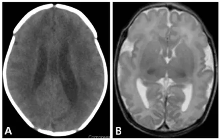
A major important issue in congenital CMV infection is cental nervous system (CNS) involvement including menin- goencephalitis, calcification, microcephaly, disturbance of neuronal migration, germinal matrix cysts, ventriculomegaly and cerebellar hypoplasia
6). CNS disease usually results in following symptoms and signs; lethargy, hypotonia, seizure, hearing deficit, or chorioretinitis. Some examples of abnor- mal magnetic resonance imaging (MRI) findings in infants with congenital CMV infection are suggested (Fig. 2).
Sensorineural hearing deficit (SNHD) may present at birth, either unilateral or bilateral, or can become later in childhood. Some patients have normal hearing for the first 6 years of life, but they can subsequently develop sudden or fluctuating hearing loss. Among children with hearing deficit, further deterioration of hearing occurs in 50%, with the median age of first progression at 18 months of age (range, 2 to 70 months of age)
8, 24, 25). Hearing loss appa- rently result from viral replication in the inner ear
26).
Infants with symptomatic congenital CMV infection have a mortality rate of 20-30%
4, 9). Death are usually due to hepatic dysfunction, bleeding, disseminated intravascular coagulopathy or secondary bacterial infection
4).
2. Perinatal infection
The incubation period of perinatal CMV infection ranges between 4 and 12 weeks (mean, 8 weeks)
1, 15). The quan- tity of virus excreted by infants with perinatal infection is less than that developed in congenital infection, but this infection is also chronic, with viral excretion persisting for years
1).
Most term infants with perinatal CMV infection are asy-
mptomatic, because the babies have maternal-transmitted
CMV IgG antibody. In contrast, 15-25% of infected preterm
infants may develop clinical illnesses such as pneumonia,
hepatitis or sepsis-like illness that present with apnea,
bradycardia, hepatosplenomegaly, distended bowel, anemia,
thrombocytopenia and abnormal hepatic function
12, 15).
Transfusion-acquired CMV infection in preterm babies
with very low birth weight may develop severe symptoms
Fig. 2. Some examples of magnetic resonance imaging findings in infants with congenital cytomegalovirus infection. (A) Periventricular calcification and mild ventriculomegaly. (B) Diffuse polymicrogyria of cerebral hemispheres.
that resembled CID
1).
Diagnosis
Congenital CMV infection is diagnosed by isolation of virus from infants within the first 3 weeks of life
3, 13, 15). So it is difficult to differentiate whether CMV infection was acquired congenitally or perinatally in infants more than 3 weeks of age, unless clinical features of the former such as chorioretinitis, intracranial calcification, microcephaly and hearing loss, are present
13, 15).
CMV is usually isolated from urine and saliva but may be isolated other body fluids including breast milk, cervicova- ginal secretions, amniotic fluid, white blood cell, cerebro- spinal fluid, stool and biopsy specimens. The best test for diagnosis of congenital or perinatal CMV infection is virus isolation or demonstration of CMV genetic material by poly- merase chain reaction (PCR) from urine or saliva of new- born infants
1, 15). The sensitivity of PCR with urine speci- mens is 89%, and the specificity is 96%
1, 27). Urine speci- mens should be refrigerated (4℃) but never frozen or stored at room temperature. The recovery rate of viruses remains at 93% in urine after 7 days of refrigeration, de- creasing to 50% after 1 month
5). Although a fourfold rise in IgG titer in paired sera or a strong positive IgM anti- CMV may be a useful finding suggesting infection, serologic assays are not recommended for diagnosis of CMV infection in neonates. Because IgG anti-CMV detected in newborn infants reflects transplacentally acquired maternal antibody
and that can persist for up to 18 months of age
15). And also IgM assay has both false positive and false negative
28, 29). Computed tomography (CT) is most sensitive to detect intracranial calcification. And MRI is particularly valuable for detection of the disorders of neuronal migration and other cerebral parenchymal lesions complicated with con- genital CMV infection
6).
Amniocentesis may be the most valuable single antenatal diagnostic test, and viral culture or PCR test of amniotic fluid are equally specific and sensitive
2, 5). Quantitative PCR demonstrating 10
5genome equivalent per mL of amniotic fluid may be a predictor of symptomatic congenital infec- tion
2). Ultrasonographic fetal abnormalities in pregnant women with primary or recurrent CMV infection generally indicate symptomatic fetal infection. Reported sonographic abnormalities of the fetus include oligohydroamnios, IUGR, microcephaly, ventriculomegaly, intracranial calcification, hypoplastic corpus callosum, ascites, hepatosplenomegaly, hypoechogenic bowel, pleural and pericardial effusion, and hydrops
16).
Treatment
There are now two drugs for systemic treatment of congenital and perinatal CMV infection: ganciclovir and valganciclovir (oral prodrug of ganciclovir).
In a randomized controlled, multicenter, phase III study
with ganciclovir (6 mg/kg/dose intravenously every 12 hr
for a 6 week course)
30), it was carried out to treat newborn
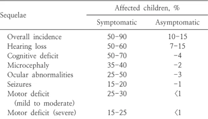
Table 2. Long-term Sequelae of Congenital Cytomegalovirus In- fection in Children
*Sequelae Affected children, %
Symptomatic Asymptomatic Overall incidence
Hearing loss Cognitive deficit Microcephaly Ocular abnormalities Seizures
Motor deficit (mild to moderate) Motor deficit (severe)
50-90 50-60 50-70 35-40 25-50 15-20 25-30
15-25
10-15 7-15 -4 -2 -3 -1
<1
<1
*
Rates reflect a range of incidence data reported in the pedi- atric literature.
Data from Schleiss MR
32)infants who have congenital CMV infection involving the
CNS, as defined by microcephaly, intracranial calcification, abnormal findings of cerebrospinal fluid, chorioretinitis and/
or hearing deficit. When tested at 6 months, 84% of treated infants maintained normal hearing or improved their hearing, in contrast to 59% of control infants. At 6 months follow- up, none of the treated infants had hearing deterioration, while 41% of control patients did. When tested 1 year of age or beyond 21% of treated and 68% of control infants had hearing deterioration. Primary ganciclovir-related toxi- city was neutropenia. 63% of treated infants developed neutropenia, compared with 21% of control infants.
In a pharmacokinetic and pharmacodynamic study of oral valgancyclovir in treatment of infants with congenital CMV infection
31), it showed that a 6 mg/kg intravenous ganciclo- vir dose and 16 mg/kg oral valganciclovir provides similar systemic exposure to ganciclovir. In addition, treated pa- tients with a 6 week course decreased in viral load of 0.7 log viral DNA copies/mL in urine. Toxicity of valganciclovir is similar to that of ganciclovir with 38% of subjects de- veloping neutropenia.
Treatment with ganciclovir of a 6 week course can be considered for symptomatic infants with congenital CMV infection involving the CNS, but can not be routinely re- commended
15). A 2 week course of ganciclovir may be be- neficial in critically ill infants with symptomatic perinatal CMV infection. And additional 1-2 weeks of ganciclovir can be considered if symptoms and signs have not been re- solved
12).
Prognosis
Infection during early pregnancy more often result in more severe sequelae than infection later in pregnancy.
Approximately 50-90% of symptomatic survivors have long-term sequelae such as microcephaly, hearing loss, motor deficit (paresis/paralysis), cerebral palsy, mental retardation, seizure, ocular abnormalities (chorioretinitis, optic atrophy), autism and learning disabilities
32)(Table 2).
In contrast, 10-15% of infant with congenital CMV infection who are asymptomatic at birth have long-term neuro- developmental injury.
Factors associated with poor cognitive prognosis are microcephaly and abnormalities detected on brain CT
4, 6). In contrast, children with normal findings on brain CT and normal head circumference have been revealed to have a
good cognitive outcome
33). So radiologic findings of brain with MRI and CT may be the most specific and sensitive predictors of neurodevelopmental outcomes
6). Microcephaly dose not always persist, and dose not always result in later handicap. Infants who have microcephaly alone without other neurologic findings have relatively a good prognosis in early childhood
34). Chorioretinitis is nearly always indi- cative significant mental retardation and often lead to optic atrophy. It can also develop on several weeks after birth
8). SNHD, the most common sequelae of congenital CMV infec- tion may occur in both symptomatic and asymptomatic in- fected infants. Although about 60% of infants with hearing loss is developed at birth or in neonatal period, at least 40
% may have delayed-onset loss
6). Additionally progression of hearing loss in patients who had already SNHD has also been observed in about 60% of infants with through child- hood and into adolescence
15, 25, 35). So all babies with con- genital CMV infection should be followed at least school entry, and hearing test regularly performed to detect late deficit
6, 8). Patient with disseminated symptomatic infection including petechiae or IUGR at birth and those with higher viral load at birth appear to have an increased risk of hea- ring loss
1, 35).
Premature and ill full-term infants with serious perinatal
CMV infection may be an increased risk of neurologic se-
qulae and psychomotor retardation, even though there dose
not appear to be a higher rate of SNHD, microcephaly and
chorioretinitis
1, 2).
Prevention
CMV is usually not very contagious, and its horizontal transmission requires direct contact with infected materials, such as different secretions that contain the virus, and less likely, formites. So adherence to standard precautions with good hand hygiene is effective to prevent CMV infection
1, 13).
Preexisting maternal immunity to CMV affords significant protection to the fetus. In contrast, with maternal primary infection, the overall risk for delivering a symptomatic neonate is about 5%
3, 4, 9). So women of childbearing age should have CMV serologic tests, especially if they provide care for young children who are potential CMV excreters
1,2, 36)
. And pregnant women who are CMV seronegative should be provided with information on prevention mea- sures and reassured that common sense steps such as hand washing and avoiding contact with secretions should pre- vent this infection
1, 37). Although newborn hearing screening programs may miss the late-onset hearing loss complicated in congenital CMV infection, routine screening of all new- borns for CMV is not yet recommended
38, 39).
Pasteurization (72℃ for 5 seconds) of breast milk can inactivate CMV completely without affecting nutritional and immunologic properties of milk, although freezing (-20℃
for 3-7 days) will significantly decrease viral titers
12, 40,41)