성상세포종에서 혈관내피세포 성장인자의 발현*
한림대학교 의과대학 신경외과학교실, 해부병리학교실**
박세혁·장인복·김창현·조용준·조병문 신동익·오세문·김덕환**·남은숙**
= Abstract =
Expression of Vascular Endothelial Growth Factor Protein in Astrocytic Tumors
Se-Hyuck Park, M.D., In-Bok Chang, M.D., Chang-Hyun Kim, M.D.
Young-Jun Cho, M.D., Byung-Moon Cho, M.D., Dong-Ik Shin, M.D.
Sae-Moon Oh, M.D., Duk-Whan Kim, M.D.,** Eun-Sook Nam, M.D.**
Department of Neurosurgery, Pathology,** Hallym University College of Medicine, Seoul, Korea
bjective:Angiogenesis, the proliferation of capillary endothelial cells, is a vital component in the development, progression, and metastasis of many human tumors. Vascular endothelial growth factor(VEGF) is an endothelial cell-specific mitogen and induces angiogenesis and vascular permeability. The features of glioblastoma, distinct from low grade astrocytomas, are the presence of necroses and vascular endothelial proliferation. In this study, we investigated VEGF expression in the different grades of astrocytomas and determined whether VEGF expression correlates with development of glioblastoma and progression of astrocytomas.
Patients and Methods:Forty seven patients with astrocytic tumors(24 males and 23 females), aged 3 to 65 years, were evaluated. Immunohistochemical staining was carried out using labelled streptavidin biotin method and primary antibody was a antirabbit polyclonal Ab against N-terminus region of VEGF165(Oncogene research product, MA, USA). Immunoreactivity(IR) was classified into no IR(absent or a trace of stain), moderate IR and intense IR by level of staining amount and intensity.
Results:Six pilocytic astrocytomas showed 3 no IR and 3 moderate IR, 10 astrocytomas showed 2 no IR, 6 moderate IR and 2 intense IR, 12 anaplastic astrocytomas showed I no IR, 7 moderate IR and 4 intense IR and 19 glioblastomas showed 1 no IR, 11 moderate IR and 7 intense IR. Immunoreactivity was significantly different between low and high grade of tumors but there was no significant difference between anaplastic astrocytomas and glioblastomas. Gemis- tocytic tumor cells represented the predominent VEGF-immunoreactive cell types, as compared with compactly- arranged small tumor cells. In glioblastomas VEGF IR was observed in both perinecrotic and vital tumor areas.
Conclusion:VEGF seems to be a important angiogenic factor in anaplastic astrocytomas and glioblastomas and VEGF expression may contribute to neovascularization of human astrocytomas.
KEY WORDS:Vascular endothelial growth factor・Angiogenesis・Astrocytoma.
서 론
혈관신생(angiogenesis)은 모세혈관내피세포의 증식으로, 여러 종양의 발생, 성장뿐만 아니라 전이에까지 중요한 영
향을 미친다6). 종양세포에서 분비되는 혈관신생물질은 pa- racrine manner로 혈관내피세포에 작용하여 내피세포의 증 식, 종양세포의 침윤을 유도하여 종양혈관형성에 관여하는 데 신경교종의 증식에 관여하는 혈관신생요소(angiogenic factor)로서 platelet-derived growth factor(PDGF), basic fibroblastic growth factor(bFGF) 및 vascular endothelial
OOOO
*본 논문의 요지는 1999년 대한신경외과 추계학술대회에서 발표되었음.
growth factor(VEGF), epidermal growth factor 및 tra- nsforming growth factor β 등이 알려져 있다5). 신경교종 중 교모세포종(glioblastoma)은 뇌에서 가장 흔하고 악성 인 종양으로 세포괴사와 혈관내피세포증식 등이 저등급(low grade) 성상세포종과의 병리적 차이점이며 악성 성상세포 종의 일부는 저등급 성상세포종의 악성변형(malignant tr- ansformation)으로 발생된다고 알려져 있다15). 그러나 저 등급 성상세포종의 악성변형에 대한 예측인자는 아직까지 확실히 밝혀져 있지 않으나 혈관신생이 성상세포종의 악성 변형 및 악성 성상세포종의 발생에 관여할 수 있다고 추정 된다. 여러 조직학적 단계의 성상세포종에서 VEGF발현을 통한 혈관내피세포증식의 정도와 VEGF 발현이 성상세포 종의 악성변형 및 예후에 영향을 미칠 수 있는지를 알아보 고자 본 연구를 하였다.
대상 및 방법
1. 대 상
1994년부터 1999년까지 5년 동안 본원 신경외과에 입 원하여 조직학적으로 성상세포종으로 진단 받은 47명의 환 자를 대상으로 하였다. 남자는 24명, 여자는 23명이었고 연령분포는 3세에서 65세로 평균연령은 36세였다. 조직학 적으로 모낭세포성 성상세포종 6예, 성상세포종 10예, 역 형성 성상세포종 12예 및 교모세포종 19예였다.
2. 면역조직화학적 염색
포르말린고정 파라핀 포매 조직을 대상으로 DAKO LS- AB(labelled streptoavidin-biotin) Kit(DAKO Corporation, Carpinteria, U.S.A.)를 사용하여 면역염색을 하였다. 파라 핀 블록을 4μm 두께로 박절하여 슬라이드에 부착시킨 후 xylene에 5분간 3회 처리하여 파라핀을 제거하고, 계열별 ethanol에 처리한 후 증류수로 함수시켰다. 내인성 과산화 효소의 활동을 억제하기 위해서 3% H2O2가 포함된 meth- anol 용액에 20분간 처리하였다. 일차 항체의 비특이적 결 합을 감소시키기 위하여 정상 염소혈청에 30분간 처리한
후 일차 항체인 anti-VEGF(purified rabbit polyclonal an- tibody against N-terminus region of VEGF165, Oncogene, Cambridge, USA)를 5% 소혈청 알부민을 함유한 희석액 을 이용하여 1:20으로 희석하여 4℃에서 16시간 동안 반응시켰다. Tris 완충액으로 세척한 후 biotin이 부착된 anti-rabbit 항체(2차 항체, K684, DAKO, Carpinteria, U.S.A.)를 30분간 반응시키고 tris 완충액으로 세척한 후 streptovidin-peroxide를 30분간 실온에서 반응시켰다. 다 시 tris 완충액으로 세척한 후 발색제인 AEC(3-amino- 9-ethylcarbazole)를 이용하여 5분간 반응시킨 다음 세 척하여 Mayer’s hematoxylin으로 대조염색 후 wet mou- nting하여 광학 현미경으로 관찰하였다.
3. 판정 및 통계분석
음성 대조군으로 정상 뇌조직을 사용하였고 immunore- activity(IR) 정도는 염색된 세포의 비율과 염색강도에 따 라 다음과 같이 3단계로 나누었다. 염색이 되지 않거나 약 하게 염색되면 no IR, 중간 정도 염색되면 moderate IR(a moderate amount(10~50% of cells) of diffuse stain)로 구분하였고 강하게 염색되면 intense IR(the largest am- ount(>50% of cells) of diffuse stain)으로 구분하여 측정 하였다.
통계처리는 VEGF 발현 정도와 조직학적 분류에 따른 성상세포종과의 관계를 Wilcoxon rank sum test를 이용하 여 분석하였으며 p<0.05를 의의 있는 것으로 하였다.
결 과
47예의 성상세포종 중 저등급 성상세포종은 16예였고
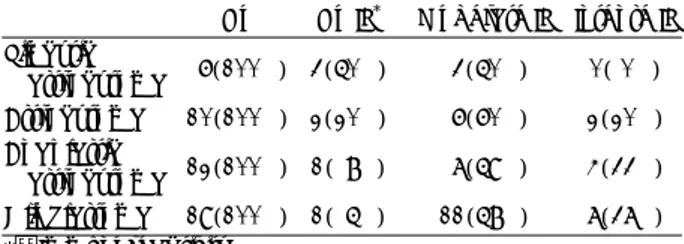
Table 1. VEGF immunoreactivity of astrocytomas
No No IR* Moderate IR Intense IR Pilocytic
astrocytoma 6(100%) 3(50%) 3(50%) 0( 0%) Astrocytoma 10(100%) 2(20%) 6(60%) 2(20%) Anaplastic
astrocytoma 12(100%) 1( 8%) 7(59%) 4(33%) Glioblastoma 19(100%) 1( 5%) 11(58%) 7(37%)
*:Immunoreactivity
Fig. 1. Photomicrograph showing VEGF immunostaining of ge- mistocytic tumor cells, rather than compactly-arranged small cells in glioblastoma. Original magnification ×400.
악성 성상세포종은 31예였다. 모낭세포성 성상세포종 6예 는 no IR 및 moderate IR가 각각 3예였고 성상세포종 10 예는 no IR 2예, moderate IR 6예 및 intense IR 2예였다 (Table 1). 역형성 성상세포종 12예는 no IR 1예, moderate IR 7예 및 intense IR가 4예였고 교모세포종 19예는 no IR 1예, moderate IR 11예 및 intense IR 7예였다(Table 1).
교모세포종 중 no IR 1예는 거구세포성 교모세포종(giant cell glioblastoma)이었다. 저등급 성상세포종 16예 중 11 예(69%)가 moderate IR 이상의 염색반응을 보였고 악성 성상세포종 31예 중 29예(94%)가 moderate IR 이상의 염색반응을 보여 조직학적 분류에 따른 성상세포종의 종류 와 VEGF 염색정도의 차이는 통계학적 유의성이 있었다 (p<0.05). 악성 성상세포종 뿐만 아니라 저등급 성상세포 종에서도 빽빽하게 배열된 작은 종양세포보다는 세포질이 풍부하고 큰 종양세포인 gemistocyte에서 VEGF 양성반 응을 잘 볼 수 있었다(Fig. 1). 교모세포종에서 괴사주위 pseudopalisading cell들에서 VEGF 양성반응을 볼 수 있 었으며 또한 괴사되지 않은 종양세포들에서도 VEGF 양성 반응을 관찰할 수 있었다(Fig. 2).
고 찰
혈관신생은 새로운 혈관이 기존혈관으로부터 생기는 것 으로 종양이 수 mm이상의 크기로 자라기 위해서 필요하 며 1971년 Folkman3)에 의해서 이 혈관신생의 개념이 도 입되었다. 악성 종양에서 새로운 혈관이 형성되는 것은 혈 관신생인자와 혈관신생억제인자들에 의해서 조절되며 이 들 인자들은 종양세포와 혈관 내피세포 등에서 분비된다8). 혈관신생은 여러 단계를 거쳐 이루어지는데 종양에 인접 한 혈관내피세포가 혈관신생인자의 자극을 받아 활성화되 면 단백분해효소(type Ⅳ collagenase, matrix metallop- orotienase 및 serine proteinase)가 분비되고 이들 단백 분해효소들에 의해서 혈관내피세포의 기저막과 세포외기
질(extracellular matrix)이 분해된다8). 이때 생긴 틈새로 내피세포가 이동하여 증식하며 처음에는 코드형으로 성장 한 혈관원형이 곧 내부에 관이 생겨 모세혈관을 형성하며 종양주위에 분포하여 망조직을 형성하게 된다. 인체내에서 혈관신생은 정상적으로는 태아 발육때 일어나며 비정상적 인 상태로는 종양발생, 당뇨병성 망막증 및 창상치유때 일 어난다4).
여러 종류의 혈관신생인자들 중 VEGF와 bFGF가 신경 교종의 혈관신생에 중요한 역할을 한다고 알려져 있다8). 그 중 VEGF는 혈관투과성인자(vascular permeability fa- ctor)라고도 하며, 34~43kDa의 당단백으로 PDGF와 구조 적으로 동종(homology)으로 4개의 isoform(VEGF121, VE- GF165, VEGF189 및 VEGF206)이 있는데 이들은 mRNA의 alternative splicing에 의해서 형성되며7) 그 중 VEGF165
가 가장 흔하고 종양혈관신생에 중요역할을 한다8). 본 연 구에서도 VEGF165의 N-terminus region 에 대한 항체를 사용하였다. VEGF는 혈관내피세포에 유사분열물질(mit- ogen)로 직접 작용할 뿐만 아니라 혈관의 투과성을 증가시키 고 기저막과 세포외기질을 분해시켜 혈관신생에 관여하며8) VEGF와 그 수용체는 정상 뇌조직에서 소량 발견되는데 그것은 VEGF의 정상혈관 유지기능 때문으로 추정된다2).
VEGF는 신경교종, 뇌수막종 및 전이성 뇌종양 등에서 발현된다12). 본 연구에서 VEGF발현정도의 차이는 있었지 만 악성 성상세포종 뿐만 아니라 저등급 성상세포종에서 도 VEGF는 발현되었는데 악성 신경교종에서는 저등급 신경교종보다 많은 예에서 VEGF가 중정도 이상으로 발 현되었다. 이것은 악성신경교종의 병리적 특성인 혈관내피 세포의 증식 때문이라 생각된다. 그러나 일부 악성 성상세 포종에서 중정도 이상의 VEGF발현없이 혈관내피세포의 증식을 보여 VEGF외의 인자가 일부의 성상세포종 혈관 신생과정에 관여할 수 있다고 추정된다. Pietsch 등10)의 보고에 의하면 일부 모양세포성 성상세포종을 제외한 전 성상세포종에서 VEGF에 양성면역반응을 보였으나 악성
Fig. 2. Photomicrographs showing VEGF immunostaining of pseudo- palisading tumor cells adjacent to necrotic area(A) and tumor cells in non-necrotic area(B) in gliobl- astoma. Original magnification A:
×100, B:×200.
AAA
A BBBB
도가 높을수록 발현정도가 높다고 하여 본 연구와 유사한 결과를 보였다. 본 연구에서 저등급 성상세포종에서 악성 성상세포종으로 변형한 예들은 없었으나 악성 성상세포종 의 VEGF 발현정도가 저등급 성상세포종의 그것보다 의 의 있게 높은 것으로 보아 VEGF발현정도가 저등급 성상 세포종의 악성변형에 대한 예측인자로 작용할 수도 있다 고 추정된다. 또한 빽빽하게 배열된 작은 종양세포보다는 세포질이 풍부하고 큰 종양세포인 gemistocyte에서 VE- GF 발현이 잘 된 것으로 보아 VEGF 발현은 gemistocyte 와 관계가 있을 것으로 추정되며 이에 대한 추가연구가 필요하다고 생각된다. Takekawa 등14)은 VEGF labelling index가 악성 성상세포종에서 저등급 성상세포종보다 의 의 있게 높은 것으로 보아 VEGF발현이 성상세포종의 악 성변형에 관여 할 수도 있다 하였고 Pietsch 등10)도 VEGF양성 저등급 성상세포종에서 gemistocytic differ- entiation이 발견되는 것으로 보아 이런 종양에서 악성변 형의 위험도가 높다 하였다. 또한 Abdulrauf 등1)도 VE- GF양성 저등급 성상세포종환자의 생존율이 VEGF음성 저 등급 성상세포종환자에 비해 의의 있게 높다고 하여 VEGF 가 저등급 성상세포종환자의 예후인자가 될 수 있다고 하 였다. 그러므로 VEGF양성 저등급 성상세포종은 VEGF 음성 저등급 성상세포종에 비해 보다 적극적인 치료가 필 요하다고 생각된다.
신경교종을 포함한 여러 종류의 세포에서 저산소증(hy- poxia)은 VEGF의 분비를 유도한다고 알려져 있으며 교모 세포종의 괴사주위 pseudopalisading cells에서 VEGF 양 성반응을 보이는 결과들이 이 사실을 뒷받침한다11)13). 본 연구에서도 교모세포종의 괴사주위 pseudopalisading cell 들에서 VEGF 양성반응을 볼 수 있었으며 또한 괴사되지 않은 종양세포들에서도 VEGF 양성반응을 관찰할 수 있었 고 Takekawa 등14)과 Pietsch 등10)도 유사한 보고를 하 였다. 그러므로 저산소증 외의 다른 인자 혹은 기전에 의해 서도 VEGF발현이 조절될 수 있다고 생각되었다. 그러나 악성 신경교종에서 저산소증이 VEGF발현과 유관하지 않 다는 보고도 있다9).
결 론
VEGF는 성상세포종, 특히 악성 성상세포종에서 발현되 며 이들 종양에서 VEGF는 혈관신생의 주요 인자로 관여 한다고 생각된다. 또한 성상세포종에서 VEGF발현은 환자, 특히 저등급 성상세포종 환자의 예후를 예측할 수 있는 인 자가 될 수도 있다고 생각된다. 그러나 환자들의 장기추적
에 의한 생존결과와 악성변형을 일으킨 저등급 성상세포종 의 분석 뿐만 아니라 분자생물학적 분석이 앞으로 필요하 다고 생각된다.
•논문접수일:2001년 1월 21일
•심사완료일:2001년 5월 11일
•책임저자:박 세 혁
134-701 서울 강동구 길동 445 한림대학교 의과대학 신경외과학교실
전화:02) 2224-2236, 전송:02) 473-7387 E-mail:[email protected]
References
1) Abdulrauf SI, Edvardsen K, Ho KL, Yang XY, Rock JP, Rose- nblum ML:Vascular endothelial growth factor exprression and vascular density as prognostic markers of survival in patients with low-grade astrocytoma. J Neurosurg 88:513- 520, 1998
2) Alon T, Hemo I, Itin A, Pe’er J, Stone J, Keshet E:Vascular endothelial growth factor acts as a survival factor for newly formed retinal vessels and has implication for retinopathy of prematurity. Nat Med 1:1024-1028, 1995
3) Folkman J:Tumor angiogenesis:therapeutic implications. N Engl J Med 285:1182-1186, 1971
4) Folkman J, Shing Y:Angiogenesis. J Biol Chem 267:10931- 10934, 1992
5) Hinton DR:Growth factors and proliferation potential, in Apuzzo MLJ(ed):Benign cerebral glioma, Park Ridge: American Association of Neurosurgical Surgeons, 1995, vol1, pp149-162
6) Lee AH, Dublin EA, Bobrow LG, Poulsom R:Invasive lo- bular and invasive ductal carcinoma of the breast show distinct patterns of vascular endothelial growth factor expression and angiogenesis. J Pathol 185:394-401, 1998
7) Leung DW, Cachianes G, Kaung WJ. Goeddel DV, Ferrara N:
Vascular endothelial growth factor is a secreted angiogenic mitogen. Science 246:1306-1309, 1989
8) Lund EL, Spang-Thomasen M, Skovgaard-Poulsen H. Kristj- ansen PEG:Tumor angiogenesis - a new therapeutic target in gliomas. Acta Neurol Scand 97:52-62, 1998
9) Parliament MB, Allalunis-Turner MJ, Franko AJ, Olive PL Mandyam R, Santos C, Wolokoff B:Vascular endothelial gr- owth factor expression is independent of hypoxia in human malignant glioma spheroids and tumors. British Journal of Cancer 82:635-641, 2000
10) Pietsch T, Valter MM, Wolf HK, Deimling A von, Huang HJS, Cavenee WK, Wiestler OD:Expression and distribution of vascular endothelial growth factor protein in human brain tumors. Acta Neuropathol 93:109-117, 1997
11) Plate KH, Breier G, Weich HA, Risau W:Vascular endothelial growth factor is a potent tumor angiogenesis factor in human
gliomas in vivo. Nature 359:845-848, 1992
12) Samoto K, Ikezaki K, Ono M, Shono T, Kohno K, Kuwano M, et al:Expression of vascular endothelial growth factor and its possible relation with neovascularization in human brain tumors. Cancer Res 55:1189-93, 1995
13) Shweiki D, Itin A, Soffer D, Keshet E:Vascular endothelial growth factor induced by hypoxia may mediate hypoxia-ini-
tiated angiogenesis. Nature 359:843-845, 1992
14) Takekawa Y, Sawada T:Vascular endothelial growth factor and neovascularization in astrocytic tumors. Pathol Int 48: 109-114, 1998
15) Vertosick FT, Selker RG, Arena VC:Survival of patients with well-differentiated astrocytomas diagnosed in the era of com- puted tomography. J Neurosurg 28:496-501, 1991