Y.-S. Kwak ・ K. S. Han ・ J. H. Lee ・ Y. S. Kwon ・ H. K. Kim (Department of Applied Biology & Enviromental Sciences, Research Institute of Life Science, Gyeongsang National University, Jinju 660-701, Korea)
K. h. Lee
(The Aging-associated Disease Research Center and Department of Microbiology, Yeungnam University College of Medicine, Daegu 705-717, Korea)
W. S. Chung
(Enviromental Biotechnology National Core Research Center and Division of Applied Life Science(BK21 program), Plant Molecular Biology and Biotechnology Research Center, Gyeongsang National University, Jinju 660-701, Korea) K. S. Mysore
(Plant Biology Division, Samuel Roberts Noble Foundation, Ardmore, OK 73401, USA)
D.-W. Bae ( )
(Central Instrument Facility, Gyeongsang National University, Jinju 660-701, Korea)
e-mail: [email protected]
Different oxidative burst patterns occur during host and nonhost resistance responses triggered by Xanthomonas campestris in pepper
Youn-Sig Kwak ・ Ki Soo Han ・ Jung Han Lee ・ Kyunghee Lee ・ Woo Sik Chung ・ Kirankumar S. Mysore ・ Young Sang Kwon ・ Hee Kyu Kim ・ Dong-Won Bae
Received: 30 July 2009 / Accepted: 24 August 2009
ⓒKorean Society for Plant Biotechnology
Abstract The hypersensitive reaction (HR) is the most common plant defense reaction against pathogens. HR is produced during both host- and nonhost-incompatible in- teractions. Several reports suggest that similarities exist between host and nonhost resistances. We assayed the pattern of generation of reactive oxygen species (ROS) and scav- enging enzyme activities during nonhost pathogen-plant interactions (Xanthomonas campestris pv. campestris/Capsicum annuum L.) and incompatible host pathogen-plant interactions (Xanthomonas campestris pv. vesicatoria race1/Capsicum annuum L.). Both O2- and H2O2 accumulated much faster during nonhost resistance when compared to the host resistance. The scavenging enzyme activities of superoxide dismutase (SOD), catalase (CAT) and peroxidase (POX) were also different during the host- and nonhost-incompatible interactions. CAT activity was much higher during nonhost
resistance, and several new isozymes of SOD and POX were detected during nonhost resistance when compared to the host resistance. Lipoxygenase (LOX) activity was higher in host resistance than nonhost resistance during the early stages of infection. Interestingly, the nitric oxide (NO) radical accumulated equal amounts during both host and nonhost resistance at early stages of infection. Further studies are needed to determine the specific pathways underlying these differences between host and nonhost resistance responses.
Introduction
The hypersensitive reaction (HR) that leads to rapid tissue necrosis at the site of infection is a typical plant response to an incompatible pathogen (Lam et al., 2001). This rapid tissue necrosis induced in both nonhost and disease resistant plants (due to the presence of a resistance gene) often localizes the pathogen to its infection site and thus restricts its spread (Keppler et al., 1987; Alvarez et al., 1998; Wojtaszek, 1997).
It is speculated that HR deprives the pathogen of nutrients and/or releases toxic molecules, thereby confining pathogen growth. There is also evidence suggesting that the reduction of water potential during HR could limit pathogen growth (Wright and Beattie, 2004). The HR provides resistance to a great majority of potential host and nonhost pathogens (Grant and Mansfield, 1999; Mysore and Ryu, 2004). For a given plant species, only a limited number of pathogens have the ability to evade the plant-nonhost defense system (Thordal -Christensen, 2003; Mysore and Ryu, 2004). If a pathogen can overcome the plant-nonhost defense system (nonhost resistance), the plant becomes a host for the pathogen and hence will be susceptible to the disease. However, certain cultivars or genotypes of the host plant species have evolved Research Article
to recognize certain races within the pathogenic species to trigger HR and this is termed host resistance. Host resistance is often governed by a single resistance (R) gene, the product of which directly or indirectly interacts with a specific elicitor(s) produced by the avirulence (avr) genes of pathogens (Keen, 1990; Schornack et al., 2006). These observations indicate that there is an ongoing evolution of the plant’s ability to recognize pathogen races that were previously unrecognized while the pathogen evolves to avoid recognition by a previously resistant plant. Even though HR is associated, in most cases, with plant responses to an incompatible in- teraction with a potential pathogen, its actual role in host and nonhost resistances is not very clear.
Reactive oxygen species (ROS) has been shown to rapidly accumulate in plants attacked by incompatible races of pathogen and also when treated with certain fungal elicitors (Lamb et al., 1997; Huckelhoven and Kogel, 2003). Accu- mulation of ROS in plants is popularly known as the oxidative burst (Alvarez et al., 1998; Huckelhoven and Kogel, 1993).
Many reports suggest that the oxidative burst is a putative endogenous signal to induce HR (Baker et al., 1995; Keppler et al., 1989). The ROS generating system responsible for the oxidative burst has been shown to be coupled with oxidation of reduced nicotinamide adenine dinucleotide phosphate (NADPH) in the microsomal fraction isolated from elicitor- treated sliced potato tubers (Park et al., 1998). In Nicotiana benthamiana, respiratory burst oxidase homologs (rboh genes), which have been implicated in ROS generation, are required for resistance to Phytophthora infestans (Yoshioka et al., 2003). ROS are toxic intermediates of molecular O2- reduced by successive one-electron steps. The predominant ROS de- tected during the oxidative burst in infected or elicitor-treated plants are O2-, H2O2 and OH radicals. The oxidative burst in potato tuber inoculated with an incompatible race of a pathogen was demonstrated to be due to an enhanced O2-
generating NADPH oxidase activity in the plasma membrane (Reviewed by Lamb et al., 1997; Hammond-Kosack and Jones, 1996; Lote and Geiszt, 2006). O2- generated by the NADPH oxidase enzymatically produces H2O2. H2O2, thereby formed, is further metabolized by catalysis or peroxidation.
Fe++ ion present in the plant cell can lead to the H2O2-de- pendent formation of OH radicals. The OH radical initiates chain reactions including lipid peroxidation, enzyme inac- tivation and so forth that eventually cause cell damage (Ham- mond-Kosack and Jones, 1996). Nevertheless, some recent reports question the role of ROS during hypersensitive cell death in plants (Tada et al., 2004; Zhang et al., 2003).
In plants, NO is produced non-enzymatically through light mediated conversion of NO2 by carotenoids or enzymatically by NADPH-nitrate reductase (Beligni and Lamattina, 2000;
Del Rio et al., 2004). Many reports suggest that NO radical induces phytoalexins, phenylalanine ammonia-lyase (PAL) and pathogenesis-related (PR) proteins (Durner et al., 1998;
Delledonne et al., 1998; Wendehenne et al., 2004).
Many studies have proposed a role for lipid peroxidation in plant defense (reviewed by Feussner and Wasternack, 2002;
Fan et al., 2009, Cacas et al., 2009). Lipoxygenase (LOX) has been shown to contribute to defense reactions in plants by synthesizing antimicrobial and signal molecules. The products of the LOX reaction, 9- and 13-hydroperoxylinole(n)ic acid (9- and 13-HPOD/HPOT), are substrates for several LOX pathway enzymes that catalyze the synthesis of hydro- xyoctadecadienoic acid (HOD) or hydroxyoctadecatrienoic acid (HOT), fatty acid containing divinyl ether as colnele(n)ic and etherole(n)ic acid, 12-oxo-phytodienoic acid (OPDA), jasmonic acid (JA), Ω-oxo fatty acids, aldehydes, and tri- hydroxy fatty acids (Feussner and Wasternack 2002).
Xanthomonas campestris pv. vesicatoria causes bacterial spot disease in tomato and pepper plants. Pepper plants car- rying the Bs3 resistance gene, when inoculated with X. cam- pestris pv. vesicatoria strains expressing avrBs3, express gene -for-gene resistance that leads to HR (Marois et al., 2002). X.
campestris pv. campestris causes disease on Brassica plants but not on pepper. Pepper is a nonhost for X. campestris pv.
campestris and also produces a typical nonhost HR upon inoculation with this nonhost pathogen (Conrads-Strauch et al., 1990). It is still not clear whether the HRs produced by gene-for-gene and nonhost resistances reflect the same molec- ular events. There is extensive evidence suggesting that nonhost and host resistances have similar mechanisms and may share a common pathway (Thordal-Christensen, 2003;
Mysore and Ryu, 2004). However, recently it has been shown that there are consistent differences in the execution of host and nonhost hypersensitive cell death elicited by biotrophic fungi (Christopher-Kozjan and Heath, 2003)
In this paper we investigated whether the resistance responses produced in pepper during the host and nonhost resistances elicited by a bacterial pathogen are similar. The main objective of this paper is to compare difference reactive oxygen burst between host HR (gene-for-gene resistance) and nonhost HR (general HR). Strikingly, we show that the pattern of generation of the ROS is different during host and nonhost resistances.
In addition to ROS, the scavenging enzymes and octadecanoid pathway activities also differed between the host and nonhost resistances.
Materials and methods
Plant, Bacterial strains and Inoculation
Red Pepper (Capsicum annuum L.) cv. Kalmi 25-11-3-2(Bs3) was used in this study. The red pepper seeds were sown in plastic tray (55x 30x 5 cm, 50 holes) containing vermiculite.
The red pepper plants were grown up to 45 days in green- house. Xanthomonas campestris pv. campestris and Xantho- monas campestris pv. vesicatoria race 1 were grown at 30°C on LB broth for 24 h. Bacterial suspensions were then centrifuged at 7,000 rpm for 10 min. The bacterial cells were harvested and re-suspended in sterilized water and diluted to an absorbance of 1 (~108 cfu/ml) at 600 nm prior to inoc- ulation. Forty five-days-old pepper plants were inoculated by infiltrating the bacterial cell suspension using needle-less plastic syringe into the abaxial side of completely expanded leaves.
Electron paramagnetic resonance (EPR)
Levels of O2- were determined by EPR measurements of the 1,2-dihydroxybenzene-3,5-disulphonic acid (Tiron; Sigma- Aldrich, MO, U.S.A), a semiquinone radical which is formed from the oxidation of Tiron by O2-. The measurements were carried out as described earlier (Park et al., 1998; Valgimigli et al., 2001). The leaf samples (2 × 2 cm) and EPR spectra were recorded at 20°C with a Bruker B200 spectrometer (Bruker, Germany) at a field setting of 3450 G, microwave frequency of 9.67 GHz, modulation of 100 KHz, time constant of 163 ms, and total scan time of 335 s with a variable gain.
Measurement of hydrogen peroxide
The concentration of H2O2 was measured with a colorimetric method using O-dianisidine (3,3’-dimethoxybenzidine).
This compound is colorless in the reduced form and changes color when oxidized by H2O2 (Desagher et al., 1997). Fresh pepper leaf samples (1 g) were homogenized in liquid nitrogen and 0.1 M phosphate buffer (pH 6.8). Each sample was cen- trifuged at 12,000 rpm for 20 min at 4°C. An aliquot of 0.5 ml supernatant was mixed with 2.5 ml peroxide solution (83 mM phosphate, 0.005% O-dianisidine, 40 µg/ml peroxidase).
The mixtures were incubated at 30°C for 10 min, prior to adding the stop reagent (0.5 ml 1 N perchloric acid), and centrifuged at 12,000 rpm for 10 min at 4°C. The supernatant contained red color O-dianisidine. The absorbance of the samples was determined at 436 nm using a spectrophotom- eter (Hitachi, Japan). The concentrations of H2O2 were de- termined using the standard concentration solution.
Protein extraction and ROS enzyme activity staining Plant materials were harvested directly into liquid nitrogen
and 1 g of frozen tissue was ground in 5 ml of 0.1 M potassium phosphate buffer, pH 7.0, containing 1 mM EDTA, 1 mM PMSF and 1% polyvinylpolypyrrolidone. Insoluble material was removed by centrifugation at 12,000 rpm for 20 min at 4°C. Protein content was determined using BSA as a standard, according to the method of Bradford (Bradford, 1976).
SOD activity in-gel assay: Electrophoresis samples containing 50 µg was separated by anodic electrophoresis on 10% non- denaturing polyacrylamide vertical slab gels incorporated with a 5% stacking gel. Following electrophoresis (25 mA, at 4°C for 4 h), gels were stained for SOD activity. Activity of SOD was assayed using a previously reported method (Fath et al., 2001). The gels were pre-equilibrated in a solution of 50 mM potassium phosphate buffer (pH 7.8) and 0.1 mM EDTA for 30 min and then immersed in 0.25 mM nitroblue tetrazolium chloride, 33.2 μM riboflavin, and 0.2% N,N, N,`N`-tetramethylethylenediamine (TEMED) for 30 min in the dark. Gels were rinsed twice in distilled water, placed on a glass sheet and illuminated for 10 min under a 200 W lamp placed 40 cm above the gel.
CAT activity in-gel assay: Total native protein (50 µg) was separated on 8% nondenaturing polyacrylamide gels at 15 mA at 4°C for 4 h. The 6X non-denature loading buffer (60%
Glycerol, 300 mM Tris pH 6.8, 12 mM EDTA, 0.05%
bromophenol blue) also contained 60 mM DTT. Gels were then soaked in 3.27 mM H2O2 for 25 min, rinsed in water, and stained in a solution of 1% (w/v) potassium ferricyanide, 1% (w/v) ferric chloride (equal volumes of 2% [w/v] solutions of each component, added sequentially). Color development was continued for 4 min and reaction was stopped with a brief wash in double-distilled water (Zou and Schrempf, 2000).
POD activity in-gel assay: Samples (50 µg proteins) were loaded onto 10% nondenaturing polyacrylamide gels at 20 mA, at 4°C. After the electrophoresis (at 15 mA at 4°C for 4 h), gels were incubated in 0.1 M sodium acetate buffer, pH 4.5, containing 2 mM benzidine, and initiating the reaction by the addition of 3 mM H2O2 (Zou and Schrempf,2000). When max- imum contrast was achieved, the reaction was stopped by rinsing the gel with water.
Lipoxygenase Assay
0.5 g of each pepper leaf samples was frozen in liquid nitrogen, ground with a mortar and pestle, homogenized with 5 ml of 50 mM potassium phosphate buffer (pH 9.0) and filtered through 4 layers of gauze. 2 ml of filtrate was added to the same volume of linolenic acid suspension (0.5% of 0.1 M Tris-HCl buffer containing a trace of Tween-80, pH 9.0) and the mixture was incubated at 22°C for 1 h with stirring. After
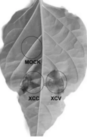
Fig. 1 HR symptoms produced by Xanthomonas campestris pv.
campestris (nonhost-incompatible) and Xanthomonas campestris pv. vesicatoria race1 (host-incompatible) on pepper leaf. X.
campestris pv. campestris and X. campestris pv. vesicatoria were infiltrated with a needle-less syringe into symmetrical sides of a pepper leaf at a concentration of 1 × 10⁸ cfu/ml. HR developed on both the inoculated sites approximately around the same time.
Photograph was taken 72 h after inoculation.
incubation, 3.0 ml of 0.1 N HCl was added to stop the enzyme reaction and the mixture was extracted with diethyl ether.
Then the extracted solution was frozen at -30°C to isolate the ether phase from the mixture emulsion. After thawing the frozen mixture at room temperature, 0.5 ml of the ether phase was picked up and added to 9.5 ml of ethanol. The mixture solution was monitored for LOX activity with a spectropho- tometer (Hitachi, Japan) at 234 nm absorbance.
Nitric oxide assay
The hemoglobin-trapping technique, based on the conversion of the ferrous form of hemoglobin (HbO2) into the ferric form, methemoglobin (metHb), by NO (Murphy and Noack, 1994) was used for the detection of NO. Briefly, 25 mg/ml of hemoglobin (HbO2, Sigma, MO, USA) was transferred to 50 mM phosphate (pH 7.4) and was gently swirled to dissolve hemoglobin, to which 1-2 mg of sodium hydrosulfate was added. The container was gently swirled to provide a light stream of O2 continuously into the container. The resulting HbO2 solution was desalted and purified by passing it through a Sephadex G-25 column at a flow rate of 1 ml/min so that the HbO2 elutes from the column after about 10 min. The con- version of metHb to HbO2 was monitored by the change of color as the metHb was reduced by the sodium dithionite to deoxyhemoglobin and then from purple to a bright orange-red due to the reaction of deoxyhemoglobin with the oxygen contained in the PO4 buffer. Appropriate amount of samples were added to NO reaction mixture containing 50 mM Tris- HCl (pH 7.5), 0.33 mM HbO2, and to the final volume to 1 ml. The purity and concentration of the desalted HbO2 stock was checked using a spectrophotometer (Hitachi, Japan) at 415 nm absorbance.
Results
The timing of occurrence of nonhost and gene-for-gene HRs induced in pepper by Xanthomonas campestris pathovars X. campestris pv. vesicatoria and X. campestris pv. campestris strains were used in this study. Both bacterial strains had the same number of colony forming units (cfu)/ml at an OD600 of 1.0 and exhibited the same growth kinetics in vitro (data not shown). Red pepper plants (Kalmi 25-11-3-2) carrying the Bs3 resistance gene were inoculated with X. campestris pv.
vesicatoria strains expressing avrBs3. HR (due to gene-for- gene resistance) started to appear 60 h after inoculation and was more evident after 72 h of inoculation (Fig. 1). When the same pepper plants were inoculated with X. campestris pv.
campestris, nonhost HR appeared at the same time as that of the gene-for-gene HR (Fig. 1).
Oxidative burst during host and nonhost resistance responses We determined the amount of ROS accumulated in pepper plants at different times after inoculation by X. campestris pv. campestris (nonhost-incompatible) or X. campestris pv.
vesicatoria race1 (host-incompatible). Levels of O2- per 2 × 2 cm leaf samples were determined by electron paramagnetic resonance (EPR) measurement of the Tiron semiquinone radical which is generated from the inoculated leaf sample.
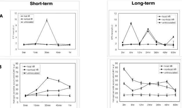
A very strong EPR signal for nonhost-incompatible inter- action was seen as early as 30 min after nonhost pathogen inoculation (Fig. 2A). A second and a third peak of EPR signals were seen at 6 h and 24 h after inoculation, respec- tively. Interestingly, in the host-incompatible interaction the first peak of signal was not seen until 24 h after inoculation and a second peak was seen at 60 h after inoculation (Fig. 2A).
We also determined the amount of H2O2 accumulation during host and nonhost resistance responses using the O-dianisidine method (Desagher et al., 1997). The H2O2 burst pattern during a plant-microbe interaction has been reported to have two phases (Wojtaszek, 1997). In our experiment, we also found two phases of H2O2 burst during both host and nonhost re- sistance responses (Fig. 2B). However, the two phases of H2O2 burst were less distinct in the host-incompatible in- teraction compared to the nonhost-incompatible interaction.
Interestingly, the H2O2 burst was much quicker during the nonhost-incompatible interaction (peaks observed at 30 min
Fig. 2 Pattern of O2- and H2O2 generation during host and nonhost resistance. (A). EPR measurements of the 1,2-dihydroxy- benzene-3,5-disulphonic acid. Leaf samples were cut by 2 cm2 at each sample collection time after both host and nonhost pathogens for superoxide detection. EPR spectra were recorded at a field setting of 3450 G, microwave frequency of 9.67 GHz, modulation of 100 kHz time constant of 163 m, and total scan time of 335 s with a variable gain. Xanthomonas campestris pv. vesicatoria race 1-pepper (◆, host-incompatible interaction), Xanthomonas campestris pv. campestris-pepper (■, nonhost-incompatible interaction), and water-pepper (▲, mock) interactions are depicted. (B) Measurement of H2O2 generation by O-dianisidine method. 1 g of fresh leaves that were inoculated with host or nonhost pathogen was homogenized for H2O2 detection. 0.5 ml of the supernatant was mixed with 2.5 ml peroxide solution and the absorbance was determined at 436 nm. X. campestris pv. vesicatoria race 1-pepper (◆, host-incompatible interaction), X. campestris pv. campestris-pepper (■, nonhost-incompatible interaction), and water-pepper (▲, mock) interactions are depicted. The data are the mean values ± standard deviations of three replicates.
and 3 h after inoculation) when compared to the host-incom- patible interaction (peaks observed at 3 h and 48 h after inoculation; Fig. 2B). When visible tissue necrosis appeared (≈ 60 h), the level of H2O2 decreased in both interactions as mock treated sample level. These results suggest that the ac- cumulation of ROS happens a lot quicker during nonhost resistance responses when compared to the host resistance responses.
Scavenger enzyme activities during host and nonhost resistances
Plants have several superoxide dismutase (SOD) that are constitutively expressed under natural conditions (Bowler.
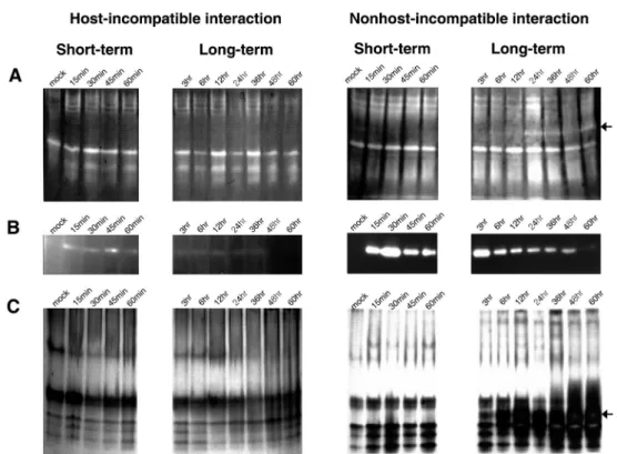
et al., 1994). When plants are attacked by incompatible pathogens, O2- is generated by membrane bound NADPH- oxidase. The generated O2- is the initiation molecule of the oxidative burst in the plant cell. SOD converts the O2- to H2O2. Several SOD isozymes (Fig. 3A) were resolved from extracts of pathogen inoculated pepper plants. As shown in Fig. 3A, the nonhost-incompatible interaction induced an additional SOD isozyme (see arrow on Fig. 3A) that was not
detected for the host-incompatible interaction.
The H2O2 generated during oxidative burst could be detox- ified by scavenging enzymes such as catalases (CATs) and peroxidases (POXs). We determined the CAT activity in the inoculated leaf area by an in-gel activity staining method (Zou and Schrempf, 2000) during both the host- and nonhost- incompatible interactions. In the case of the host-incompatible interaction, weak CAT activity was detected from 15 min to 1 h after inoculation (Fig. 3B). In contrast, for the nonhost- incompatible interaction, strong CAT activity was observed from 15 min to 60 h after inoculation (Fig. 3B). Highest CAT activity was observed at 30 min (phase I) and 3 h (phase II) after inoculation and subsequently the CAT activity decreased from 6 h after inoculation (Fig. 3B). Strikingly, the CAT activity patterns during the nonhost-incompatible interaction nearly matched the H2O2 generation pattern (Fig. 2B). Overall, the CAT activity was higher in the nonhost-incompatible interaction than in host-incompatible interaction, as was observed for H2O2 generation. The in-gel activity assay did not resolve different CAT isozymes in either interaction.
Activity staining for peroxidase (Zou and Schrempf, 2000) revealed five major isozymes for both the host- and nonhost-
Fig. 3 Scavenger enzyme activities of the host- and nonhost-incompatible interactions during various times after pathogen inoculation.
(A) Superoxide dismutase in gel assay. 50 µg protein, extracted from pathogen inoculated pepper leaves, on each lane was separated on 10% non-denaturing PAGE. The color (shown as white) was developed by 0.25 mM nitroblue tetrazolium chloride. The arrow and numbers indicate superoxide dismutase isozymes. Note a new isozyme (arrow #4) that is induced specifically during the nonhost- incompatible interaction. (B) Measurement of catalase activity by in gel assay. Each lane contains 50 µg of protein, extracted from pathogen inoculated pepper leaves, separated on 8% non-denaturing PAGE with 3% stacking gel. Color development (shown as white) was continued for 4 min and reaction was stopped with a brief wash in double-distilled water. (C) Peroxidase activity during the host- and nonhost-incompatible interactions by in gel activity assay. Each lane contains 50 µg of protein, extracted from pathogen inoculated pepper leaves, separated on 8% non-denaturing PAGE with 3% stacking gel. The gels were incubated in 0.1 M sodium acetate buffer (pH 4.5) containing 2 mM benzidine, and initiated the reaction by the addition of 3 mM H2O2. The arrow and the numbers indicate peroxidase isozymes that have differential migration on the gel. The star represents a peroxidase isozyme induced only during the nonhost-incompatible interaction and not during the host-incompatible interaction.
incompatible interactions (Fig. 3C). The peroxidase activity during the nonhost-incompatible interaction significantly increased 6 h after pathogen inoculation in contrast to the host-incompatible interaction where the activity did not appear to change. Interestingly, for the nonhost-incompatible interaction, a new peroxidase isozyme appeared at 3 h after inoculation and its activity further increased until 60 h after inoculation (marked with arrow on Fig. 3C). Collectively, these results suggest that scavenging enzyme activities differ between the host- and nonhost-incompatible interactions and in some cases the pattern of activity is dependent on the pattern of ROS production.
Lipoxygenase activity and production of nitric oxide during host and nonhost resistance responses
Lipoxygenase (LOX) activity was measured spectropho- tometrically at 234 nm (Macias. et al. 1991; see Materials
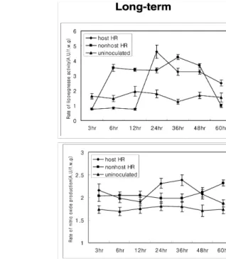
and Methods) during the host- and nonhost-incompatible interactions. Interestingly the pattern of LOX activity was opposite to the pattern of H2O2 generation (Figs. 2B and 4A).
At initial time points after inoculation, the LOX activity was much higher during the host-incompatible interaction, and the enzyme activity was seen as early as 30 min after in- oculation. Distinctive peaks of activity were seen at 30 min, 1 h and 24 h after inoculation (Fig. 4A). During the nonhost -incompatible interaction, no increase in LOX activity was detected until 6 h after inoculation and subsequently a higher LOX activity was maintained until 48 h after inoculation (Fig. 4A). These results suggest that LOX activity is inde- pendent of AOS production and is triggered more quickly in the host-incompatible interaction when compared to the nonhost-incompatible interaction.
Nitric oxide (NO) production patterns during both the host- and nonhost-incompatible interactions were measured spec- trophotometrically with the hemoglobin NO detection
Fig. 4 Lipoxygenase activity and nitric oxide generation during the host- and nonhost-incompatible interactions. (A) Lipoxygenase activity during the host-incompatible interaction (◆, Xanthomonas campestris pv. vesicatoria race 1-pepper), nonhost-incompatible interaction (■, Xanthomonas campestris pv. campestris-pepper) and mock treated control(▲). 0.5 g of pathogen inoculated pepper leaves were ground for each treatment and the samples were monitored for lipoxygenase activity with a spectrophotometer at 234 nm as described in Materials and Methods section. (B) Determination of nitric oxide generation during the host-incompatible interaction (◆) and a nonhost-incompatible interaction (■) by hemoglobin method. ▲ represent mock treated control. 1 g of pathogen inoculated pepper leaves were homogenized for the assay. Absorbance at 415 nm was measured by spectrophotometer. Error bars represent the standard deviation of three repetitions.
method (Murphy and Noack, 1994). Surprisingly, the pattern of NO production was initially similar (up to 12 h after inoc- ulation) during both the host- and nonhost-incompatible in- teractions. The NO production abruptly increased from 15 min after inoculation for both the host- and nonhost-inco- mpatible interactions (Fig. 4B). The pattern of NO production was slightly different from 12 h after inoculation between the host- and nonhost-incompatible interactions (Fig. 4B). These results suggest that, at initial stages of infection, both the host- and nonhost-incompatible interactions trigger NO pro- duction, presumably using the same pathway.
Discussion
Similarities exist between the host and nonhost resistance responses (Thordal-Christensen, 2003; Mysore and Ryu, 2004) but it is still not clear, if the same mechanism is involved in producing these resistance responses. For ex- ample the HR produced during type II nonhost resistance (Mysore and Ryu, 2004) has similar properties to that of the HR produced during host/gene-for-gene resistance. However,
the signal transduction pathway leading to HR development may be different between the two interactions. Here we used the interaction between the pepper and the two very similar Xanthomonas campestris pathovars. As a model system to investigate differences between the host and nonhost re- sistance responses.
Rapid generation of superoxide (oxidative burst) and accu- mulation of H2O2 in plants are characteristics of an early plant response during the hypersensitive reaction due to perception of an incompatible pathogen. Superoxide dismutase (SOD), which catalyzes the conversion of O2- to H2O2, is a metal-containing enzyme that is found in plant cytosol, chloroplast and mitochondria. Phase I of the oxidative burst is a relatively short lived non-specific response that occurs immediately after the inoculation with either compatible or incompatible pathogens (Baker and Orlandi, 1995). Phase II of the oxidative burst is a relatively long-lived response occurring 1.5 h to 3 h after pathogen attack and appears to be specific to incompatible interactions (Adam et al., 1995; Lamb and Dixon, 1997). Grant et al. reported that cytosolic Ca2+
concentration should be increased within 60 min by Pseu- domonas syringae pv. tomato avrRpm1/RPM1 interaction
(Grant et al., 2000). This data suggest that even phase I oxidative burst also related gene-for-gene interaction. Because increases Ca2+ concentration in cytosol is essentially required to trigger oxidative burst when the plant has interaction with a pathogen (Grant et al., 2000). However, more direct evi- dence is not available to explain phase I oxidative burst is a specific response or not. We presumably assume phase I oxidative burst could be related pathogen-associated molecular patterns (PAMPs) recognition.
Our results confirm the two distinct phases of oxidative burst during both the host- and nonhost- incompatible interactions.
However, the timing of oxidative burst differed. During the nonhost-incompatible interaction the EPR signal was am- plified in 30 min and 6 h and in the host-incompatible interaction the EPR signal was increased at 24 h after inoc- ulation. In spite of the fact that the increase of EPR signal was much faster during the nonhost-incompatible interactions (Fig. 2), an additional SOD isozyme was expressed only during the nonhost-incompatible interaction (Fig. 3A). This additional SOD enzyme could be using the rapidly generated (6 h after inoculation) superoxide as a substrate in the nonhost-incompatible interaction, but not in the host-inco- mpatible interaction. We detected new inducible SOD enzyme activity exist until 60 hour after inoculation, even O2- generation level goes down as normal level. We suggest that perhaps plant keep defense system as turn on or the other possible signal trigger to keep SOD expression 6 hour after inoculation. Adam et al. (1995) previously elucidated the activity of SOD isozyme during a host-incompatible inter- action of bean/Pseudomonas syringae pv. phaseolicola.
Our results are in agreement with their results for the host- incompatible interaction.
H2O2, formed from O2-, can stimulate phytoalexin accumu- lation and can induce expression of defense-related genes like PAL and CHS (Thomas et al., 2003; Jabs et al., 1997;
Baker et al., 1995; Alvarez et al., 1998). H2O2 has been im- plicated to play an active role in plant defense responses against pathogens (Levine et al., 1994; Mittler et al., 1996;
Chamnongpol et al., 1998). H2O2 induces benzoic acid 2-hy- droxylase (BA2H) activity and salicylic acid (SA) accumulation at concentrations above 30 mM. However, defense gene ex- pression and visible cell death due to H2O2 application are dependent on plant species. For example, in tobacco leaves infiltrated with 300 mM H2O2, BA2H activity increased 2.3- fold within 1 h when compared to the non-infiltrated leaf (Leon et al., 1995). Transgenic tobacco deficient in the H2O2
scavenger enzyme, catalase (antisense lines), has been used as an inducible and noninvasive system to study the role of H2O2 as an activator of pathogenesis-related proteins in plants (Chamnogpol et al., 1998). These results suggest that
H2O2 is probably involved in plant defense. But sensitivity to various concentrations of H2O2 depends on plant species. We infiltrated 40 mM 1,3-dimethyl-2-thiourea (DMTU) along with the bacteria to trap the H2O2 that is produced during the host- and nonhost-incompatible interactions. Interestingly, the application of DMTU did not abolish the HR during both the host- and nonhost-incompatible interactions indicating that H2O2 production is most likely not required for HR (data not shown).
When tobacco leaves were infiltrated with Pseudomonas syringae pv. glycinea, H2O2 accumulation consisted of two distinct phases, I and II, that appeared 30 min and 2-4 h after inoculation (Baker et al., 1995). Our results confirm the two distinct phases of H2O2 accumulation during both the host- and nonhost-incompatible interactions (Fig. 2). However, the timing and the amount of H2O2 accumulation varied between the host- and nonhost-incompatible interactions (Fig. 2), although both interactions produced HR at almost the same time (Fig 1). More and rapid H2O2 accumulation was observed during the nonhost resistance response when compared to the host resistance response. Baker et al. (1995) have previously emphasized that H2O2 accumulation does not necessarily lead to HR formation in plants. It has also been shown that the hypersensitive cell death of tobacco suspension cells was not directly attributed to ion flux and H2O2 generation pattern of phase I and II elicited by hrp- bacterium (Chamongpol et al., 1998). Our results also confirm that the accumulation of H2O2 and ROS is most likely not directly related to the HR.
During nonhost resistance the activity of catalase (CAT), a H2O2 scavenger, appeared within 15 min and sustained until 60 h with maximum activity at 30 min and 3 h after inoc- ulation (Fig. 3B). Activity staining of peroxidase (POX) during nonhost resistance revealed an additional POX isozyme at 3 h after inoculation and the activity subsequently increased until 60 h after inoculation (Fig. 3C). These results suggested that during early stages H2O2 is most likely scavenged by CAT and during the late stages by POX. Interestingly, the hypersensitive cell death from neither the host-incompatible interaction nor the nonhost-incompatible interaction was ob- served before 60 h after inoculation, even though the gen- eration of superoxide and accumulation of H2O2 were of distinctly different pattern. Do et al. (2003) showed that during the host-incompatible interaction of pepper with X.
campestris pv. vesicatoria, the peroxidase-like enzyme activity decreased 18 h after inoculation. We did not observe such a drastic decrease in our study. The discrepancy between these data is probably due to the different inoculation methods used. Do et al. used vacuum infiltration and observed HR by 18 h after inoculation whereas in our study the leaves were
infiltrated with a needle-less syringe and we observed HR by 60 h after inoculation. Different methods used to measure the enzyme activity and different pepper cultivars used between the two studies may also have contributed to this discrepancy.
If the reactive oxygen species are involved in the early phase of HR, suggested by others (Jabs et al., 1997; Levine et al., 1998 Mittler et al., 2002), the timing of cell death during the host- and nonhost-incompatible interactions should have been different. However, we did not see any significant differences in the timing of cell death during the host- and nonhost-incompatible interactions (Fig. 1). These results led us to consider the involvement of the octadecanoid pathway for HR development. Many reports have proposed that enzymes, such as lipoxygenase (LOX), of the octadecanoid pathway act as a putative endogenous signal to develop HR (Veronesi et al., 1996; Koch et al., 1992; Titarenko et al., 1997; Bohland et al., 1997; Rance et al., 1998; Leyen et al., 1998). LOX activities in the leaves of rice were rapidly activated by inoculation with an incompatible race but not with a compatible race of the rice blast fungus Magnaporthe grisea (Chta et al., 1991). Our results indicated increase in LOX activity during early stages (30 min and 1 h) of the host-incompatible interaction while the increase of LOX activity was observed 6 h after inoculation during the nonhost -incompatible interaction (Fig 4A). Interestingly, these results were in contrast to the pattern for superoxide and H2O2 accumulation that we observed during the same host and nonhost resistance responses.
Nitric oxide (NO) is known to be involved synergistically with ROS in induction of plant defense mechanism (Del- ledonne et al., 1998). Application of nitric oxide synthase and its substrate arginine has been shown to activate PR-1, cGMP and PAL genes (Durner et al., 1998). Interestingly in our assays, unlike ROS, we did not see any significant dif- ference in the NO accumulation pattern between the host- and nonhost-incompatible interactions. These results suggest ROS and NO generation could result from different pathways after a plant recognizes a pathogen attack. Several recent studies suggest that reactive oxygen and nitric oxide species do not elicit hypersensitive cell death during plant-microbial interaction (Tada et al., 2004; Zhang et al., 2003; Christopher- Kozjan and Heath, 2003). Based on our results regarding the pattern for ROS accumulation, H2O2 accumulation, LOX activity, and NO concentration during host and nonhost re- sistance responses, we propose three possible hypotheses. 1) Oxidative burst does not play an important role in triggering the plant hypersensitive cell death during host and nonhost resistance responses. 2) Host and nonhost resistance responses in pepper, due to Xanthomonas campestris, are triggered by distinct pathways. 3) Possibly, ROS has synergetic effect
with octadecanoid pathway to trigger HR in plant. Early stages of a nonhost-incompatible interaction are NADPH- oxidase complex dependent, while that of a host-incompatible interaction are dependent on LOX activity. Interestingly the NO signal is being activated in both interaction systems in a similar pattern. It warrants further studies to determine which pathways are activated during host and nonhost resistance responses.
Acknowledgement We thank Dr. Byung-Su Kim, Kyungpuk National Univ., S. Korea for the pepper seeds and Dr. John Damicone, Oklahoma State Univ., OK, USA, for providing the bacterial pathogens (Xanthomonas campestris pv. cam- pestris and Xanthomonas campestris pv. vesicatoria race1).
We thank Dr. Rick Dixon for critical reading of the manuscript. This work was partly supported by the Noble Foundation (K. S. Mysore) and funds from Gyeongsang National University to H. K. Kim.
Literature cited
Adam, A.L., Bestwick, C.S., Barna, B., Mansfield, J.W. (1995) Enzymes regulating the accumulation of active oxygen species during the hypersensitive reaction of bean to Pseudomonas syringae pv. phaseolicola. Planta 197:240-249
Alvarez, M.E., Pennell, R.I., Meijer, P., Ishikawa, A., Dixon, R., Lamb, C. (1998) Reactive oxygen intermediates mediate a sys- temic signal network in the establishment of plant immunity.
Cell 92:773-784
Baker, C.J., Orlandi, E.W. (1995) Active oxygen in plant path- ogenesis. Annu. Rev. Phytopathol. 33:299-321
Beligni, M.V., Lamattina, L. (2000) Nitric oxide stimulates seed germination and deetiolation, and inhibits hypocotyl elongation, three light-inducible responses in plants. Planta 210:215-221 Bohland, C., Balkenhohl, T., Loers, G., Feussner, I., Grambow,
H.J. (1997) Differential induction of lipoxygenase isoform in wheat upon treatment with rust fungus elicitor, chitin oligo- saccharides, chitosan and methyl jasmonate. Plant Physiol.
114:679-685
Bowler, C. Van-Camp, W., Van-Montagu, M., Inze, D. (1994) Superoxide dismutase in plants. Crit. Rev. Plant Sci. 13:
199-218
Bradford. (1976) A rapid and sensitive methods for the quantitation of microgram of protein utilizing the principle of protein dye binding. Anal. Biochem. 72:248-254
Cacas JL, Marmey P, Montillet JL, Sayegh-Alhamdia M, Jalloul A, Rojas-Mendoza A, Clérivet A, Nicole M. (2009). A novel patatin-like protein from cotton plant, GhPat1, is co-expressed with GhLox1 during Xanthomonas campestris-mediated hy- persensitive cell death. Plant Cell Rep. 28(1):155-164 Chamnongpol, S., Willekens, H., Moeder, W, Langebartels, C.
Sandermann, H., Montagu, M., Inze, D., Camp, W. (1998)
Defense activation and enhanced pathogen tolerance induced by H2O2 in transgenic tobacco. Proc. Natl. Acad. Sci. USA.
95:5818-5823
Christopher-Kozjan, R., and Heath, C.M. (2003) Cytological and pharmacological evidence that biotrophic fungi trigger dif- ferent cell death execution processes in host and nonhost cells during the hypersensitive response. Physiol. Mol. Plant Pathol.
62:265-275
Chta, H., Shida, K., Peng, Y., Furusawa, I., Shishiyama, J., Aibara, S., Morita, Y. (1991) A lipoxygenase pathway is activated in rice after infection with the rice blast fungus Magnaporthe grisea. Plant Physiol. 97:94-98
Conrads-Strauch, J., Dow, J.M., Milligan, D.E., Parra, R., Daniels, M.J. (1990) Induction of hydrolytic enzymes in Brassica cam- pestris in response to pathovars of Xanthomonas campestris.
Plant Physiol. 93:238-243
Del Rio, L.A., Corpas, F.J., Barroso, J.B. (2004) Nitric oxide and nitric oxide synthase activity in plants. Phytochemistry 65:
783-792
Delledonne, M., Xia, Y., Dixon, R.A., Lamb, C. (1998) Nitric oxide functions as a signal in plant disease resistance. Nature 394: 585-588
Desagher, S., Glowinski, J., Prémont, J. (1997) Pyruvate protects neurons against hydrogen peroxide-induced toxicity. J. Neurosci.
17:9060-9067
Do, H.M., Hong, J.K., Jung, H.W., Kim, S.H., Ham, J.H., Hwang, B.K. (2003) Expression of peroxidase-like genes, H2O2 pro- duction, and peroxidase activity during the hypersensitive re- sponse to Xanthomonas campestris pv. vesicatoria in Capsicum annuum. Mol. Plant-Microbe Interact. 16:196-205
Durner, J., Wendehenne, D., Klessig, D.F. (1998) Defense gene induction in tobacco by nitric oxide, cyclic GMP, and cyclic ADP-ribose. Proc. Natl. Acad. Sci. USA. 95:10328-10333 Fan XW, Li FM, Song L, Xiong YC, An LZ, Jia Y, Fang XW.
(2009) Defense strategy of old and modern spring wheat varieties during soil drying. Physiol Plant. 136(3):310-323 Feussner, I., Wasternack, C. (2002) The lipoxygenase pathway.
Annu. Rev. Plant Biol. 53:275-297
Grant, M., Mansfield, J. (1999) Early events in host-pathogen interactions. Curr. Opin. Plant Biol. 2:312-319
Grant, M., Brown, I., Adams, S., Knight, M., Ainslie, A. and Mansfield, J. (2000) The RPM1 plant disease resistance gene facilitates a rapid and sustained increase in cytosolic calcium that is necessary for the oxidative burst and hypersensitive cell death. The Plant Journal, 23:441-450
Hammond-Kosack, K.E., Jones, J.D.G. (1996) Resistance gene- dependent plant defense responses. Plant Cell 8:1773-1791 Huckelhoven, R., Kogel, K. (2003) Reactive oxygen intermediates
in plant-microbe interactions: Who is who in powdery mildew resistance? Planta 216:891-902
Jabs, T., Tschope, M., Colling, C., Hahlbrock, K., Scheel, D.
(1997) Elicitor-stimulated ion fluxes and O2- from the oxidative burst are essential components in triggering defense gene activation and phytoalexin synthesis in parsley. Proc.
Natl. Acad. Sci. USA. 94:4800-4805
Keen, N.T. (1990) Gene-for-gene complimentarity in plant-pathogen interactions. Annu. Rev. Genet. 24:447-463
Keppler, L.D., Baker, C.J., Atkinson, M.M. (1989) Active oxygen production during a bacteria-induced hypersensitive reaction in tobacco suspension cells. Phytopathol. 79:974-978 Koch, E., Meier, B.M., Eiben, H., Slusarenko, A. (1992) A Lipoxy-
genase from leaves of tomato (Lycopersicon esculentum Mill.) is induced in response to plant pathogenic Pseudomonas.
Plant Physiol. 99:571-576
Lam, E., Kato, N., Lawton, M. (2001) Programmed cell death, mitochondria and the plant hypersensitive response. Nature 411:848-853
Lamb, C., Dixon, R.A. (1997) The oxidative burst in plant disease resistance. Annu. Rev. Plant Physiol. Plant Mol. Biol. 48:
251-275
Leon, J., Lawton, M.A., Raskin, I. (1995) Hydrogen peroxide stimulates salicylic acid biosynthesis in tobacco. Plant Physiol.
108:1673-1678
Levine, A., Tenhaken, R., Dixon, R., Lamb, C. (1994) H2O2 from the oxidative burst orchestrates the plant hypersensitive disease resistance response. Cell 79:583-593.
Leyen, K., Duvoisin, R.M., Engelhardt, H., Wiedmann, M. (1998) A function for lipoxygenase in programmed organelle degra- dation. Nature 395:392-395
Leto TL, Geiszt M. (2006) Role of Nox family NADPH oxidases in host defense. Antioxid Redox Signal. 8(9-10):1549-1561 Marois, E,, Van den Ackerveken, G,, Bonas, U. (2002) The
Xanthomonas type III effector protein AvrBs3 modulates plant gene expression and induces cell hypertrophy in the susceptible host. Mol. Plant-Microbe Interact. 15:637-646 Mittler, R., Seskar, V.S.M., Lam, E. (1996) Inhibition of pro-
grammed cell death in tobacco plants during a pathogen- induced hypersensitive response at low oxygen pressure.
Plant Cell 1991-2001
Murphy, M.E, Noack, E. (1994) Nitric oxide assay using hemoglobin method. Methods Enzymol. 233:240-250
Mysore, K.S., Ryu, C. (2004) Nonhost resistance: how much do we know? Trends Plant Sci. 9:97-104
Park, H.J., Miura, Y., Kawakita, K., Yoshioka, H., Doke, N. (1998) Physiological mechanisms of a sub-systemic oxidative burst triggered by elicitor-induced local oxidative burst in potato tuber slices. Plant Cell Physiol. 39:1218-1225
Rance, I., Fournier, J., Esquerre-Tugaye, M. (1998) The incompatible interaction between Phytophthora parasitica var. nicotianae race 0 and tobacco is suppressed in transgenic plants expressing antisense lipoxygenase sequences. Proc. Natl. Acad. Sci. USA.
95:6554-6559.
Schornack S, Meyer A, Römer P, Jordan T, Lahaye T. (2006) Gene-for-gene-mediated recognition of nuclear-targeted AvrBs3 -like bacterial effector proteins. J Plant Physiol. 163(3):256-272 Tada, Y., Mori, T., Shinogi, T., Yao, N., Takahashi, S., Betsuyaku,
S., Sakamoto, M., Park, P., Nakayashiki, H., Tosa, Y., Mayama, S. (2004) Nitric oxide and reactive oxygen species do not elicit hypersensitive cell death but induce apoptosis in the adjacent cells during the defense response of oat. Mol.
Plant-Microbe Interact. 17:245-253
Thomas, I., Loeffler, C., Sinha, A.K., Gupta, M., Krischke, M., Steffan, B., Roitsch, T., Mueller, M.J. (2003) Cyclopentenone isoprostanes induced by reactive oxygen species trigger defense gene activation and phytoalexin accumulation in plants. Plant J. 34:363-375
Thordal-Christensen, H. (2003) Fresh insights into processes of nonhost resistance. Curr. Opin. Plant Biol. 6:351-357 Titarenko, E., Rojo, E., Leon, J., Sanchez-Serrano, J.J. (1997)
Jasmonic acid-dependent and -independent signaling pathway control wound-induced gene activation in Arabidopsis thaliana.
Plant Physiol. 115:817-826
Valgimigli, L., Pedulli, G.F., Paolini, M. (2001) Measurement of oxidative stress by EPR radical-probe technique. Free Radical Biology & Medicine. 31:708-716
Veronesi, C., Rickauer, M., Fournier, J., Pouenat, M., Esquerre- Tugaya, M. (1996) Lipoxygenase gene expression in the tobacco-Phytophthora parasitica Nicotianae interaction.
Plant Physiol. 112:997-1004
Wendehenne, D., Durner, J., Klessig, D.F. (2004) Nitric oxide: a new player in plant signaling and defense responses. Curr.
Opin. Plant Biol. 7:449-455
Wojtaszek, P. (1997) Oxidative burst: an early response to path- ogen infection. Biochem. J. 322:681-692
Wright, C., Beattie, G.A. (2004) Pseudomonas syringae pv.
tomato cells encounter inhibitory levels of water stress during the hypersensitive response of Arabidopsis thaliana. Proc.
Natl. Acad. Sci. USA. 101:3269-3274
Yoshioka, H., Numata, N., Nakakima, K., Katou, S., Kawakita, K., Rowland, O., Jones, D.G.J., Doke, N. (2003) Nicotiana ben- thamiana gp91phox homologs NbrbohA and NbrbohB participate in H2O2 accumulation and resistance to Phytophthora infestans.
Plant Cell 15:706-718
Zhang, C., Czymmek, K.J., Shapiro, A.D. (2003) Nitric oxide does not trigger early programmed cell death events but may contribute to cell-to-cell signaling governing progression of the Arabidopsis hypersensitive response. Mol. Plant-Microbe Interact. 16: 962-972
Zou, P., Schrempf, H. (2000) The heme-independent manganese- peroxidase activity depends on the presence of the C-terminal domain within the Streptomyces reticuli catalase-peroxidase CpeB. Eur. J. Biochem. 267:2840-2849