성균관대학교 의과대학 내과학교실 마산삼성병원 호흡기내과 김우열, 강구현, 이진호, 박순효, 강경우
A Case of Primary Endobronchial Leiomyosarcoma Noticed by Massive Hemoptysis
Woo Youl Kim, M.D., Gu Hyun Kang, M.D., Jin Ho Lee, M.D., Sun Hyo Park, M.D., Kyung Woo Kang, M.D.
Division of Pulmonary and Critical Care Medicine, Department of Internal Medicine, Masan Samsung Hospital, Sungkyunkwan University School of Medicine, Masan, Korea
A primary pulmonary leiomyosarcoma is a very rare pulmonary malignancy that arises from smooth muscle of either the bronchial or arterial walls. Common symptoms of the tumor are cough, dyspnea, chest pain and hemoptysis. The diagnosis of a primary pulmonary sarcoma can be established only after extensive clinical and radiologic examinations have failed to identify an alternative primary source. The only effective treatment for the tumor is a complete surgical resection when feasible. The type of resection is dictated by the local anatomic extent of the tumor.
We report a case of a 21-year-old male with a primary endobronchial leiomyosarcoma who presented with massive hemoptysis. A necrotic ulcerative endobronchial lesion was observed in the orifice of left lower lobe bronchus on a bronchoscopic examination. He was treated with a complete sleeve resection of the left lower lobe. Three months later, local recurrence of the tumor was noticed on the follow up bronchoscopy and a then left pneumonectomy was then performed. Fifteen months later, the patient died from empyema with a bronchopleural fistula that was associated with tumor recurrence at the stump of the pneumonectomy. (Tuberc Respir Dis 2006; 61: 484-489)
Key words: Endobronchial leiomyosarcoma, Surgical resection.
Address for correspondence: Kyung Woo Kang, M.D.
Division of Pulmonary and Critical Care Medicine, Department of Internal Medicine, Masan Samsung Hospital, Sungkyunkwan University School of Medicine, 50 Habsung 2-dong, Masan 630-723, Korea
Phone: (8255)290-6399 Fax: (8255)290-6654 E-mail: [email protected]
Received: Aug. 7. 2006 Accepted: Sep. 15. 2006
서 론
호흡기관에 발생하는 원발성 평활근육종은 매우 드 문 종양으로서1-5 초기 임상증상이 비특이적이고 통상 의 경기관지 생검이나 경피적 세침흡인 생검으로는 육종의 진단에 필요한 충분한 검체를 얻기가 힘들어 폐생검의 목적을 포함한 시험적 수술(exploratory operation)로 진단되는 경우가 많다6,7.
원발성 폐평활근육종의 치료로는 종양의 근치적 외 과적 절제를 통해서만 완치를 기대할 수 있으며8-10,수 술 후의 보조적 항암 화학요법이나 보조적 방사선 치 료는 그 효과가 미미한 것으로 알려져 있다1,11.
저자들은 대량객혈을 주소로 내원한 환자에서 시험 적 개흉술을 통해 원발성 기관지내 평활근육종을 진 단하고 좌하엽 수상절제술(sleeve resection)을 시행 하였다. 수술 후 3개월 째 문합부위에서 종양이 재발 하여 좌측 전폐절제술을 시행하였는데, 2차 수술 후 11개월이 경과하여 다시 수술부위에 종양이 재발하고 이와 연관된 기관지흉막루와 농흉으로 사망한 증례를 경험하였기에 문헌 고찰과 함께 보고하는 바이다.
증 례
환 자: 21세 남자
주 소: 400ml 정도의 대량객혈
현병력: 내원 2년 전부터 수개월에 한번씩 간헐적 기침과 혈담이 있었으나 특별한 검사나 치료는 받지 않은 상태로 군입대 후 훈련도중 400ml이상의 대량 객혈이 발생하였다. 객혈 발생 후 상급 군병원으로 이 송되어 객혈에 대한 치료와 검사 후 객혈이 멎은 상태 로 객혈 발생 7일 째 기관지내시경 검사를 위해 전원 되었다.
(A) (B)
Figure 1. (A) Chest radiograph shows no abnormal mass or infiltration. (B) High resolution computed tomographic scan of the chest demonstrates endobronchial occupying soft tissue mass which nearly obstructs the lumen of left lower lobe bronchus.
(A) (B)
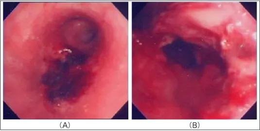
Figure 2. (A) Bronchoscope shows ulcerative mucosal lesions with necrosis on the distal portion of the left main bronchus and the left lower lobe bronchus. (B) The left lower lobe bronchus is narrowed with necrotic materials.
과거력: 특이사항 없음 가족력: 특이사항 없음 흡연력: 비흡연자
진찰소견: 내원 당시 혈압은 140/90mmHg, 맥박수 는 100회/분, 호흡수는 20회/분, 체온은 36.6℃이었으 며 의식은 명료하였다. 결막 관찰에서 빈혈 징후는 보 이지 않았으며 경부 림프절 비대는 없었다. 흉부 청진 에서 좌측 폐 상부에 국한된 천명음이 청진되었고 심 음은 정상이었다. 복부 촉진과 청진에서는 특이소견
이 없었다.
방사선 소견: 단순 흉부 X-선 촬영에서는 특이소 견이 관찰되지 않았으나 군부대에서 촬영한 고해상도 흉부 전산화단층촬영(HRCT)에서는 좌측 주 기관지 의 원위부가 다소 좁아져 있었고, 좌폐하엽 기관지 입 구의 내강이 거의 폐쇄되어 있었다(Figure 1A, B).
검사실 소견: 말초 혈액 검사에서 백혈구 10,400/mm3 (호중구 73%, 림프구 20%, 단핵구 4.8%, 호산구 2.2%), 혈색소 13.0g/dL, 혈소판 319,000/mm3
(A) (B)
(C) (D)
Figure 3. (A) Gross specimen of the left lung shows a whitish mass which grows into the lumen of bronchus and obliterates the lumen of the left lower lobe bronchus. (B) Histopathologic finding of the lesion shows irregular and infiltrating peribronchial growing of spindle cells with partial destruction of bronchial cartilage and hemorrhage necrosis. (H & E stain, ×40) (C) Plump and atypical tumor cells with intratumoral neo-vascularization and 2 mitotic figures, tumor cells have cigar to ovoid shaped cells with presence of hyperchromatism and a few prominent nucleoli in the nuclei. (H & E stain, ×400) (D) Immunohistochemical study of the tumor cells, strong intracytoplasmic positivity of the alpha-SMA(smooth muscle actin) by elongated and spindle in configuration. (α -SMA stain, ×400)
이었다. 혈청 생화학검사에서 총단백질은 7.8g/dL, 알 부민은 4.3g/dL, AST/ALT 13/12 IU/L, ALP 219 IU/L이었고, 총 빌리루빈은 0.5mg/dL, 직접 빌리루빈 은 0.1mg/dL, BUN 9.0mg/dL, Creatinine 0.9mg/dL 이었으며 LDH 292 IU/L이었다. 혈청 전해질 검사에 서 Na 143mEq/L, K 4.3mEq/L, Cl 102mEq/L로 정상 소견을 보였다. 혈액 응고 검사에서 출혈 시간은 2분 30초, 응고 시간은 8분30초였으며 프로트롬빈 시간은 11.6초, 활성 부분프로트롬빈 시간은 32.6초로 정상 범 위 내였다.
기관지내시경 소견: 기관지내시경으로 좌측 주기 관지 원위부의 혈괴를 제거한 후 관찰한 바로는 좌하 엽 기관지 입구에 변연이 불규칙한 직경 1.5cm 정도 의 암적색의 궤양성 점막병변이 관찰되었고 좌하엽의 기관지 입구는 괴사조직과 혈괴로 인해 막혀 있었다.
궤양성 점막병변에 대한 조직검사를 시행하였다 (Figure 2A, B).
임상경과 및 치료: 내원 후 곧바로 시행하였던 경 기관지생검의 결과가 평활근종으로 진단되었으나 궤 양을 동반한 점막병변의 육안적 소견이 기관지내 양
(A) (B)
(C) (D)
Figure 4. (A, B) Both Chest CT with enhance and HRCT shows that soft tissue lesion is growing around the anastomosis site. (C) 0.6cm sized pedunculated mucosal lesion is noticed on the distal trachea. (D) Abnormal mucosal irregularities and pinpoint hole of the bronchopleural fistula on the stump of the left main bronchus.
성종양에서는 흔히 볼 수 없는 소견으로 악성의 가능 성을 완전히 배제할 수 없었고 향후 대량객혈의 재발 가능성을 줄이고자 수술적 폐생검의 목적을 포함한 시험적 개흉술을 시행하기로 하였다. 내원 5일 째 수 술 전 검사로 시행한 폐기능 검사상 1초간 노력성 호 기량(Forced expiratory volume at 1 sec, FEV1)은 3.20L로 추정예측치의 84%, 노력성 폐활량(Forced vital capacity, FVC)은 3.83L로 추정예측치의 92%이 었으며 FEV1/FVC 비는 84%였다. 내원 7일 째 시험 적 개흉술을 시행하였고 수술 중 시행한 동결절편검 사에서 양성 평활근종으로 결과를 보고 받고 환자의
젊은 연령을 고려하여 폐기능의 보존을 위하여 좌하 엽에 대해 수상절제술을 시행하였다(Figure 3). 내원 14일 째 병리과에서 보고된 영구절편검사의 결과가 기관지내 평활근육종으로 나왔고 기관지 절단면에서 암종의 침윤은 없었으나 종양의 근위부 침범부위와의 거리가 8mm 정도로 짧았다. 저자들은 이미 시행하였 던 좌하엽 수상절제술로는 tumor free margin을 충분 히 확보할 수 없어 향후에 국소적 재발의 위험요소가 될 것을 염려하여 환자에게 좌측 전폐절제술을 권유 하였으나 환자는 수술을 거부하고 퇴원하였다.
환자는 퇴원 후 수술 3개월째 다시 소량의 객혈이
재발되어 흉부 전산화단층촬영과 기관지내시경을 시 행하였는데 좌측 주 기관지 원위부의 수술 문합부위 에서의 국소 재발을 확인할 수 있었다(Figure 4). 환 자는 2차 수술을 계획하였으며 수술 전에 원격전이를 확인하기 위해 골주사와 뇌 및 복부에 전산화 단층촬 영을 하였는데 전이를 의심할 만한 소견은 없었다. 1 차 수술 후 4개월 째 재발 부위를 포함한 좌 상엽을 제거하는 좌측 전폐절제술을 시행 받고 퇴원 후 외래 경과관찰을 하였다. 2차 수술 후 11개월 째 발열과 좌측 흉통을 주소로 내원하였는데 기관지내시경 검사 를 시행하여 원위부 기관에서 종양의 재발을 확인하 였고 좌측 주기관지 수술부위의 파열로 기관지흉막루 가 형성되어 농흉이 발생하였다.
환자는 농흉의 치료를 위해 입원하였고 입원 후 10 일 째 종양의 재발과 관련된 농흉으로 치료받던 중 패 혈증으로 사망하였다.
고 찰
폐에 발생하는 원발성 육종은 매우 드물며 발생빈 도는 보고마다 차이는 있으나 폐에 원발성으로 발생 하는 악성 종양의 1%미만으로 알려져 있다1-5. 조직학 적으로는 평활근육종이 가장 많으며 주로 성인에서 발견되나 일부에서는 소아에서 발견되기도 하며13, 대 개 50세 이상에서 호발하고 여자보다 남자에서 좀 더 많이 발생하는 것으로 알려져 있다4,6. 폐에서 발견되 는 육종은 실제 다른 장기로부터 전이된 육종인 경우 가 많아서 원발성 폐육종으로 진단하기 위해서는 충 분한 임상적 검토와 방사선검사를 통해 타 장기로부 터의 전이 가능성을 배제하여야 한다1,8,9,12.
임상증상은 종양의 침범부위에 따라 무증상에서부 터 발열, 체중감소, 기침, 객혈, 흉통, 호흡곤란 등 다 양하며 평활근육종의 경우는 무증상이거나 본 증례의 경우와 같이 객혈을 나타내는 경우가 많다6. 방사선검 사에서는 평활근 육종의 특징적인 소견이 없으나 폐 실질에서 발생하는 경우는 모서리가 잘 유지된 균일 한 소엽형태의 결절로 나타나거나 커다란 괴사 덩어 리로 보이는 경우가 많다3. 기관지 세척이나 기관지 솔 또는 세침흡인에 의한 세포 검사를 통해서는 진단
률이 극히 낮고 기관지내시경적 폐 생검을 통해서도 충분한 양의 표본을 얻기 어려워 수술적 폐생검이 필 요한 경우가 많다7. 병리학적 소견으로는 타원형의 핵 을 가지는 방추세포의 다발성 증식이 특징적이며, 면 역조직화학검사에서는 주로 smooth muscle actin (SMA)에 양성을 나타내며 vimentin, desmin 에 양성 을 나타내어 평활근에서 기원했음을 시사한다7,11. 양 성 평활근종과 평활근육종의 병리학적 구분의 기준은 핵의 비정형상이나 유사핵 분열상으로 구분하는데 악 성은 양성에 비해 세포의 수가 많고 핵은 크고 진하게 염색되는 경향이며 10개의 고배율 시야에서 5개 이상 의 유사핵 분열상이 있으면 육종으로 판단할 수 있는 진단적 가치가 있다14.
폐 육종의 치료는 근치적 절제술만이 유일하게 완 치를 기대할 수 있는 치료 방법이며 환자의 예후를 결 정하는 가장 중요한 인자이다8-10. 근치적 절제가 이루 어지지 못한 경우에 보조적 항암 화학요법이나 방사 선 치료를 시행해 볼 수 있으나 생명을 연장시키지는 못하는 것으로 알려져 있으며, 수술을 할 수 없었던 경우에도 항암 화학요법이나 방사선 치료는 별다른 효과가 없다1,11. 5년 생존율은 근치적 절제가 이루어 졌을 경우 15~48% 정도로 보고되고 있다5,7.
본 증례의 경우 저자들은 경기관지 생검에서는 평 활근종으로 진단되었으나 시험적 개흉술을 시행하였 는데 이유는 병변의 양상이 기관지내 양성종양에서 흔히 볼 수 없는 괴사성, 궤양성 점막병변이 주였고 평활근육종을 완전히 배제하기에는 조직검체의 양이 적었기 때문이다. 그리고 수술 중 시행한 동결절편검 사에서 양성 평활근종으로 보고되어 좌하엽 수상절제 술만을 시행하였는데 수술 후 영구절편검사에서 평활 근육종으로 변경되어 보고되었다. 본 증례에서와 같 이 동결절편검사와 영구절편검사의 진단이 상이한 경 우가 흔하지는 않은데 동결절편검사의 진단타당성에 관한 연구에서는 정확도가 88%에서 98%정도로 보고 하고 있고 부정확한 진단의 67%가 위음성(false negative)로 보고된다고 한다15. 특히 수술 중에 제공 되는 한정된 조직에서 동결절편을 취하므로 표본추출 의 오차가 있어 육종을 감별해야 할 경우에는 더욱 어려움이 있을 것으로 추정된다.
환자는 1차 수술 후 불과 3개월 만에 원격전이가 아니라 국소성 재발이 있었던 점을 미루어 보면 1차 수술에서 좌하엽을 수상절제술을 하며 남긴 tumor free margin이 충분하지 못했던 점도 적지 않은 영향 을 미쳤을 것으로 추정된다.
요 약
원발성 기관지내 평활근육종은 폐에 원발하는 매우 드문 악성 종양으로 통상적인 생검방법으로는 육종의 진단이 쉽지 않고 수술적 폐생검이 필요한 경우가 많 으며 치료는 근치적 절제술로 완치를 기대할 수 있다.
저자들은 대량객혈을 주소로 내원한 21세 남자에서 좌폐하엽 기관지에서 발생한 원발성 기관지내 평활근 육종을 진단하고 좌하엽 수상절제술을 시행하였으나 3개월 후에 국소 재발하였다. 1차 수술 후 4개월 째 좌측 전폐절제술을 시행하였으나 2차 수술 후 11개월 째 다시 국소재발과 기관지흉막루가 발생하였고 이와 관련된 농흉과 패혈증으로 사망하였던 환자를 경험하 여 이를 보고하는 바이다.
참 고 문 헌
1. Corpa-Rodriguez ME, Mayoralas-Alises S, Garcia- Sanchez J, Gil-Alonso JL, Diaz-Agero P, Casillas- Pajuelo M. Postoperative course in 7 cases of primary sarcoma of the lung. Arch Bronconeumol 2005;
41:634-7.
2. Odashiro AN, Miiji LO, Nguyen GK. Primary lung leiomyosarcoma detected by bronchoscopy cytology.
Diagn Cytopathol 2005;33:220-2.
3. Fitoz S, Atasoy C, Kizilkaya E, Basekim C, Karsli F.
Radiologic findings in primary pulmonary leiomyo- sarcoma. J Thorac Imaging 2000;15:151-2.
4. Etienne-Mastroianni B, Falchero L, Chalabreysse L, Loire R, Ranchere D, Souquet PJ, et al. Primary sarcomas of the lung: a clinicopathologic study of 12 cases. Lung Cancer 2002;38:283-9.
5. Regnard JF, Icard P, Guibert L, de Montpreville VT, Magdeleinat P, Levasseur P. Prognostic factors and results after surgical treatment of primary sarcoma of the lung. Ann Thorac Surg 1999;68:227-31.
6. Gladish GW, Sabloff BM, Munden RF, Truong MT, Erasmus JJ, Chasen MH. Primary thoracic sarcomas.
Radiographics 2002;22:621-37.
7. Conner WC, Fink GW, McGinnis KM, Alfieris GM.
Surgical management of leiomyosarcoma of the mediastinum. Ann Thorac Surg 2004;77:334-6.
8. Magne N, Porsin B, Pivot X, Tchiknavorian X, Marcy PY, Foa C, et al. Primary lung sarcoma: long survivors obtained with iterative complete surgery.
Lung Cancer 2001;31:241-5.
9. Porte HL, Metois DG, Leroy X, Conti M, Gosselin B, Wurtz A. Surgical treatment of primary sarcoma of the lung. Eur J Cardiothorac Surg 2000;18:136-42.
10. Janssen JP, Mulder JJ, Wagenaar SS, Elbers HR, van den Bosch JM. Primary sarcoma of the lung: a clinical study with long-term follow-up. Ann Thorac Surg 1994;58:1151-5.
11. Takeda F, Yamagiwa I, Ohizumi H, Shiono S.
Leiomyosarcoma of the main bronchus in a girl: a long-time survivor with multiple lung metastases.
Pediatr Pulmonol 2004;37:368-74.
12. Suster S. Primary sarcomas of the lung. Semin Diagn Pathol 1995;12:140-57.
13. Moran CA, Suster S, Abbondanzo SL, Koss MN.
Primary leiomyosarcomas of the lung: a clinico- pathologic and immunohistochemical study of 18 cases. Mod Pathol 1997;10:121-8.
14. Crum CP. Chapter 22. The female genital tract. In:
Kumar V, Abbas AK, Fausto N, editors. Pathologic basis of disease. 7th ed. Philadelphia: W.B. Saunders 2005. p. 1059-118.
15. Rosai J. Chapter 1. Introduction. In: Rosal J, Acker- man LV, editors. Surgical pathology. 9th ed. New York: Mosby; 2004. p. 9-12.