• 검색 결과가 없습니다.

Gallic Acid Inhibits STAT3 Phosphorylation and Alleviates DDS-induced Colitis via Regulating Cytokine Production

N/A
N/A
Protected

Academic year: 2021

Share "Gallic Acid Inhibits STAT3 Phosphorylation and Alleviates DDS-induced Colitis via Regulating Cytokine Production"

Copied!
9
0
0

로드 중.... (전체 텍스트 보기)

전체 글

(1)

Introduction

Signal transducer and activator of transcription 3 (STAT3), a member of STAT family transcription factors, is phosphorylated by receptor-associated kinases in response to various cytokines and growth factors, such as interleukin (IL)-5, IL-6, IL-10, leukemia inhibitory factor, epidermal growth factor, and bone morphogenetic protein 2

1)

. Activation of STAT3 is associated with diverse human diseases, including cancer, diabetes, auto-immune thyroiditis, and intestinal inflammation

2-4)

. Among these diseases, inflammatory bowel disease (IBD) is characterized by recurrent and progressive inflammation of the intestines

5,6)

.

Diverse inflammatory mediators, such as cytokines and chemokines, are involved in the development of colitis

7,8)

. The expressions of cytokines related to colitis were

regulated by Janus kinase (JAK)/Signal Transducer and Activator of Transcription (STAT) signaling pathways

9,10)

. Among the STATs, the crucial role of STAT3 in IBD has been reported

3,9)

. In addition, conventional therapies for ulcerative colitis, such as aminosalicylic acid, glucocorticosteroids, immunosuppressants, and antibiotics

11)

, still show a high recurrence rate and severe unwanted side effects

12,13)

. Therefore, the small molecule inhibitor of JAKs including tofacifinib (CP-690,550) is reported as an efficacy drug candidate in ulcerative colitis

14,15)

. However, as tofacifinib can inhibit other tyrosine kinases including tyrosine kinase 2 (TYK2), diverse side and adverse effects were reported

9)

. Therefore, novel and specific inhibitors against STAT3 signaling are needed for developing effective and safe medications for IBD.

Gallic acid (GA), 3,4,5-trihydroxybenzoic acid, is a polyhydroxy phenolic compound, which is widely distributed

Gallic Acid Inhibits STAT3 Phosphorylation and Alleviates DDS-induced Colitis via Regulating Cytokine Production

Ji Hyun Jeong

1,2

, Eun Yeong Kim

1,2

, Hee Jung Choi

2

, Tae Wook Chung

2

, Keuk Jun Kim

3

, So Yeon Kim

1,4

*, Ki Tae Ha

1,2

*

1 : School of Korean Medicine, 2 : Korean Medicine Research Center for Healthy Aging, Pusan National University, 3 : Detment of Clinical Pathology, TaeKyeung University,

4 : Department of Internal Medicine, Korean Medical Hospital of Pusan National University Hospital

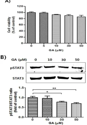
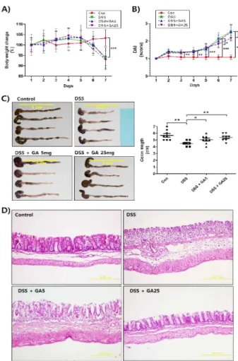
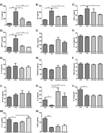
Signal transducer and activator of transcription 3 (STAT3) is associated with various human diseases, such as cancer, auto-immune disease, and intestinal inflammation. The limited and inadequate effect of standard approaches for treating inflammatory bowel disease (IBD) has prompted to develop alternative anti-colitis agents through inhibition of STAT3. Here, we show that gallic acid (GA), a 3,4,5-trihydroxybenzoic acid, markedly reduced phosphorylation of STAT3. Among the derivatives of benzoic acids, GA showed significant inhibition on STAT3 phosphorylation. In addition, GA ameliorated the dextran sodium sulfate (DSS)-induced acute colitis as determined by the measurement of symptomatic and histological indices. The suppression of DSS-induced acute colitis by GA treatment may be related to the regulation of cytokines and growth factors. Furthermore, GA inhibited phosphorylation of STAT3 in the colon tissue of DSS-treated mice. These findings may be useful in comprehending the molecular action of GA on STAT3 phosphorylation and provide novel insights into the potential application of GA in the treatment of STAT3-related inflammatory disease, such as IBD.

keywords : STAT3, Gallic acid, Inflammatory bowel disease, Dextran sodium sulfate

* Corresponding author

So-Yeon Kim, School of Korean Medicine, Pusan National University and Department of Internal Medicine, Korean Medical Hospital of Pusan National University Hospital, Yangsan, Gyeongsangnam-do, 50612 Republic of Korea

·E-mail : [email protected] ·Tel : +82-55-360-5954

Ki-Tae Ha, School of Korean Medicine and Korean Medicine Research Center for Healthy Aging, Pusan National University, Yangsan, Gyeongsangnam-do, 50612 Republic of Korea

·E-mail : [email protected] ·Tel : +82-51-510-8464

·Received : 2016/07/03 ·Revised : 2016/09/07 ·Accepted : 2016/09/23

ⓒ The Society of Pathology in Korean Medicine, The Physiological Society of Korean Medicine pISSN 1738-7698 eISSN 2288-2529 http://dx.doi.org/10.15188/kjopp.2016.10.30.5.338 Available online at http://www.hantopic.com/kjopp/KJOPP.htm

(2)

in various herbal plants

16,17)

. GA was previously reported to have various biological activities including antioxidant, antibacterial, antitumor, and anti-inflammatory

18-21)

. In addition, the natural compounds harboring the galliate moiety such as epigallocatechin-3-gallate, gallotannin corilagin, penta-O-galloyl-β-D-glucose, and theaflavin-3,3ʹ -digallate were reported as potent candidates for ameliorating colitis

22-25)

. Moreover, natural products harboring gallate residues such as epigallocatechin-3-gallate and penta-O-galloyl-β-D-glucose

26-28)

, were reported as STAT3 inhibitors.

In this study, we examined the inhibitory effect of GA and its analogous compounds on the activation of STAT3.

In addition, anti-inflammatory effect of GA on the dextran sodium sulfate (DSS)-induced experimental acute colitis mouse model was evaluated. In addition, the inhibitory effect of GA on the production of cytokines and activation of STAT3 in colon tissues were analyzed to explain the molecular mechanisms underlying its anti-colitis effect.

Materials and Methods

1. Chemicals and reagents

Antibodies against phospho-STAT3 and STAT3 were purchased from Cell Signaling (Danvers, MA, USA). The DSS salt (molecular weight 36,000–50,000) was supplied by MP Biomedicals (Santa Ana, CA, USA). GA and its derivatives, such as benzoic acid (BA), 3-hydroxybenzoic acid (3-HBA), 4-hydroxybenzoic acid (4-HBA), procatechuic acid (PA), 3,5-dihydroxybenzoic acid (3,5-DBA), and methyl gallate (MG), was purchased from Sigma-Aldrich (St. Louis, MO, USA) and the purity (over 98%) was guaranteed by the supplier. All other chemicals and reagents were obtained from Sigma-Aldrich, unless otherwise indicated.

2. Cell culture and viability assay

Human colorectal carcinoma HCT116 cells were purchased from the American Type Culture Collection (ATCC, Rockville, MD, USA). The cells were grown in Dulbecco’s modified Eagle’s medium (DMEM, Invitrogen, Carlsbad, CA, USA) containing L-glutamine (200 mg/L, Invitrogen) supplemented with 10% (v/v) heat-inactivated fetal bovine serum (FBS, Sigma-Aldrich), 100 U/mL penicillin, and 100 μg/mL streptomycin (Invitrogen) and maintained in a humidified incubator at 37℃ and 5% CO

2

prior to the experiments. The cytotoxicity of GA was examined in a formazan colorimetric assay using 3-(4,5-dimethylthiazol-2-yl)-2,5-diphenyltetrazolium bromide

(MTT, Sigma-Aldrich). In brief, the cells were cultured in 24-well plates with GA for 24 h and the MTT solution (2.0 mg/mL) was added to each well containing cells. After 4h incubation at 37℃ in a cell culture incubator, the supernatants were removed. The formazan crystals formed in the viable cells were solubilized in dimethyl sulfoxide and the absorbance was measured at 540 nm using a microplate reader (Victor 3, Perkin-Elmer, Waltham, MA, USA). The percentage of live cells was calculated relative to the untreated cells.

3. Western blot analysis

Equal amounts (30 μg) of proteins were separated using sodium dodecyl sulfate (SDS)-polyacrylamide gel electrophoresis (PAGE) and transferred onto a nitrocellulose membrane (Bio-Rad Laboratories). The blots were blocked for 1 h with 5% nonfat dry milk prior to incubation with antibodies against p-STAT3 and STAT3 at 4℃ overnight.

After incubating the blots with the HRP-conjugated secondary antibodies at room temperature for 1 h, the bands of interest were visualized using chemiluminescence (Pierce enhanced chemiluminescence, ECL Plus western blotting substrate, Invitrogen). The band intensities obtained from the western blot analysis were quantified using the ImageJ software (NIH, Bethesda, MA, USA).

4. Animals

Seven-week-old male C57BL/6 mice (weight, 20–24 g) were purchased from Orient Bio Inc. (Sungnam, Korea). The animals were housed in certified standard laboratory cages, fed with a standard diet, and provided drinking water ad libitum prior to the experiment. All the mice were kept in rooms maintained at a constant temperature (22 ± 1℃), humidity (50 ± 5%), and 12 h dark/light cycles. All experimental procedures followed the Guidelines for the Care and Use of Laboratory Animals of the National Institutes of Health of Korea, and were approved by the Institutional Animal Care and Use Committee of Pusan National University, Pusan, Republic of Korea.

5. Induction of colitis and treatment

For the experiments, mice were divided into four

groups (n = eight). The control group mice were provided

drinking water and orally (p.o.) administered normal saline

as a vehicle for 7 days. Experimental colitis was induced by

administering 4% (w/v) DSS in drinking water ad libitum for

7 days, as previously reported

29)

. The volume of

DSS-containing water consumed by the treated mice was

(3)

carefully monitored daily. The mice in the DSS group were administered 4% DSS-containing water and normal saline for 7 days. The mice in other two groups were administered 4% DSS-containing water and the indicated concentrations of GA (5 and 25 mg·kg

-1

·day

-1

, p.o.) for 7 days. The drug administration was started on the same day as DSS treatment.

6. Evaluation of disease activity index (DAI)

The mice were identified by ear tags and checked daily for body weight, stool consistency, and gross bleeding. The DAI was determined by combining the scores of body weight loss, stool consistency, and gross bleeding. Each score was determined using the following scale: change in body weight loss (1, none; 2, 1–5%; 3, 10–20%; and 4, > 20%), stool consistency (1, normal; 2, loose; 3, diarrhea; and 4, watery diarrhea), and bloody stool (1, none; 2, slight bleeding; and 4, gross bleeding). At the end of the experiment, the mice were killed and the colons dissected from the proximal rectum. The colon length was measured from the ileocecal junction to the anal verge.

7. Histological assessment

The mouse colons were washed with syringe injections of ice-cold phosphate-buffered saline (PBS), opened longitudinally, and separated into two segments. They were then fixed in 4% paraformaldehyde overnight and embedded in paraffin. Then 4 μm sections were cut, mounted on slides, cleared, and hydrated. The colonic damage was assessed by staining the slides with hematoxylin and eosin (H&E) according to the standard procedure.

8. Measurements of cytokines, chemokines, and growth factors

The colon tissues samples were homogenized in ice-cold 1% NP-40 lysis buffer containing 150 mM sodium chloride (NaCl), 10 mM HEPES, 1% NP-40, 5 mM sodium pyrophosphate (Na

4

O

7

P

2

), 5 mM sodium fluoride (NaF), 2 mM sodium orthovanadate (Na

3

VO

4

), and a protease inhibitor cocktail tablet (Roche, Mannheim, Germany). After homogenization, the samples were centrifuged at 15,000 rpm at 4℃ for 15 min. The supernatants were collected and stored at -80℃ before use for the cytokine measurement and western blot analysis. The protein amounts were measured using the Bradford method (Bio-Rad protein assay, Bio-Rad Laboratories, Hercules, CA, USA). The 14 analytes including the GM-CSF, interferon (IFN)-γ, interleukin (IL)-10, IL-1α, IL-1β, IL-4, IL-6, KC, MCP-1,

M-SCF, RANTES, TNF-α, VEGF, and IL-13, were measured using the commercially available Luminex multiplexing system (Koma Biotech, Seoul, Korea). The level of each cytokine evaluated in the samples were expressed as pg/mg.

9. Statistical analysis

The data from all the experiments were calculated as fold increases compared to the control, and are expressed as the mean ± SD. The differences between two groups were determined using a student t-test (for Fig. 3B), two-way ANOVA with Bonferroni post-test (for Fig. 4A and 4B), or one-way ANOVA with Turkey’s post-hoc (for other Fig.s) assisted with the GraphPad Prism (GraphPad Software, San Diego, CA, USA). The minimum significance level was set at a p value of 0.05 for all analyses.

Results

1. GA inhibits phosphorylation of STAT3 in HCT116 cells Previously, various cytokines and growth factors were shown to communicate with the Janus kinase (JAK)/STAT signaling pathways

9)

, which also play key roles in the development of IBD

9,10)

. Among the STATs, the crucial role of STAT3 in IBD has been reported

3,9)

. Several natural products harboring gallate residues, including epigallocatechin-3-gallate, gallotanin corilagin, penta-O-galloyl-β-D-glucose, and theaflavin-3,3ʹ -digallate

26-28)

, were reported that inhibit STAT3 activation or DSS-induced colitis. It is difficult to directly compare the effect of gallic acid with these tannins owing to its different physiochemical properties and different structural formula.

However, due to similarities in pharmacophore features shown in Fig. 1, it may be possible that free GA has an inhibitory effective on STAT3 activation and colitis. Thus, we examined the direct inhibitory effects of GA on the phosphorylation of STAT3 in HCT116 colorectal carcinoma cells, which are established screening model of STAT3 inhibition owing to express high endogenous levels of constitutively activated STAT3

30)

. As shown in Fig. 2, the highly phosphorylated STAT3 was reduced by non-toxic dose of GA treatment, dose-dependently.

Since GA is a derivative of benzoic acid (BA), we

assumed that the inhibitory effect on STAT3 phosphorylation

of GA can be replaced by BA or its derivatives. Thus we

evaluated the effects of GA, BA, 3-hydroxybenzoic acid

(3-HBA), 4-hydroxybenzoic acid (4-HBA), procatechuic acid

(PA), 3,5-dihydroxybenzoic acid (3,5-DBA), and methyl gallate

(MG) on STAT3 phosphorylation using constitutively STAT3

(4)

activated-HCT116 colon cancer cells. The results showed that among these compounds, only GA has inhibitory effects on STAT3 activation at the dose of 50 μM. BA slightly suppressed the phosphorylation of STAT3, but there is no statistical significance (Fig. 3). These result showed that three hydroxyl residues attached on the 3,4,5-carbon of BA are important for modulating STAT3 phosphorylation. In addition, we supposed that GA has the structural uniqueness and is sufficient for inhibition of STAT3 activation.

Fig. 1. Similarities in pharmacophore features of several known inhibitors on the STAT3 activation were shown. Gallate residues are shown as dashed-circles.

Fig. 2. Effects of GA on STAT3 activation in HCT116 human colon cancer cells. A) HCT116 cells were treated with indicated concentrations of GA for 24 h. Cell viability was measured using the MTT assay. B) Cells were treated with indicated concentrations of GA for 30 min. STAT3 phosphorylation was analyzed by three independent Western blot and densitometry results are shown as mean ± SD. *p < 0.05 and **p < 0.01, comparing indicated two groups.

Fig. 3. Comparative effects of GA and its derivatives on STAT3 activation in HCT116 human colon cancer cells. A) Structure of GA and its derivatives, including BA, 3-HBA, 4-HBA, PA, 3,5-DBA, and MG. B) Cells were treated with 50 μM of indicated compounds for 30 min.

Phosphorylation of STAT3 was estimated by three different Western blot analysis and densitometry data were presented as mean ± SD. **p < 0.01, comparing indicated two groups.

2. GA ameliorates the symptoms and histological changes in DSS-induced murine acute colitis

The DSS-induced acute colitis model is widely used for simulating human ulcerative colitis because of its several relevant characteristics including weight loss, diarrhea, fecal bleeding, superficial ulceration, and mucosal damage

5,31)

. In this study, mice were treated with 4% DSS to induce acute colitis and predetermined doses of GA were simultaneously administered. As shown in Fig. 4A, the body weights of the DSS-treated mice were significantly decreased by day 6.

Although the body weights of the DSS- and GA-treated mice did not significantly change compared with those of the DSS-treated mice, the DAI scores of the mice treated with DSS and GA (25 mg/kg) were more significantly reduced than those of the mice treated with only DSS were (Fig. 4B). In addition, the colon length, which is another symptomatic parameter of DSS-induced acute colitis, was shortened following DSS treatment. The colon length of the GA-treated mice increased dose-dependently (Fig. 4C). The histological observations also showed that GA reduced the DSS-induced damage to the colon tissue such as the crypt destruction, lesions, and inflammatory cell infiltration (Fig.

4D). These results clearly indicate that GA has a potent

(5)

therapeutic effect against DSS-induced acute colitis.

Fig. 4. Effects of GA on symptoms, indices, colon length, and histological changes of DSS-induced acute colitis in mice. A) Change in body weight is presented as a percentage of body weight before induction of colitis. Data are expressed as means ± SD, ***p < 0.001 comparison between two groups. B) DAI was determined by evaluating body weight loss, stool consistency, and stool bleeding scores. Data are expressed as means ± SD, *p<0.05, **p<0.01, and ***p < 0.001 comparison between two groups. C) At day 7 following treatment with DSS and GA, mice were killed and colon length measured. Data are expressed as means ± SD, *p < 0.05, **p < 0.01, comparison between two groups. D) Representative images of hematoxylin and eosin (H&E) staining (100× magnification).

3. GA modulates colitis-induced production of cytokines, chemokines, and growth factors

Numerous inflammatory mediators including cytokines, chemokines, and growth factors are involved in the pathogenesis of colitis and they control multiple aspects of the inflammatory response

7-9)

. Therefore, drug candidates that target pro-inflammatory cytokines such as tumor necrosis factor-α (TNF-α), interferon-γ (INF-γ), IL-12, IL-13, and IL-23 are currently being tested in clinical trials

8,32)

. However, because of alternative compensatory cytokine pathways, anti-cytokine therapies have some limitations, which include being beneficial to only certain

groups of patients

8)

. Therefore, alternative approaches to treating IBD by blocking multiple cytokines or cytokine signaling pathways are needed. Thus, we next examined the production of cytokines, chemokines, and growth factors using the Luminex multiplex assay. The results showed that GA decreased the DSS-induced production of pro-inflammatory T-helper cells type 1 (Th1) cytokines including IL-6 and TNF-α (Fig. 5A, B, and C). In addition, the DSS-induced expression of the chemokines keratinocyte-derived chemokine (KC) and monocyte chemoattractant protein-1 (MCP-1) were also decreased following GA treatment (Fig. 5D). However, the production of other Th1 cytokines and chemokines including IFN-γ, IL-1β, IL-13, and regulated on activation normal T cell expressed and secreted (RANTES) was not significantly changed following treatment with DSS, GA, or both (Fig. 5E, F, G, and H). The expression of granulocyte-macrophage colony-stimulating factor (GM-CSF) and macrophage colony-stimulating factor (M-CSF) was also not changed (Fig. 5I and J). In addition, the production of the anti-inflammatory Th2 cytokines including IL-4 and IL-10 9, and wound-healing mediators IL-1α

33)

and vascular endothelial growth factor (VEGF)

34)

, were reduced in the colon tissues of DSS-treated mice. The production of IL-1α and IL-4 was significantly increased by GA treatment.

However, the production of IL-10 and VEGF was not significantly recovered following GA treatment(Fig. 5K, L, M, and N).

Previously, the levels of multiple cytokines were examined in the serum of mice from two different experimental acute colitis models

7)

. The profiles of cytokines from serum of children with IBD showed similar results

35)

. However, the production of multiple cytokines in colon tissue samples has not been evaluated until now. The cytokine profiles from previous studies and this study are consistent with the acute inflammatory responses as shown by the elevation of TNF-α, IL-6, and KC. Taken together, these results suggest that the amelioration of the DSS-induced acute colitis by GA treatment may be associated with the regulation of cytokines, chemokines, and growth factors.

4. GA inhibits phosphorylation of STAT3 in colon tissue

Therefore, we evaluated the phosphorylation of STAT3

in colon tissue of mice treated with DSS, GA, or both. The

results showed that STAT3 was highly phosphorylated in the

colon tissue of mice treated with DSS and this effect was

reduced following GA treatment at a dose of 25

(6)

mg·kg

-1

·day

-1

(Fig. 6). Several previous researches reported that STAT3 activates the expression of cytokines such as IL-1β, IL-6, IL-11, TNF-α, and MCP-1, and can itself be activated by the pro-inflammatory cytokine IL-6

4,36)

. Although GA treatment may inactivate the phosphorylation of STAT3 via a decreased production of pro-inflammatory cytokines such as IL-6, the results from HCT116 cells (Fig.

2B) suggest that GA inhibits the phosphorylation of STAT3, at least partly.

The limited efficacy and serious toxic effects associated with conventional treatments for IBD, necessitates the continued search for alternative treatments

12,14)

. Therefore, other strategies have been developed including monoclonal antibodies targeting TNF-α, IL-6R, IL-12/IL-23, IL-13, and IL-17A, as well as recombinant proteins of IL-10 and IL-11

8,12)

. Recently, the small molecule inhibitor of JAKs tofacifinib (CP-690,550), showed efficacy in ulcerative colitis and a tolerable safety profile in clinical trials

14,15)

. However, tofacifinib has side and adverse effect including diarrhea, headache, infection, neutropenia, and elevated low-density lipoprotein (LDL) cholesterol levels, in addition to the inhibition of other tyrosine kinases including tyrosine kinase 2 (TYK2)

9)

. Therefore, many researchers are still trying to find novel and specific inhibitors of JAK/STAT signaling pathways for developing effective and safe medications for IBD.

Fig. 5. Effects of GA on cytokine and chemokine production in the colon of DSS-induced acute colitis mouse model. A)-N) Day 7 following treatment with DSS and GA and total proteins were extracted from colon tissue homogenates of sacrificed mice. Cytokine and chemokine production was measured using the Luminex multiplexing analysis. Data are expressed as means ± SD, *p < 0.05, **p < 0.01, and ***p < 0.001, comparing two groups.

Fig. 6. Effects of GA on STAT3 activation in the colon of DSS-induced acute colitis mouse model. A) At day 7 following treatment with DSS and GA, total proteins were extracted from colon tissue homogenates of sacrificed mice. Phosphorylation of STAT3 was measured using Western blot analysis. Representative images of Western blots are shown. B) Densitometric analysis of results from Western blot analysis are expressed as means ± SD. *p < 0.01 and ***p < 0.001, comparing two groups.

Discussion

In Traditional Medicine in Korea and China, IBD is recognized as a type of diarrhea or dysentery. According to its symptoms, in Traditional Medicine, IBD can be originated by blocking normal circulation of gastrointestinal tract by the damp heat

37)

. The accumulated damp heat damage the small vessels and surface lining of gastrointestinal tract, thus induce bleeding, ulceration, and purulent secretion. At the terminal course of IBD, the functions of digestive systems are much deteriorated

38,39)

. For treating or for ameliorating the symptoms of the disease, diverse medical options have been tried

37,40)

. The effects and underlying mechanisms of several herbal formulae including pulsatilla decoction

41)

and Shenling Baizhu San

42)

, compounds from medicinal herbs, such as purslane polysaccharide

43)

, fucoidan

44)

, naringin

45)

, and curcumin

46)

, on the IBD have been studied. In addition, several reports have demonstrated that acupuncture also can be the effective treatment on

IBD

47,48)

. Thus, herbal medicines and its ingredient

compounds may be good resources to develop the novel and effective agents for treatment of IBD.

Natural products from herbal plants including boswellic

acid, curcumin, sophocarpine, and anthocyanins

49-52)

were

(7)

previously reported as potent therapeutic agents for IBD. In addition, natural products harboring gallate residues such as epigallocatechin-3-gallate and penta-O-galloyl-β -D-glucose

26-28)

, were reported as STAT3 inhibitors. A previous report suggested possible therapeutic roles for free gallic acid contained in Quercus brantii Lindl. in murine colitis. However, there is no direct evidence supporting the effect of gallic acid for colitis

53)

. Recently, Pandurangan et al.

54,55)

reported that gallic acid suppresses DSS-induced colitis through inhibiting phosphorylation of NF-κB and STAT3 and through inducing Nrf2-related defensive enzymes. In this study, we also confirmed the anti-colitis effect of gallic acid and involvement of STAT3 as a mode of action. Previous study by Pandurangan et al.

54,55)

presented that anti-colitis effect of GA might be mediated by reducing the expression of pro-inflammatory cytokines.

However, in addition to this, here we firstly demonstrated that anti-inflammatory cytokines including IL-4 and IL-1α were induced by GA treatment. Thus, we assumed that anti-colitis effect of gallic acid may be related with induction of anti-inflammatory cytokines as well as with inhibition of pro-inflammatory cytokines. Moreover, among the several derivatives of benzoate (BA), only GA has sufficient inhibitory action against STAT3 activation. Thus, we suggest that GA is structurally unique and sufficient compound for the inhibition of STAT3 phosphorylation.

In conclusion, we showed that GA ameliorated the DSS-induced acute colitis using symptomatic and histological indices. The molecular mechanism underlying the effects of GA on the DSS-induced colitis may be related to the regulation of pro- and anti-inflammatory cytokines as well as with the inhibition of STAT3 activation.

Considering the importance of STAT3 in the development of IBD, our finding may be useful for comprehending the molecular actions of GA on colonic inflammation and provide novel insights into the potential application of GA and its derivatives in the treatment of IBD.

Acknowledgements

This work was supported by a 2-Year Research Grant of Pusan National University.

References

1. Atreya, R., Atreya, I., Neurath, M.F. Novel signal transduction pathways: analysis of STAT-3 and Rac-1 signaling in inflammatory bowel disease. Annals of the

New York Academy of Sciences 1072: 98-113, 2006.

2. Milner, J.D., Vogel, T.P., Forbes, L., et al. Early-onset lymphoproliferation and autoimmunity caused by germline STAT3 gain-of-function mutations. Blood 125:

591-599, 2015.

3. Li, Y., de Haar, C., Peppelenbosch, M.P., et al. New insights into the role of STAT3 in IBD. Inflammatory bowel diseases 18: 1177-1183, 2012.

4. Li, N., Grivennikov, S.I., Karin, M. The unholy trinity:

inflammation, cytokines, and STAT3 shape the cancer microenvironment. Cancer cell 19: 429-431, 2011.

5. Chassaing, B., Aitken, J.D., Malleshappa, M., et al.

Dextran sulfate sodium (DSS)-induced colitis in mice.

Current protocols in immunology. edited by John E Coligan [et al] 104: Unit 15 25, 2014.

6. Ordas, I., Eckmann, L., Talamini, M., et al. Ulcerative colitis. Lancet 380: 1606-1619, 2012.

7. Alex, P., Zachos, N.C., Nguyen, T., et al. Distinct cytokine patterns identified from multiplex profiles of murine DSS and TNBS-induced colitis. Inflammatory bowel diseases 15: 341-352, 2009.

8. Neurath, M.F. Cytokines in inflammatory bowel disease.

Nature reviews Immunology 14: 329-342, 2014.

9. Coskun, M., Salem, M., Pedersen, J., et al. Involvement of JAK/STAT signaling in the pathogenesis of inflammatory bowel disease. Pharmacological research : the official journal of the Italian Pharmacological Society 76: 1-8, 2013.

10. Plevy, S. A STAT need for human immunologic studies to understand inflammatory bowel disease. The American journal of gastroenterology 100: 73-74, 2005.

11. Adams, S.M., Bornemann, P.H. Ulcerative colitis.

American family physician 87: 699-705, 2013.

12. Nielsen, O.H. New strategies for treatment of inflammatory bowel disease. Frontiers in medicine 1: 3, 2014.

13. Atreya, R., Neurath, M.F. New therapeutic strategies for treatment of inflammatory bowel disease. Mucosal immunology 1: 175-182, 2008.

14. Sandborn, W.J., Ghosh, S., Panes, J., et al. Tofacitinib, an oral Janus kinase inhibitor, in active ulcerative colitis. The New England journal of medicine 367:

616-624, 2012.

15. Panes, J., Su, C., Bushmakin, A.G., et al. Randomized trial of tofacitinib in active ulcerative colitis: analysis of efficacy based on patient-reported outcomes. BMC gastroenterology 15: 14, 2015.

16. Jayamani, J., Shanmugam, G. Gallic acid, one of the

components in many plant tissues, is a potential

(8)

inhibitor for insulin amyloid fibril formation. European journal of medicinal chemistry 85: 352-358, 2014.

17. Wang, K., Zhu, X., Zhang, K., et al. Investigation of gallic acid induced anticancer effect in human breast carcinoma MCF-7 cells. Journal of biochemical and molecular toxicology 28: 387-393, 2014.

18. Bravo, H.R., Copaja, S.V., Figueroa-Duarte, S., et al.

1,4-benzoxazin-3-one, 2-benzoxazolinone and gallic acid from Calceolaria thyrsiflora Graham and their antibacterial activity. Zeitschrift fur Naturforschung C, Journal of biosciences 60: 389-393, 2005.

19. Kim, Y.J. Antimelanogenic and antioxidant properties of gallic acid. Biological & pharmaceutical bulletin 30:

1052-1055, 2007.

20. Zhao, B., Hu, M. Gallic acid reduces cell viability, proliferation, invasion and angiogenesis in human cervical cancer cells. Oncology letters 6: 1749-1755, 2013.

21. Hsiang, C.Y., Hseu, Y.C., Chang, Y.C., et al. Toona sinensis and its major bioactive compound gallic acid inhibit LPS-induced inflammation in nuclear factor-kappaB transgenic mice as evaluated by in vivo bioluminescence imaging. Food chemistry 136: 426-34, 2013.

22. Ukil, A., Maity, S., Das, P.K. Protection from experimental colitis by theaflavin-3,3'-digallate correlates with inhibition of IKK and NF-kappaB activation. British journal of pharmacology 149: 121-31, 2006.

23. Jang, S.E., Hyam, S.R., Jeong, J.J., et al.

Penta-O-galloyl-beta-D-glucose ameliorates inflammation by inhibiting MyD88/NF-kappaB and MyD88/MAPK signalling pathways. British journal of pharmacology 170:

1078-1091, 2013.

24. Xiao, H.T., Lin, C.Y., Ho, D.H., et al. Inhibitory effect of the gallotannin corilagin on dextran sulfate sodium-induced murine ulcerative colitis. Journal of natural products 76: 2120-2125, 2013.

25. Abboud, P.A., Hake, P.W., Burroughs, T.J., et al.

Therapeutic effect of epigallocatechin-3-gallate in a mouse model of colitis. European journal of pharmacology 579: 411-417, 2008.

26. Wang, Y., Ren, X., Deng, C., et al. Mechanism of the inhibition of the STAT3 signaling pathway by EGCG.

Oncology reports 30: 2691-2696, 2013.

27. Lee, H.J., Seo, N.J., Jeong, S.J., et al. Oral administration of penta-O-galloyl-beta-D-glucose suppresses triple-negative breast cancer xenograft growth and metastasis in strong association with JAK1-STAT3 inhibition. Carcinogenesis

32: 804-811, 2011.

28. Hu, H., Lee, H.J., Jiang, C., et al.

Penta-1,2,3,4,6-O-galloyl-beta-D-glucose induces p53 and inhibits STAT3 in prostate cancer cells in vitro and suppresses prostate xenograft tumor growth in vivo.

Molecular cancer therapeutics 7: 2681-2691, 2008.

29. Shin, J.S., Cho, E.J., Choi, H.E., et al. Anti-inflammatory effect of a standardized triterpenoid-rich fraction isolated from Rubus coreanus on dextran sodium sulfate-induced acute colitis in mice and LPS-induced macrophages. Journal of ethnopharmacology 158 Pt A:

291-300, 2014.

30. Lin, L., Deangelis, S., Foust, E., et al. A novel small molecule inhibits STAT3 phosphorylation and DNA binding activity and exhibits potent growth suppressive activity in human cancer cells. Molecular cancer 9: 217, 2010.

31. Axelsson, L.G., Landstrom, E., Goldschmidt, T.J., et al.

Dextran sulfate sodium (DSS) induced experimental colitis in immunodeficient mice: effects in CD4(+) -cell depleted, athymic and NK-cell depleted SCID mice.

Inflammation research : official journal of the European Histamine Research Society [et al] 45: 181-191, 1996.

32. Bantel, H., Schulze-Osthoff, K. TNF Antagonists in IBD:

Novel Antiinflammatory Mechanisms Beyond Cytokine Inhibition. Inflammatory bowel diseases 19: E51-2, 2013.

33. Bersudsky, M., Luski, L., Fishman, D., et al.

Non-redundant properties of IL-1alpha and IL-1beta during acute colon inflammation in mice. Gut 63:

598-609, 2014.

34. Sandor, Z., Deng, X.M., Khomenko, T., et al. Altered angiogenic balance in ulcerative colitis: a key to impaired healing? Biochemical and biophysical research communications 350: 147-150, 2006.

35. Toptygina, A.P., Semikina, E.L., Bobyleva, G.V., et al.

Cytokine profile in children with inflammatory bowel disease. Biochemistry Biokhimiia 79: 1371-1375, 2014.

36. Fukuda, A., Wang, S.C., Morris, J.P.4th., et al. Stat3 and MMP7 contribute to pancreatic ductal adenocarcinoma initiation and progression. Cancer cell 19: 441-455, 2011.

37. Rahimi, R., Nikfar, S., Abdollahi, M. Induction of clinical response and remission of inflammatory bowel disease by use of herbal medicines:A meta-analysis. World Journal of Gastroenterology, 2013.

38. Han, J., Wang, J., Wang, J.H. How to achieve deep remission in treatment of inflammatory bowel disease.

Journal of Traditional Chinese Medicine, 2013.

39. Lee, S.W., Ryu, B.H., Park, J.W. Effects of Sargassum

(9)

pallidum on 2,4,6,-Tritrobenzene Sulfonic Acid-Induced Colitis in Mice. Korean J Orient Int Med 31: 224-241, 2010.

40. Ling, X.H., Yu, X., Kong, D.J., et al. Treatment of inflammatory bowel disease with Chinese drugs administered by both oral intake and retention enema.

Chin J Integr Med 16: 222-228, 2010.

41. Lu, S.W., Liu, H.J., Zhao, W., et al. Molecular mechanisms involved in the treatment of inflammatory bowel disease by Pulsatilla decoction. Zhongguo ying yong sheng li xue za zhi = Zhongguo yingyong shenglixue zazhi = Chinese journal of applied physiology 27: 106-109, 2011.

42. You, Y., Liu, Y.H., Gao, S.L. Effect and Mechanism of Shenling Baizhu San on the Murine Model of Inflammatory Bowel Disease Induced by Dextran Sodium Sulfate in Mice. Chinese Journal of Experimental Traditional Medical Formulae 18: 136-140, 2012.

43. Qian, W., Wentao, F. Effect of Purslane Polysaccharide on Ulcerative Colitis Associated Colorectal Cancer and IL-6/STAT3 Pathway. World Chinese Medicine 8:

1256-1260, 2013.

44. Iraha, A., Chinen, H., Hokama, A., et al. Fucoidan enhances intestinal barrier function by upregulating the expression of claudin-1. World Journal of Gastroenterology, 2013.

45. Kumar, V.S., Rajmane, A.R., Adil, M., et al. Naringin ameliorates acetic acid induced colitis through modulation of endogenous oxido-nitrosative balance and DNA damage in rats. Journal of Biomedical Research, 2014.

46. Rajasekaran, S.A. Therapeutic potential of curcumin in gastrointestinal diseases. World Journal of Gastrointestinal Pathophysiology, 2011.

47. Chuan-zi, D., Hui, F., Xiao, Z., et al. Thinking on

Functional Mechanism of Acupuncture for Inflammatory Bowel Diseases Based on Metabolomics. Journal of Acupuncture and Tuina Science, 2014.

48. Schneider, A., Streitberger, K., Joos, S. Acupuncture treatment in gastrointestinal diseases: A systematic review. World Journal of Gastroenterology, 2007.

49. Piberger, H., Oehme, A., Hofmann, C., et al. Bilberries and their anthocyanins ameliorate experimental colitis.

Molecular nutrition & food research 55: 1724-1729, 2011.

50. Liu, L., Liu, Y.L., Liu, G.X., et al. Curcumin ameliorates dextran sulfate sodium-induced experimental colitis by blocking STAT3 signaling pathway. International immunopharmacology 17: 314-320, 2013.

51. Wang, X.J., Deng, H.Z., Jiang, B., et al. The natural plant product sophocarpine ameliorates dextran sodium sulfate-induced colitis in mice by regulating cytokine balance. International journal of colorectal disease 27:

575-581, 2012.

52. Anthoni, C., Laukoetter, M.G., Rijcken, E., et al.

Mechanisms underlying the anti-inflammatory actions of boswellic acid derivatives in experimental colitis.

American journal of physiology Gastrointestinal and liver physiology 290: G1131-G1137, 2006.

53. Khanavi, M., Sabbagh-Bani-Azad, M., Abdolghaffari, A.H., et al. On the benefit of galls of Quercus brantii Lindl. in murine colitis: the role of free gallic acid.

Archives of medical science : AMS 10: 1225-1234, 2014.

54. Pandurangan, A.K., Mohebali, N., Mohd Esa, N., et al.

Gallic acid suppresses inflammation in dextran sodium sulfate-induced colitis in mice: Possible mechanisms.

International immunopharmacology 28: 1034-1043, 2015.

55. Pandurangan, A.K., Mohebali, N., Norhaizan, M.E., et al.

Gallic acid attenuates dextran sulfate sodium-induced

experimental colitis in BALB/c mice. Drug Des Devel

Ther 9: 3923-3934, 2015.

수치

Fig.  3.  Comparative  effects  of  GA  and  its  derivatives  on  STAT3  activation  in HCT116 human colon cancer cells
Fig.  4.  Effects  of  GA  on  symptoms,  indices,  colon  length,  and  histological  changes  of  DSS-induced  acute  colitis  in  mice
Fig. 6. Effects of GA on STAT3 activation in the colon of DSS-induced  acute  colitis  mouse  model

참조

관련 문서

Changes in blood components decreased in total cholesterol, low density lipoprotein cholesterol, and triglyceride and increased in high density

Micro- and nano-sized pores were formed on the surface of the alloy using PEO and anodization methods, and the pore shape change according to the Zr

This same argument can be used to show that the complementary virtual work of redundant member forces is zero not only for truss elements but for members of any other

skeleton bond energy differences of monomer, oligomers, and polymer. Skeletal bond energy difference

FBG: fasting blood glucose, TC: total cholesterol, TG: triglyceride, HDL-C: high density lipoprotein-cholesterol, LDL-C: low density lipoprotein-cholesterol,

Effect of amino acid addition in culture media on the mycelial growth and g-aminobutyric acid (GABA) production of Sparassis latifolia..

Second, the total cholesterol, low density lipoprotein cholesterol, and triglyceride of the exercise group significantly decreased after 8 weeks of Ju-do

Effect of mulberry leaf powder containing the lactic acid bacteria on body weight gain, food intake and food efficiency ratio in a constipation model