Original Article
원고 접수일 2012년 5월 22일, 원고 수정일 2012년 6월 13일, 게재 확정일 2012년 7월 24일
책임저자 이백수
(130-701) 서울시 동대문구 경희대로 26, 경희대학교 치의학전문대학원 구강악안면 외과학교실
Tel: 02-958-9440, Fax: 02-966-4572, E-mail: [email protected]
RECEIVED May 22, 2012, REVISED June 13, 2012, ACCEPTED July 24, 2012
Correspondence to Baek-Soo Lee
Department of Oral and Maxillofacial Surgery, Kyung Hee University School of Dentistry
26, Gyeongheedae-ro, Dongdaemun-gu, Seoul 130-701, Korea Tel: 82-2-958-9440, Fax: 82-2-966-4572, E-mail: [email protected]
CC This is an open access article distributed under the terms of the Creative Commons Attribution Non-Commercial License (http://creativecommons.org/licenses/
by-nc/3.0) which permits unrestricted non-commercial use, distribution, and reproduction in any medium, provided the original work is properly cited.
양악전돌증 환자에서 하악 6전치 후방 이동 시 치료 방법에 따른 하악 경조직과 연조직의 변화:
하악 전방분절골절단술과 발치 교정 치료
김영주ㆍ김경아1ㆍ유용재ㆍ유경선ㆍ유정민ㆍ오주영ㆍ김수정1ㆍ김성훈1ㆍ이백수
경희대학교 치의학전문대학원 구강악안면외과학교실, 1치과교정학교실
Abstract
The Evaluation of Soft and Hard Tissue Change for Retraction of Lower Anterior Tooth in Bimaxillary Protrusion Patients according to Two Different Therapeutic Methods: Mandibular Anterior Segmental
Osteotomy and Orthodontic Treatment with Teeth Extraction
Young-Joo Kim, Kyung-A Kim
1, Yong-Jae Yu, Kyung-Sun Ryu, Jeong-Min Ryu, Joo-Young Ohe, Su-Jung Kim
1, Seoung-Hun Kim
1, Baek-Soo Lee
Departments of Oral and Maxillofacial Surgery,
1Orthodontics, Kyung Hee University School of Dentistry
Purpose: The purpose of the study is to evaluate the effectiveness of anterior segmental osteotomy (ASO) in bimaxillary protrusion (BP) patients by comparison between the mandibular soft and hard tissue changes from orthodontic treatment and ASO.
Methods: All 44 patients were diagnosed with BP in Kyung-Hee Medical Center. Orthodontic treatment with teeth extractions were underwent by 23 patients (Group A) and 21 patients underwent ASO (Group B). Mandibular soft and hard tissue changes were measured and evaluated, which were based on the vertical and horizontal reference line in lateral cephalometric radiographs.
Statistical significance between the changes and correlation between each measurement were analyzed.
Results: The amount of B point movement was lesser than that of the lower incisal tip (LIT) retraction, and LIT was tilted lingually in group A. The posterior movement discrepancy between LIT and B point was insignificant, and the inclination of lower incisor was not changed in group B. The soft tissues, including the lower lip, showed a posterior movement and reduction in the depth of mento-labial groove. According to the correlation analysis, the movement of the lower incisor was significantly related to the movement of the lower lip in group A, and the movement of the lower incisor was significantly related to that of the movement of lower lip, B point and Pog in group B.
Conclusion: The orthodontic treatment in BP patients results in posterior tilting movement of the lower incisor, but ASO results in the bodily movement of the lower incisor. Consequently, ASO is more effective in BP cases because it ensures the controlled movement of the lower incisors.
Key words: Bimaxillary protrusion, Anterior segmental osteotomy, Orthodontic treatment
서 론
양악전돌증은 골성, 치성문제로 인해 입술이 돌출되어 보이는 1급 부정교합을 말하며[1-3], 전치와 입술의 과도한 돌출로 인해 입술 기능부전, 외번된 vermilion border를 동반한 두꺼워 보이 는 입술, 입술폐쇄를 위해 과활성화된 이부와 같은 심미적, 기능적 문제가 존재한다[4]. 또한, 양악전돌증은 서양인에 비해 아프리카 와 아시아인에게 두드러진 경향을 보인다[1,5,6]. 과거에는 양악 전돌증이 동양인의 정상적인 안모형태로 간주되어 치료를 받지 않았지만 최근에는 미적 기준이 서양인의 안모형태로 바뀌면서 기능적, 심미적인 이유로 치료 받으려는 환자가 늘고 있다[7].
양악전돌증은 일반적으로 4개의 제1 소구치 발치 후 발치된 공간에 최고의 고정력으로 6전치를 후방 이동시켜 치료를 한다.
이러한 치료는 후방 이동량에 따라 치근흡수, 전치부의 심한 설측 경사, 순측골의 열개나 천공, 고정력의 상실로 인한 불충분한 후방 이동, 상악 전치부의 불필요한 하방 이동 같은 부작용이 발생할 수 있다[5,8]. 최근 교정용 miniscrew와 straight wire appliance bracket 같은 교정기술의 발달로 정교한 치아이동과 치료 기간의 단축을 이룰 수 있었다[9-12]. 그러나 교정적 후방 이동량에는 한계가 있으며 성인환자에 있어 해면골 감소와 혈액공 급의 감소와 같은 구조적 변화는 치료 기간의 연장을 가져올 수 있다[1]. 또한 교정치료는 치아만을 이동시킨다는 한계로 인해 악골의 전돌로 생긴 심미적인 문제를 해결하는 데 한계가 있다[8].
이러한 교정적 한계를 극복하기 위해 전방분절골절단술(anterior segmental osteotomy)이 추천되고 있다. 전방분절골절단술은 1921년 Cohn-stock[13]에 의해 상악에 처음으로 시술된 이래로 Wassmund[14], Wunderer[15]와 Bell과 Condit[16], Park과 Hwang[17]에 의해 연조직 절개와 골절단 방법이 수정, 발전되어 왔으며, 이로 인해 재위치된 전방분절 괴사, 비익저 폭의 증대, 비첨의 반시계 방향으로의 회전과 같은 문제점이 많이 개선되었 다. 그럼에도 불구하고 여전히 술 후 합병증인 견치의 치근절단과 강직, 골절단 부위의 열개, 일시적인 하순 감각마비, 견치와 소구 치부의 교합평면의 불일치 등이 발생하는 등의 문제점이 있다[18].
하지만, 전방분절골절단술은 치료기간의 단축과 안모형태의 즉시 개선, gummy smile의 개선, 치아 후방이동 시 전치부 하방 이동 의 방지와 같은 장점으로 일반 교정치료로 해결할 수 없는 많은 문제를 해결할 수 있다. 특히 하악의 경우, 상악과는 달리 얇은 치조골로 인한 교정적 후방이동의 한계가 있으며, 얇은 치근으로 인해 급격한 교정력이 가해질 경우 치근 흡수와 골천공 열개가 발생할 확률이 더 높아 전방분절골절단술로 치료 시 많은 장점을 가질 수 있다. 또한 이부형태가 부족하거나 또는 이근이 과활성을 보이는 경우 하악 전방분절의 후방이동을 통해 이부의 상대적 돌출을 유도할 수 있어 이부 성형효과를 기대할 수 있다[19].
양악전돌증에서 전방분절골절단술 시행 후 상악 경조직과 연조
직의 변화에 관한 여러 연구가 있어왔다[8,17,19,20]. 그러나 전방 분절골절단술 시행 시 발치공간의 폐쇄 전후에 대한 하악의 경조 직과 연조직의 변화에 대한 연구는 적은 편이다. 본 연구는 상악의 경우 발치 후 통상적인 교정 치료 혹은 피질골 절단술을 행한 환자 중, 하악에서 발치를 동반한 교정치료를 한 경우와 전방분절 골절단술을 동반한 교정 치료를 시행한 경우에 있어서 발치공간 폐쇄 전, 후 측모방사선사진을 비교, 분석하여 치료 방법에 따른 하악의 경조직 및 연조직의 형태 변화를 비교 평가하고자 한다.
연구방법
1. 연구 대상
본 연구는 후향적 연구로 연구대상은 2007년부터 2011년까지 경희의료원 치과병원에 내원하여 양악전돌증으로 진단 받고 상악 은 발치 혹은 피질골 절단술 후 교정적 견인으로 발치 공간을 폐쇄하였고, 하악은 발치교정을 하거나 전방분절골절단술을 동반 한 교정치료를 시행하여 제1 소구치 공간이 폐쇄된 시점의 45명 한국인 성인을 대상으로 하였으며, 하악의 치료법에 따른 분류만 시행하였다. 양악전돌증 환자 중 구치부는 Angle 분류 1급 관계 에 있고 3 mm 이하의 총생 관계에 있으며 하악 평면과 하악 전치 사이 각도가 90도 이하인 환자를 대상으로 하였으며, 안면비 대칭(이부에서 안면중앙선에 3 mm 이상 이개)과 측두하악장애 의 증상과 징후를 가진 환자군은 제외하였다. 발치 교정치료를 시행한 군(Group A)과 동일한 술자가 전방분절골절단술을 시행 한 군(Group B)으로 분류하였다. 최종적으로 발치를 동반한 교 정치료를 시행한 군은 23명, 전방분절골절단술을 동반한 교정 치료를 시행한 군은 21명이었으며, Group A의 평균 연령은 24±6.54세, Group B의 평균 연령은 28±9.18세였다.
2. 계측방법
측모두부방사선사진은 자연두부위치에 중심 교합위에서 CX
90SP (Asahi, Tokyo, Japan)를 사용하여 촬영하였다. 측모두부
방사선 사진은 초진 시(T0), 발치공간 폐쇄 후(T1)에 촬영하였으
며, 동일한 계측자에 의해 V-ceph program Version 4.0
(CYBERMED Inc., Seoul, Korea)으로 투사도 작성 및 계측을 하였
다. 기준선은 Burstone 등[21]과 Moore[22]가 제시한 Sella-Nasion
(SN) plane에서 Sella 중심으로 시계방향으로 7도 회전시킨 수평
선을 수평기준선(horizontal reference line, HRL)으로 설정하고
Sella에서 수평기준선에 수직으로 내린 선을 수직기준선(vertical
reference line, VRL)으로 정하였으며, 경조직(Fig. 1)과 연조직
(Fig. 2)의 각각의 계측점을 다음과 같이 설정하였다. 경조직 및
경조직의 측정치는 0.05
o와 0.05 mm 오차로 계산되었다.
Fig. 1. Hard tissue landmarks and measurements. LIT, lower
incisor tip; B, the deepest point between pogonion and the lower incisor alveolus in the symphysis, supramentale; Pog, the most protrusive point in the symphysis, Pogonion; Me, the most inferior point in the symphysis, Menton; IMPA, angle between lower incisor to mandibular plane (Go-Gn); LIT-Me, distance between lower incisior tip and Menton; HRL, horizontal reference line; VRL, vertical reference line; S, sella; N, nasion.Fig. 2. Soft tissue landmarks and measurements. LL, the most
anterior point of the lower lip; B’, the point of greatest concavity in the midline between the lower lip (LL) and chin (Pog’), inferior labial sulcus, Mentolabial sulcus; Pog’, soft tissue pogonion; Sn, the point at which the columella merges with the upper cutaneous lip in the midsagittal plane, Subnasale; MLS-depth (Mentolabial sulcus-depth), distance between B’ to LL-Pog’; Sn-Pog’-LL, distance between LL to Sn-Pog’; HRL, horizontal reference line;VRL, vertical reference line; S, sella; N, nasion.
3. 통계처리
각 군에서의 초진 시와 발치공간 폐쇄 후의 차이 비교 시 Paired t-test를 사용하여 유의성을 검정하였고 두 군의 변수 항목 간 통계적 유의성 평가에는 two sample t-test로 검정하였다. 각 변수 간의 상관관계는 Pearson’s correlation method를 통해 평가하였다.
결 과
1. 발치를 동반한 교정치료군(A군)과 전방분절골절단술군(B군) 의 치료 전, 후의 하악 경, 연조직 변화 비교
1) 수평이동
경조직의 변화로 VRL에 대해 lower incisal tip (LIT), B, Pog은 A군에서 각각 6.54 mm, 1.70 mm, 0.88 mm 후방이동을 보였으며 B군에서는 5.80 mm, 6.02 mm 후방이동을 보였다.
Pog은 통계적으로 유의한 변화를 보이지 않았다. 연조직의 변화 로 VRL에 대해 lower lip (LL), B’는 A군에서 5.06 mm, 2.43 mm 후방이동을 보였으며 B군에서 4.69 mm, 3.40 mm 후방이 동을 보였으며 Pog’은 두 군 모두에서 통계적으로 유의한 변화를 보이지 않았지만 A군에서 0.36 mm 후방이동을 보이고 B군에서 는 0.14 mm 전방이동을 보였다.
2) 수직이동
통계적으로 유의한 변화를 보이지 않았지만 HRL에 대해 A군에 서 Pog, Pog’가 하방으로 각각 1.09 mm, 0.57 mm 이동한 것에 비해 B군에서는 상방으로 0.50 mm, 0.42 mm 이동하였다.
LIT는 두 군에서 각각 0.38 mm, 0.78 mm 상방으로 이동하였지 만 LL은 A군에서 하방으로 0.23 mm 이동한 것에 비해 B군에서 는 상방으로 0.14 mm 이동하였다.
3) 각도변화 및 길이변화
A군에서는 ∆IMPA (
o)가 11.11
o감소를 보였으나 Group B에 서는 유의한 차이를 보이지 않았다. 하순돌출도(∆Sn_Pog'-LL)는 A군에서 3.64
o감소를 보이고 B군에서는 4.04
o감소를 보였다.
A군에서는 ∆Me_LIT가 1.18 mm 감소를 보였으며 Group B는 2.85 mm 감소를 보였다. 이순구 깊이(∆MLS_depth)는 A군에 서는 0.25 mm 감소로 통계적으로 유의한 차이는 없었지만 B군은 0.83 mm 감소를 보였다.
2. 치료 전, 후 두 군 간에 변화량 비교(Table 1) 1) 경조직 변화량
HRL에서 LIT, B점까지 거리(∆HRL_LIT, ∆HRL_B)는 통계
적인 차이는 없지만 B군이 A군보다 하방으로 0.39 mm, 0.43
mm 하방으로 이동하였다. HRL에서 Pog까지 거리(∆HRL_Pog)
는 통계적으로 유의한 차이는 없었지만 A군이 B군보다 평균값에
Table 1. Comparison of the change from pre- to post-treatment
between two groups (Group A, B)(∆=∆A∆B)
P
Meandifference Std. error difference Hard tissue (mm)
∆HRL_LIT ∆HRL_B ∆HRL_Pog ∆VRL_LIT ∆VRL_B ∆VRL_Pog ∆IMPA ∆Me_LIT Soft tissue (mm) ∆HRL_LL ∆HRL_B' ∆HRL_Pog' ∆VRL_LL ∆VRL_B' ∆VRL_Pog' ∆Sn_Pog'_LL ∆MLS_depth
0.651 0.740 0.068 0.209 0.000*
0.535 0.000*
0.000*
0.669 0.765 0.405 0.613 0.334 0.688 0.484 0.088
0.39935
0.43939 1.59091
0.73593 4.31926
0.50541
11.32792 1.67532
0.37013 0.27814 0.95130
0.37771 0.97294
0.50649 0.40216
0.58333
0.87563 1.31248 0.84873 0.57608 0.60749 0.80524 1.75401 0.40665 0.86083 0.92406 1.13039 0.74178 0.99539 1.24961 0.57001 0.33366 HRL, horizontal reference line; LIT, lower incisal tip; B, the deepest point between pogonion and the lower incisor alveolus in the symphysis, supramentale; Pog, the most protrusive point in the symphysis, Pogonion; VRL, vertical reference line; IMPA, angle between lower incisor to mandibular plane; Me, the most inferior point in the symphysis, Menton; LL, the most anterior point of the lower lip; Sn, the point at which the columella merges with the upper cutaneous lip in the midsagittal plane, Subnasale; MLS, Mentolabial sulcus; Std, standard.
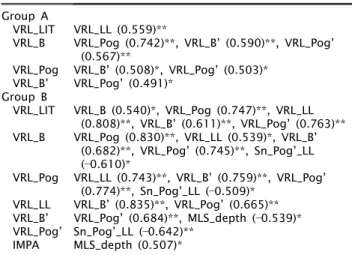
Table 2. The correlation between variables in two groups
Group AVRL_LIT VRL_B VRL_Pog VRL_B’
Group B VRL_LIT VRL_B
VRL_Pog VRL_LL VRL_B’
VRL_Pog’
IMPA
VRL_LL (0.559)**
VRL_Pog (0.742)**, VRL_B’ (0.590)**, VRL_Pog’
(0.567)**
VRL_B’ (0.508)*, VRL_Pog’ (0.503)*
VRL_Pog’ (0.491)*
VRL_B (0.540)*, VRL_Pog (0.747)**, VRL_LL (0.808)**, VRL_B’ (0.611)**, VRL_Pog’ (0.763)**
VRL_Pog (0.830)**, VRL_LL (0.539)*, VRL_B’
(0.682)**, VRL_Pog’ (0.745)**, Sn_Pog’_LL (0.610)*
VRL_LL (0.743)**, VRL_B’ (0.759)**, VRL_Pog’
(0.774)**, Sn_Pog’_LL (0.509)*
VRL_B’ (0.835)**, VRL_Pog’ (0.665)**
VRL_Pog’ (0.684)**, MLS_depth (0.539)*
Sn_Pog’_LL (0.642)**
MLS_depth (0.507)*
Pearson’s correlation coefficient, *P <0.05, **P < 0.01.
VRL, vertical reference line; LIT, lower incisal tip; B, the deepest point between pogonion and the lower incisor alveolus in the symphysis, supramentale; Pog, the most protrusive point in the symphysis, Pogonion; LL, the most anterior point of the lower lip; IMPA, angle between lower incisor to mandibular plane; Sn, the point at which the columella merges with the upper cutaneous lip in the midsagittal plane, Subnasale; MLS, Mentolabial sulcus.
있어 1.59 mm 하방으로 이동하였다. VRL에서 B까지의 거리(∆
VRL_B, P <0.05)는 A군보다 B군에서 4.31 mm 후방으로 이동 하였으며 통계적으로 유의성을 보였다. VRL에서 Pog까지 거리 (∆VRL_Pog)는 통계적으로 유의한 차이는 없었지만 A군이 B군 보다 0.50 mm 후방으로 이동하였다. ∆IMPA (
o)는 A군이 B군 보다 11.32
o더 설측으로 통계적으로 유의한 경사이동을 보였다.
∆Me_LIT는 B군에서 1.67 mm 유의한 감소를 보였다.
2) 연조직 변화량
HRL과 VRL에서 두 군 간에 통계적으로 유의한 변화는 없었지 만 Pog’이 B군에 비해 A군에서 더 하방과 후방으로 0.95 mm, 0.50 mm 이동하였다. ∆VRL_B’는 경조직 변화와 비슷하게 B군 이 A군보다 0.97 mm 후방으로 이동하였다. 하순돌출도(∆
Sn_Pog’-LL)와 이순구 깊이(∆MLS_depth)는 A군에 비해 B군 에서 0.40 mm, 0.58 mm 감소하였다.
3. A군과 B군에 변수별 상관관계 분석(Table 2) 두 군을 비교하였을 때 LIT의 수평 변화량에 대해 A군과 B군에 서 LL에서 상관관계를 보이지만 B군에서 더 강한 상관관계를 보이며 B점과 pog에서도 높은 상관관계를 보였다. B점의 수평 변화량에 대해 하순 돌출도가 A군에서는 상관관계를 보이지 않지 만 B군에서는 역 상관관계를 보였고 LL의 수평변화량과 상관관계
를 보였다. Pog의 수평 변화량에 대해 A군에서는 연조직 계측점 인 B’, Pog’만 상관관계를 보였지만 B군에서는 B’, Pog’에서 더 높은 상관관계를 보이며 LL과 상관관계를 보였다. B’점의 수평 이동량은 A군에서 Pog’에만 상관관계를 보이지만 B군에서는 더 높은 상관관계를 보이며 이순구 깊이(∆MLS_depth)에 역상관관 계를 보였다. B군에서는 Pog’의 수평 이동량과 하순돌출도 (Sn_Pog’_LL)와 역상관관계를 보였다.
고 찰
전방분절골절단술은 전방부 치아의 이동이 요구되지만 많은
치아 이동량, 나쁜 치주환경과 같은 객관적인 요인과 환자 연령,
치료기간, 경제적 조건 같은 주관적 요인에 의해 교정치료만으로
불가능한 부위에 사용하게 되는 술식이다[8]. 특히 양악전돌증
환자는 전돌된 전치 모두를 후방 이동시켜야 하기 때문에 통상적
인 교정치료보다 치료 기간이 증가하게 된다. 즉, 교정력에 의한
치아이동이 1개월에 1 mm씩 이동하는 것을 감안한다면 제 1
소구치 발치 공간만을 폐쇄하는 데 8개월 이상의 시간이 소요되게
된다[23]. 이에 비해 전방분절골절단술의 경우 치료기간의 단축은
물론 조절된 치아이동도 가능하다. Lee 등[19]의 연구에서 상악
전치는 교정 치료 시 구개측으로 경사이동을 한 것에 비해 전방분
절골절단술은 치체 이동을 하기 때문에 상악 전치의 경사가 정상
이고 치은 노출이 심한 경우 전방분절골절단술은 시행 시 결과가
더 양호하다고 보고하였다. Baek과 Kim[8]은 양악전돌증에서
교정치료와 전방분절골절단술 치료 후 비교분석에서 상악 전치의
전방경사가 심하고 전치 노출이 적고 개교합 경향이 적은 경우 교정 치료 시 양호한 결과를 얻을 수 있다고 했다. 위 연구들은 전방분절골절단술 시행 시 전치의 정교한 전후방적인 치체이동뿐 만 아니라 수직이동도 가능하다고 보고하고 있다. 하악의 경우 설측 치조골 부족으로 설측 후방이동이 힘들 뿐 아니라 양악전돌 증 환자의 경우 이부의 발달이 저조하기 때문에 교정치료 후 추가적인 이부성형술이 필요한 경우가 발생할 수 있다. 따라서 본 연구에서는 교정치료와 전방분절골절단술 치료 시 하악의 연조 직과 경조직 변화를 비교하여 전방분절골절단술 시 그 효용성에 대하여 알아보고자 하였다.
경조직의 수평 이동량을 보면 A, B군 모두에서 하악 절치와 B점이 후방이동을 보였다. B군에서 B점 이동량이 더 컸으며 교정치료에서 하악 절치 이동량이 약간 더 많았다. 이는 B군에서 는 소구치 발치 공간을 이용하여 골편 전체를 후방 이동시키지만 A군에서는 설측으로 경사이동을 하기 때문에 발치공간보다 더 많은 하악 절치이동이 가능했을 것으로 생각하며, 이에 비해 B점 의 이동은 적었을 것으로 생각된다. Pog은 A군에서 후방이동을 하였으나 B군에서 미약한 것으로 보아, A군에서는 하악 절치의 설측 경사이동으로 인한 하악골의 구부림 현상 또는 상악 절치가 설측으로 경사 이동하면서 하악이 하후방으로 회전한 결과라고 생각된다. 이에 비해 B군에서는 Pog 상방에서 골절단을 시행하여 Pog의 상대적 이동량이 적은 것으로 유추해 볼 수 있다. 연조직의 수평 이동량은 연조직의 두께로 인해 후방이동이 적었다. 또한, B군에서 Pog이 전방 이동한 것은 양악 전돌로 인한 이부 과긴장 상태가 해소됨으로 인해 전방 이동된 것으로 생각된다.
수직 이동량을 보면 큰 차이는 보이지 않지만 A군에서 하악 절치가 설측으로 경사 이동함으로써 절치는 하방으로, B점은 상 방으로 이동한 것에 비해 B군에서는 골편의 함입으로 인해 절치와 B점이 모두 하방으로 이동하였다. 이는 ∆Me_LIT가 B군에서 더 감소한 것으로 확인할 수 있었다. 이처럼 전방분절골절단술은 절치를 수직이동시킴으로서 잘못된 spee 만곡을 제한적으로 개선 시켜줄 것으로 생각하였다.
하악 평면에 대한 하악 절치의 경사는 교정 치료군에서 설측으 로 크게 이동하였고 A군과 B군 모두에서 입술돌출도가 감소하였 다. 이는 교정치료는 하악 절치의 설측 경사이동에 의해, 전방분절 골절단술은 하악 절치의 후방으로 치체 이동에 의해 양악 전돌증 이 개선되었다는 것을 보여준다. 이순구 깊이는 B군에서 더 증가 하는 것으로 보아 하악 골편의 후방이동에 의한 상대적 Pog의 돌출로 인한 것으로 생각한다. 양악전돌증에서 이부형성부전을 보일 때 전방분절골절단술을 시행하면 이순구 깊이 증가와 Pog의 상대적 돌출로 인해 이부성형효과를 보일 것으로 생각한다.
두 군 간의 술 전, 후 변화에 대한 비교평가에서는 B계측점의 변화가 B군에서 큰 이유는 치체 이동을 통한 후방이동을 하였다는 것을 보여주며 하악 평면에 대한 하악 절치 각도가 교정치료한
군에서 큰 것으로 보아 설측 경사이동을 한 것으로 보인다. Me에 서 하악 절치까지 거리가 B군에서 큰 것으로 보아 전방분절골절단 술 시 수직이동을 조절할 수 있는 것으로 보인다. 유의성은 없었지 만 B군에서 입술돌출도 감소와 이순구 깊이 증가한 것으로 보아 양악전돌증에서 더 심미적인 결과를 보인다고 생각한다.
상관관계 분석을 보면 교정 치료한 군에서는 치아와 관련된 계측치(LL, LIT)와 하악과 관련된 계측치(B, Pog, B’, Pog’)가 각각에 대해 상관관계를 보이지만 서로에 대해 상관관계를 보이지 않는다. 이에 비해 B군에서는 치아와 관련된 계측치와 하악과 관련된 계측치가 상호 상관관계를 보인다. 치아문제에 의한 양악 전돌증 치료 시 교정치료가 유용하나 악골문제에 의한 양악전돌증 의 치료에는 전방분절골절단술이 유용하다는 것을 보여준다. B군 에서는 입술 돌출도와 이순구 깊이 증가가 B점의 후방이동과 상관관계를 보이는 것으로 보아 교정치료에 비해 심미성 조절에 더 용이할 것이라 생각한다.
결 론
본 연구는 양악전돌증 치료에 있어 발치를 통한 교정치료를 시행한 군(A군)과 전방분절골절단술을 동반한 교정치료를 시행 한 군(B군)의 발치공간 폐쇄 전, 후의 하악 경, 연조직의 변화와 상관관계를 비교하였다. 두 군 모두에서 안면 돌출도 개선에 효과 가 있었다. 교정치료 시행한 군은 전치부의 설측 경사이동으로 돌출도 개선되었으며 전방분절골절단술 시행 시 군에서는 전치부 가 후방으로 치체 이동함으로써 개선되었다. 전방분절골절단술 시행 시 전치의 수직이동에 유용하며 이순구 깊이의 증가, 입술돌 출도 감소 같은 하안모 심미 개선에 효과적이었다. 이처럼 양악전 돌증에서 전치부의 전방경사가 있는 경우 교정치료 유용하나 교합 평면의 개선이나 악골전돌, 이부저형성 같은 심미적 문제가 동반 될 경우 전방분절골절단술이 더 유용할 것으로 생각한다.
References