⋅교신저자 : 김이화, 충북 제천시 세명로 117번지 세명대학교 한의과대학 경혈학교실
Tel. 043-649-1876, E-mail:[email protected]
⋅This work was supported by a grant from the Ministry of Knowledge Economy of the Republic of Korea (RIC-07-06-01)
⋅투고 : 2011/03/03 심사 : 2011/03/09 채택 : 2011/03/23
The Effect of Pharmacopuncture with Polygonum cuspidatum Sieb et Zucc. Solution on
Collagen-induced Arthritis in Wistar Rats
Joon-Hee Chung1⋅Yong-Min Kim2⋅Jong-Phil Bak2⋅In-Sun Ryu1 Kang-Hyun Leem3⋅Chang-Ju Kim1⋅Ee-Hwa Kim1
1Dept. of Meridian & Acupoint, College of Oriental Medicine, Se-Myung University
2Clinical Trial Center for Bio-Industry, Se-Myung University
3Dept. of Herbology, College of Oriental Medicine, Se-Myung University
4Dept. of Physiology, College of Medicine, Kyung-Hee University
호장근 약침이 흰쥐의 콜라겐 유발 류마티스 관절염에 미치는 영향
정준희1, 김용민2, 박종필2, 류인선1, 임강현3, 김창주1, 김이화1
1세명대학교 한의과대학 경락경혈학교실, 2세명대학교 한방바이오산업 임상지원센터
3세명대학교 한의과대학 본초학교실, 4경희대학교 의과대학 생리학교실 Abstract
목적 : 호장근(
Polygonum cuspidatum
Sieb et Zucc.)은 관절통, 만성 기관지염, 황달, 월경분순, 고협압 등의 치 료제로 사용되고 있는 약물로서, 본 연구에서는 흰쥐의 류마티스 관절염 병태모델에서 호장근 약침이 류마티스 관절염에 미치는 영향에 대해 관찰하였다.방법 : 본 연구에서는 bovine type Ⅱ collagen으로 유발된 흰쥐의 류마티스 관절염 병태모델에서 인체의 족삼리 (ST36)에 상응하는 부위에 호장근 약침액을 주입한 후, 체중변화, 족부종의 변화, 족근관절폭의 변화, cytokine의 변화, NOS 발현 양상 등을 관찰하였다.
결과 : 족부종 감소율은 고농도 호장근 약침군에서 높았고, 족근관절폭 감소율은 고농도 및 저농도 호장근 약침군 에서 대조군에 비하여 유의하게 높게 관찰되었다. 족부 삼출물 내의 TNF-α 함량은 고농도 호장근 약침군에서 높았고, IL-1β 함량은 고농도 및 저농도 호장근 약침군에서 대조군에 비하여 유의하게 높게 관찰되었다. 대뇌 피 질에서 NOS 양성 신경세포수는 고농도 및 저농도 호장근 약침군에서 대조군에 비하여 유의하게 낮게 관찰되었 다.
결론 : 이상의 결과에서 호장근 약침은 type II collagen으로 유발된 흰쥐의 류마티스 관절염 병태모델에서 염증 반응을 억제하는 효과가 있는 것으로 사려된다.
Key words : Nitric oxide synthase, Pharmacopuncture,
Polygonum cuspidatum
Sieb et Zucc.(PC), Rheumatoid arthritisⅠ. Introduction
Rheumatoid arthritis (RA) is a type of chronic
inflammtory disease which starts as a non-pyogenic
proliferative synovitis and ultimately proceeds to
ankylosing arthritis accompanied by demolition of the articular cartilage and bone. Although its prevalence is high and, the exact reason has not been clarified, but it is presumed to be an autoimmune disease influenced by environmental causes and genetic predisposition. Also it is thought to be caused by continuous damage of joint tissues by diverse infection-causing disease which is isolated by lymphocyte, macrophagocyte, synobiocyte, etc
1,2). Currently, non-steroidal anti-inflammatory drugs (NSAIDs), adrenocortical hormones, disease-modifying anti-rheumatic drugs, anti-TNF-α drugs, immunosuppressive agents, and cell cytotoxicity inhibitors are used as remedies for rheumatoid arthritis
3). However, as long-term injection of these drugs may cause serious side effects, a more effective and safer remedy is needed
4).
Pharmacopuncture is a type of acupuncture that herbal ingredients through a thin tube for the purpose of combining the effects of the herb and acupuncture
5). The agents used in pharmacopuncuture are not refined for a desired effect and not produced by sterile, standardized processes under strict medical surveillance. Currently many agents know to have a beneficial effect on rheumatoid arthritis are being tested with pharmacopuncture to determine their ability to relieve the symptoms associated with rheumatoid arthritis
6-12).
Polygonum cuspidatum
Sieb et Zucc. (PC) is a medicinal herb used as a remedy for arthralgia, chronic bronchitis, jaundice, menstrual irregularity, and hypertension, among other diseases
13). Results from current research using PC extract shows that it has antioxidant
14,15)and anti-inflammatory effects
16,17). Also, compounds derived from PC including resveratrol, quercetin, and emodin are known to have powerful anti-inflammatory effects
18-20).
The aim of this study, therefore, was to evaluate the influence of PC pharmacopuncture treatment on
in vivoand
ex vivobiomarkers such as inflammatory response and autoimmune response in a bovine type Ⅱ collagen-induced rheumatoid arthritis (RA) model.
Ⅱ. Materials and Methods 1. Animals
Wistar rats (male, 5-6 weeks old) were obtained from the Hanlym Laboratory Animal Company (Seoul, Korea). The animals were housed in a temperature-controlled environment (25 ± 3 ℃, 55
± 5 % humidity). All animals were exposed to a 12 hour light-dark cycle and had
ad libitumaccess to food and water. Procedures involving animals and their care conformed to institutional guidelines, which were in full compliance with current laws and policies (
Guide for the Care and Use of Laboratory Animals, National Academy Press, 1996).
2. Induction of collagen-induced arthritis
RA was induced by modifying a method
described by Trentham
21). Bovine type Ⅱ collagen
(Chondrex Inc., Redmond, WA, USA, 6mg/ml)
was solubilized in 0.05 M acetic acid with stirring
at 4℃ for more than 12 hours. N-acetylmuramyl
L-alanyl D-isoglutamine (MDP) (Sigma-Aldrich,
St Louis, MO, USA, 2㎎/㎖) was put in distilled
water and dissolved by stirring. The collagen
solution and MDP solution were mixed at a 1:1
ratio and kept in -80℃. In day 1 of the experiment,
this solution was made suspension by mixing it
with the same amount of Freund’s incomplete adjuvant (Sigma-Aldrich, St Louis, MO, USA), and then 0.2 ㎖ of suspension was injected into the base of the tail in all the rats except the control group. Seven days later (day 7), suspension made from similar methods was injected again in order to cause arthritis. Then, 14 days later (day 21), by measuring the width of the left and right ankle joint with a digital caliper (Mitutoyo, CD-15CP, Japan) three times and getting the average of left and right ankle joints, 32 rats with width larger than 9mm were selected and arbitrarily distributed into 4 experimental groups each with 8 rats. (Normal group : Untreated group; Control group : collagen-immunized group; PH group : collagen-immunized and 6 ㎎/㎏ PC pharmacopuncture treated group; PL group : collagen-immunized and 3 ㎎/㎏ PC pharmacopuncture treated group;
SA group : collagen-immunized and saline treated group)
3. Preparation of PC pharmacopuncture
PC was purchased from Haniyutong (Kyunggi, Korea), and the voucher specimen (SM-2010-03) was deposited at the herbarium of the Pharmacy of Oriental Medicine of Semyung University (Chungbuk, Korea). After dissolving 50g of PC in 1 L of distilled water for 2 hours in room temperature, the liquid was then heated for 2 hours at 80℃. It was then filtered three times by a filter bed, obtaining 700ml of filtrate. 1400ml of 95% EtOH was added to this filtrate and the solution was kept in 4℃ for 12 hours, precipitating the impurities in the solution. The filtrate which was filtered three times was then decompress- concentrated, refrigerated, and filtered by rotary
evaporator (EYELA, NE-1001, Japan). The resulting 150 ㎖ of filtrate was then added to 600 ㎖ of 95% EtOH and the process of precipitation, filtering, concentration, refrigeration, and filtering was repeated once more. Finally, 40 ㎖ of the filtrate was again put through the same process of precipitating, filtering, concentrating, refrigerating, and filtering with 400 ㎖ of 95% EtOH. The filtrate (25㎖) was frozen at -80℃. The powder (4.425 g) was obtained by lyophilization using the freeze dry for 7 days. This powder was dissolved in PBS (pH 7.4) and filtered by 0.8 micrometer syringe filter and then again filtered by 0.2 micrometer syringe filter. It used as the pharmacopuncture solution. The PH and PL groups were injected with 0.2 ㎖ of pharmacopuncture solution (each 6
㎎/㎏ and 3㎎/㎏) on their bilateral ST36 9 times from day 21 to day 35 using a 26 gauge syringe (Dongshinmeditec Inc., Korea). The SA group was injected with 0.2 ㎖ of saline solution on the bilateral ST36 the same day as the PH and PL groups. ST36 is located at the proximal 1/5 site of craniolateral surface of the leg distal to the head of the tibia in a depression between the muscles of the cranial tibia and the long digital extensor.
4. Measurements of the body weight, the Paw edema and the ankle thickness
After collagen was injected, the weight, paw
edema, and width of the ankle joint was measured
on days 21, 24, 26, 28, 31, 33, and 35. Paw edema
was measured three times each by pletysmometer
(UGO BASILE, 7141, Italy) to obtain the average
of right and left paw edema. The width of ankle
joint was likewise measured three times and the
average of right and left was obtained by using a
digital caliper (Mitutoyo, CD-15CP, Japan).
5. Measurements of cytokines in paw exudate
1) Measurement of tumor necrosis factor-α (TNF-α) production
In the last day of the experiment, the portion of rat right leg under the knee joint was extracted and incised several times as to show the tarsal skin. These were centrifuged in a 15 ㎖ tube and the exudates was used as a test liquid. TNF-α concentration was measured by Enzyme-Linked ImmunoSorbent Assay kit (ELISA) (Endogen, USA).
Standard solution and test liquid was incubated for 1 hour, biotinylated antibody reagent for 2 hours, streptavidin-HRP solution for 30 minutes, and TMB substrate solution for 30 minutes. All the reactions were stopped by a stop solution and was measured the optical density at 450 nm wavelength by ELISA reader (Molecular Devices, E10514, USA). Regression equation was obtained from the optical density of the standard solution, and then, TNF-α concentration was induced by applying other test liquids’ optical density to the regression equation.
2) Measurement of interleukin-1β (IL-1β) production
Using the exudation prepared above as test liquid, an ELISA kit was used to measure the interleukin concentration. Standard solution and test liquid were reacted for 1 hour, biotinylated antibody reagent for 2 hours, streptavidin-HRP solution for 30 minutes, and TMB substrate solution for 30 minutes. All the reactions were stopped by a stop solution. The optical density was measured at 450 nm wavelength by ELISA reader. Regression equation was obtained from the
optical density of the standard solution, and then IL-1β concentration was induced by applying other test liquids’ optical density to the regression equation.
6. Measurement of NADPH-diaphorase positive neuronal cells
On the last day of the experiment, under anesthesia by ether, all the blood was removed from the body by flowing 0.05 M PBS (pH 7.4) solution through the heart, and 4% PFA solution was stationed in the heart. After carefully separating the brain and soaking it in a 4% PFA solution for 24 hours, the brain was washed thoroughly by phosphate buffer and kept in 15% sucrose. The next day, the brain is transferred to 30% sucrose, and 24 hours later a microtome was used to section the part 3.3 ± 0.3 mm away from the bregma in 30 micrometer thickness. Frozen and fragmented tissues were incubated for 3 hours in 0.1 M PBS + 0.3% Triton X-100 + 0.1 ㎎/㎖
nitroblue tetrazolium + 0.1 ㎎/㎖ NADPH solution.
They were then washed 3 times by 0.1 M PBS, mounted on gelatin-coated slide, dried by ethanol, and then covered by Permount (Fisher Scientific).
Next, using an optical microscope (Olympus, Tokyo, Japan), the number of NADPH-d positive neurons in a part(which part?) which corresponds to the section’s cerebral cortex was counted.
7. Statistical Analysis
The information gained from the experiment
was analyzed by SPSS (Ver. 12.0) statistical
software. The mean and the standard deviation
were indicated. The difference between different
groups was analyzed by one-way ANOVA test,
and was tested again by Turkey test. The statistical level of significance was set as p < 0.05.
Ⅲ. Results 1. The effect of body weight
1) Changes of body weight
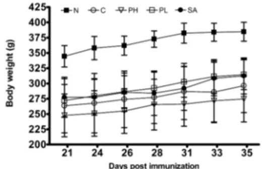
From day 21 to day 25 after the collagen injections, all C, PH, PL, SA groups’ weight decreased significantly. There was no significant difference between group C and the other three groups (Fig. 1).
Fig. 1. Changes of body weight during experiment.
N : Normal group (n=7), C : collagen-immunized group (n=8), PH : collagen-immunized and 6 ㎎/㎏
PC pharmacopuncture treated group (n=8), PL : collagen-immunized and 6 ㎎/㎏ PC pharmacopuncture (n=8), SA : collagen-immunized and saline treated group (n=8). Values are represented as mean ± SD.
2) Rate of weight gain
From day 21 to day 25 after the collagen injection, group PL's weight increase (15.63±6.09%) was somewhat higher than found in group C (12.48±5.79%). Also, while group SA's (12.62±4.97%) was similar and group PH’s (10.85±4.27%) rate was somewhat lower compared to group C, there was no significant difference between the groups
(Fig. 2).
Fig. 2. Rate of weight gain from day 21 to day 35 after treatments.
C : collagen-immunized group (n=8), PH : collagen -immunized and 6 ㎎/㎏ PC pharmacopuncture treated group (n=8), PL : collagen-immunized and 6 ㎎/㎏ PC pharmacopuncture (n=8), SA : collagen-immunized and saline treated group (n=8). Values are represented as mean ± SD.
2. The effect of paw edema
1) Changes of paw edema
From day 21 to day 25 after collagen injection, all of the groups, except group N (group C, PH, PL, SA), showed a decrease in paw edema. Group PH displayed a strong decrease (Fig.3).
Fig. 3. Changes of paw edema during experiment.
N : Normal group (n=7), C : collagen-immunized group (n=8), PH : collagen-immunized and 6 ㎎/㎏ PC pharmacopuncture treated group (n=8), PL : collagen -immunized and 6 ㎎/㎏ PC pharmacopuncture (n=8), SA : collagen-immunized and saline treated group (n=8). Values are represented as mean ± SD.
2) Rate of decreased paw edema
From day 21 to day 25 after collagen injection, the rate of paw edema decreased in group C (9.37±7.28%) and showed a significant difference from group PH (21.23±6.28%). Group PL (18.20±
7.65%) showed bigger decrease than group C, but there was no statistical significance, and group SA's (12.57±11.56%) rate of paw edma decrease was similar to group C’s (Fig. 4).
Fig. 4. Rate of decreased paw edema from day 21 to day 35 after treatments.
C : collagen-immunized group (n=8), PH : collagen -immunized and 6 ㎎/㎏ PC pharmacopuncture treated group (n=8), PL : collagen-immunized and 6 ㎎/㎏ PC pharmacopuncture (n=8), SA : collagen-immunized and saline treated group (n=8). Values are represented as mean ± SD. # : p<0.05 versus group C.
3. The effect on the width of the ankle
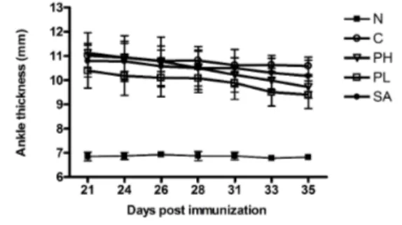
1) Changes of ankle thickness
From day 21 to day 25 after collagen injection, all of the groups, except group N (group C, PH, PL, SA), showed overall decrease in ankle thickness. Similar to the decrease of podedema, group PH showed a more significantly decrease in the width of tarsal joints compared to groups C, PL, and SA (Fig. 5).
Fig. 5. Changes of ankle thickness during experiment.
N : Normal group (n=7), C : collagen-immunized group (n=8), PH : collagen-immunized and 6 ㎎/㎏
PC pharmacopuncture treated group (n=8), PL : collagen-immunized and 6 ㎎/㎏ PC pharmacopuncture (n=8), SA : collagen-immunized and saline treated group (n=8). Values are represented as mean ± SD.
2) Rate of decreased ankle thickness
From day 21 to day 25 after collagen injection, group PH (12.66±5.55%) and PL (9.54±2.76%) showed significantly decreased tarsal joint widths compared to group C (3.84±3.24%), but group SA (5.78±3.66%) showed no significant difference from group C (Fig. 6).
Fig. 6. Rate of decreased ankle thickness from day 21 to day 35 after treatments.
C : collagen-immunized group (n=8), PH : collagen- immunized and 6 ㎎/㎏ PC pharmacopuncture treated group (n=8), PL : collagen-immunized and 6 ㎎/㎏ PC pharmacopuncture (n=8), SA : collagen-immunized and saline treated group (n=8). Values are represented as mean ± SD. ### : p<0.001 versus group C, # : p<0.05 versus group C.
4. The effect on the TNF-α level
From the paw edema exudates extracted in day 35, it could be seen that group C's (788.32±48.26 pg/㎖) TNF-α concentration increased significantly compared to that of group N’s (80.11±42.98 pg/
㎖). Although the TNF-α concentration of group PH (414.21±117.27 pg/㎖) decreased significantly compared to that of group C, groups PL (691.89
±84.96 pg/㎖) and SA (716.71±46.68 pg/㎖) did not show a significant difference from group C (Fig. 7).
Fig. 7. Effect on TNF-α in paw exudates.
N : Normal group (n=7), C : collagen-immunized group (n=8), PH : collagen-immunized and 6 ㎎/㎏ PC pharmacopuncture treated group (n=8), PL : collagen- immunized and 6 ㎎/㎏ PC pharmacopuncture (n=8), SA : collagen-immunized and saline treated group (n=8). Values are represented as mean ± SD. *** : p<0.001 versus group N, ### : p<0.001 versus group C.
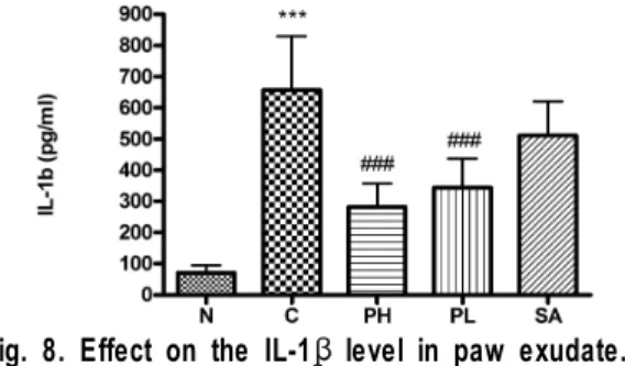
5. The effect on the IL-1β level
From the podedema exudates extracted on day 35, group C (657.35±172.89 pg/㎖)’s IL-1β concentration increased significantly compared to that of group N’s (70.86±24.06 pg/㎖). Groups PH (281.50±75.54 pg/㎖) and PL (343.58±93.19 pg/㎖) had a sharper decrease in IL-1β concentration compared to group C. Although group SA (510.99±109.82 pg/㎖) showed greater decrease in IL-1β concentration compared to group C, the difference was not statistically significant (Fig. 8).
Fig. 8. Effect on the IL-1β level in paw exudate.
N : Normal group (n=7), C : collagen-immunized group (n=8), PH : collagen-immunized and 6 ㎎/㎏ PC pharmacopuncture treated group (n=8), PL : collagen- immunized and 6 ㎎/㎏ PC pharmacopuncture (n=8), SA : collagen-immunized and saline treated group (n=8).
Values are represented as mean ± SD. *** : p<0.001 versus group N, ### : p<0.001 versus group C
6. The effect on the NOS positive cells
When the number of NADPⅡ-diaphorase positive nerve cells were observed using an optical microscope, group C (86.13±9.47) had significantly greater NOS positive cells compared to group N (43.21
±6.60). However, groups PH (51.25±10.04) and PL (66.50±6.16) decreased significantly compared to group C. Group SA did not show any significant difference (Fig. 9).
Fig. 9. Effect on the NOS positive cells.
N : Normal group (n=7), C : collagen-immunized group (n=8), PH : collagen-immunized and 6 ㎎/㎏ PC pharmacopuncture treated group (n=8), PL : collagen- immunized and 6 ㎎/㎏ PC pharmacopuncture (n=8), SA : collagen-immunized and saline treated group (n=8).
Values are represented as mean ± SD. *** : p<0.05 versus group N, # : p<0.05 versus group C, ### : p<0.001 versus group C.
Ⅳ. Discussion
RA is a chronic, inflammatory auto-immune disease which is found mostly in women around the age of 30 to 50, and is 2 to 4 times more common among women
22).
Pharmacopuncture is a new acupuncture method which injects refined oriental medicine to acupoints, achieving both the effects of acupuncture and the medicinal herbal preparation. It was first introduced in the 1960s by Nam in a book titled "meridians".
Pharmacopuncture is a unique kind of treatment using oriental medicine’s chemical stimulation rather than the original physical stimulation, such as acupuncture or moxibustion. Currently, several different studies utilizing pharmacopuncture are being conducted
23).
PC has been traditionally used for the treatment of various inflammatory diseases, hepatitis, tumors, and diarrhea
5). Recently, its extracts have been reported to possess antiviral activity against the hepatitis B virus, have potent estrogenic activities, inhibit bacterial DNA primase, and inhibit acyl- coenzyme A-cholesterol acyltransferase activity.
Current reports point out that compounds extracts form PC, such as reseveratrol, quercetin, emodin have great effects in suppressing inflammation
24).
The collagen-induced arthritis model used in our experiments is one of the most widely used experimental animal models. Histologically, it shows symptoms similar to RA. For example, synovium increase, monocyte calmness, formation of pannus, and destruction of cartilage and collagen are some of the effects seen in the collage-induced arthritis model
25-26).
Consequently, Our work used a rheumatoid arthritis model which was induced by bovine type
Ⅱ collagen, and we observed the changes in rat paw edema, width of ankle thickness joint, cytokine, and NOS expression when PC was injected to the part which correspondings to the acupuncture point ST36.
One important symptom of inflammatory reaction is edema. Edema is an excessive accumulation of inflammatory edema solution between cells and tissues. This exudation is caused by endotheliocyte’s permeable acceleration which is related to histamine, prostaglandin, and other pro-inflammatory mediators.
As the inflammation becomes worse, because of the increases in exudation, the size of the edema increase, therefore, the measurement of edema could be said to be an indirect way to measure the degree of inflammation
2). Consequently, our research confirmed that PC pharmacopuncture suppress inflammation by measuring the size of paw edema and the width of ankle thickness.
Group PH, which had PC solution (6㎎/㎏) injected from 21 to 35 days after the injection of collagen had a significant difference in the rate of decrease of size of paw edema and width of ankle thickness compared to group C. Group PL, which was injected with a lower concentration of PC solution (3㎎/㎏), also had a significant difference in the ankle thickness, but did not have a significant decrease in paw edema, although it still had a bigger decrease compare to control group.
Meanwhile, suppression of cytokine expression that causes inflammation is very important in curing RA. TNF-α and IL-1β are cytokines known to be produced in mononuclear cells are related to many acute and chronic inflammatory reactions
27).
TNF-α was first discribed as a substance which
destroys oncocytes, but it also has diverse effects
on several cells such as leukocytes and fibroblasts.
It is as type of a carrier of inflammatory response which is secreted by activated monocytes or macrophagocytes and is increased and activated by lipopolysaccharide (LPS). Along with IL-1, it has a major role in destroying articular cartilage by boosting the expression of a breakdown enzyme for collagen fibers and protein and suppressing the formation of proteoglycan in fibroblasts or cartilage cells
28). Our data shows that TNF-α concentration in exudate from rat ankle joint was higher in group C than group N, and group PH had a significantly lower production of TNF-α compared to group C.
IL-1β is a carrier which activates chemokine, cytokine, and many other substances such as synovial cells, endotheliocyte, lymphocyte, macrophagocyte which produces inflammatory medium
29,30). It is produced in several cells including the macrophagocyte, mononuclear cells, and synovial cortical cells. Our research shows that IL-1β concentration in exudates from rat ankle joints was higher in group C than group N, and groups PH and PL showed significantly reduced.
Nitric Oxide is known as a neurotransmitter of the central nervous system, has several roles such as vasolidation, sterilization, and many other functions as a neurotransmitter. The concentration of NO increases when there is an inflammatory reaction. Cells that produce NOS be identified histochemically by staining with NADPH-d.
Therefore, We dyed brain tissue sections and observed the amount of NOS production using an optical microscope, and the number of NADPH diaphorase positive neuron cells in cerebral cortex was increased in group C compared to group N.
Also, groups PH and PH had fewer NADPH
diaphorase positive neurons compared to group C.
These results demonstrate that PC pharmacopuncture has a beneficial effect in suppressing inflammation in an animal model for RA, and that further research will be benefical.
References