Problems and Solutions of Zymography Techniques
Dae-Ook Kang1 and Nack-Shick Choi1,2*
1Department of Bio Health Science, Changwon National University, 20 Changwondaejak-ro Uichang-gu, Changwon-si, Gyeongsangnam-do 51140, Korea
2KPROTEC INC., R & D Center, Gwahak-ro, Yuseong-gu, Daejeon 34141, Korea
Received December 9, 2019 /Revised December 26, 2019 /Accepted December 26, 2019
Enzymes are widely used in industrial applications such as detergents, food, feed production, pharma- ceuticals and medical applications and major contributors to clean industrial products and processes.
To screen, identify, and characterize the enzymes the zymography techniques are routinely used. The zymography technique is a simple, sensitive, and quantifiable technique that is widely used to detect functional enzymes following electrophoretic separation in sodium dodecyl sulfate (SDS)-polyacryl- amide gels. The method is a versatile two-stage technique involving protein separation by electro- phoresis followed by the detection of enzyme activity in polyacrylamide gels under non-reducing conditions. It is based on SDS-polyacrylamide gel (PAG) copolymerization with substrates, which are degraded by the hydrolytic enzymes restored in enzyme reaction buffer after the electrophoretic separation. Any kind of biological sample can be applied and analyzed on zymography, including cul- ture supernatants of microbes, plants extracts, blood, tissue culture fluids, enzymes in foods extracts and metaproteome. The advantage of zymography is that it is possible to directly detect the protein with activity on the electrophoretic gel as well as confirm the activity at the nanogram level. Thus, this zymography technology can be applied in various fields. However, these advantages are rather disadvantageous and can often lead to experimental errors. In this review, the advantages, dis- advantages, and problem solving of zymography technique are described.
Key words : Binding mode, diagonal zymography, electrotransfer, enzyme, zymography
*Corresponding author
*Tel : +82-42-710-5924, Fax : +82-42-710-5925
*E-mail : [email protected]
This is an Open-Access article distributed under the terms of the Creative Commons Attribution Non-Commercial License (http://creativecommons.org/licenses/by-nc/3.0) which permits unrestricted non-commercial use, distribution, and reproduction in any medium, provided the original work is properly cited.
Journal of Life Science 2019 Vol. 29. No. 12. 1408~1414 DOI : https://doi.org/10.5352/JLS.2019.29.12.1408
서 론
Zymography 기술은 1962년 Gross와 Lapiere에 의해 처음 소개되었는데, 이들은 올챙이 조직에서 콜라겐 분해 활성을 확인하는 분석법을 개발하고 이를 matrix metalloproteinase (MMP)를 기술할 때 zymography가 도입되었다[12]. 그리고 현재 널리 사용되는 zymography 기술(in gel zymography, IGZ)은 1978년에 plasminogen activators를 연구하기 위해 도 입되었다[11]. 당시에 사용된 IGZ의 형태는 overlay zymog- raphy 또는 indirect zymography로 현재 알려진 reverse zy- mography기술이었다. 이는 protease가 포함된 시료들을 SDS- 전기영동을 수행하여 시료 내 단백질들을 분리 한 다음, in- dicator 겔 상에 겹쳐서 기질 분해 및 검출을 가능하게 하였다.
하지만, 이 기술은 전기영동 겔과 indicator 겔의 분리된 두 개의 겔을 만들어야 하는 단점과 효소 활성의 해상도가 낮다 [22].
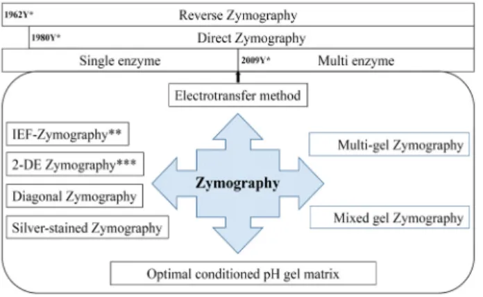
Heussen과 Dowdle (1980)가 단백질 기질(젤라틴)과 플라 스미노겐(plasminogen, 혈액 내 혈전용해효소 plasmin의 전 구체)을 SDS-전기영동 겔에 직접 첨가하여 기질-효소 전구체 acrylamide 겔 중합체를 만들어 플라스미노겐 활성인자 (plasminogen activator)를 탐색하였다. 활성은 겔 중합체 내 에서 전기영동에 의해 분리된 단백질들 중 플라스미노겐 활성 인자가 존재할 경우 플라스미노겐이 플라스민으로 전환되고 전환된 플라스민에 의한 기질 젤라틴을 분해시켰다. 이 방법 은 이전의 분리된 두 개의 겔을 만드는 번거로움을 해결함과 동시에 기술의 해상도 역시 개선하였다. 이를 직접 zymog- raphy (direct zymography)의 시초가 되었으며 현재 가장 널 리 사용되는 zymography 기술로 발전하게 되었다(Fig. 1) [15].
현재 zymography 기술은 단백질이 전기적으로 분리된 SDS-전기영동 겔 상에서 효소 활성을 검출하는 방법으로 활성 을 지닌 단백질의 분자량을 직접 측정이 가능할 뿐만 아니라 효소 활성의 민감도(sensitivity)는 나노그램(nanogram) 수준 에서 검출이 가능하기 때문에 소량의 시료로도 적용이 가능하 다[22]. 이 기술은 현재까지 암 전이와 관련된 효소인MMPs (MMP-2/MMP-9) 연구 분야[16, 28], 효소 검출 및 정제[29], MMP의 발현 및 조절[18, 28], 활성 억제[30], 가공 및 파괴 등 다양한 연구 분야에 도입되었다. 뿐만 아니라, 산업용효소 분야; 단백질분해효소(protease) [3, 13, 14, 37, 39], 섬유소분해 효소(cellulase, xylanase 등)[35, 36, 38, 40], 전분분해효소 - Review -
Fig. 1. Various zymography techniques over fifty years. Various techniques have been developed for research purposes.
(amylase) [17, 24], 및 지방분해효소(lipase) [10, 33] 등 유용효 소 연구에도 사용되고 있다.
2009년에는 zymography 기술에 전기적 이동(electrotrans- fer) 방법[2, 4, 23, 25, 41]을 접목하여 하나의 SDS-전기영동 겔을 이용하여 다종 효소의 검출이 가능하였으며(multi-gel zymography) [4], 또한, 하나의 SDS-전기영동 겔에 두 종류의 서로 다른 효소(protease와 lipase)에 대한 기질을 첨가함으로 써 하나의 SDS-전기영동 겔을 이용하여 동시에 두 종류의 효소 활성 검출이 가능하게 되었다(mixed gel zymography) (Fig.
1) [2]. 전기적 이동 방법은 효소가 포함된 단백질 시료를 SDS- 전기영동을 통해 분리한 후 하나 이상의 개별 기질 겔로 옮겨 진다. 또한, 이 기술은 다수의 2 차 기질 겔이 적층되어 다수의 효소를 동시에 정보를 얻을 수 있다. 본 리뷰에서는 zymog- raphy 기술의 장점 및 단점을 정리하고 zymography의 한계 를 새로운 zymography 기술의 도입 과정 등을 상세히 서술하 고자 한다. 그래서, zymography 기술의 장단점을 최적화하여 모든 연구자들이 쉽게 적용 가능한 보편화된 기술로 소개하고 자 한다.
본 론
Zymography 기술의 개요
Zymography 기술은 SDS-전기영동 겔 상에 기질을 첨가하 여 기질을 분해하는 효소를 직접적으로 검출하는 방법으로 이 기술의 일반적인 원리는 다음과 같다. SDS-전기영동 겔 상 에 기질을 첨가하여 겔을 만든 후 효소 단백질이 포함된 시료 를 주입하여 전기영동을 수행 한다(step 1). 분획된 단백질의 효소 활성은 2.5% (v/v) Triton X-100을 이용하여 SDS에 의해 불활성화된 효소 활성을 복원시키고 활성 반응완충용액(200 mM NaCl, 10 mM CaCl2, 0.02% NaN3가 포함된 30 mM Tris 완충용액, pH 7.4)에 겔을 침적하여 37℃ 배양기에서 12시간 방치하여 기질에 대한 효소 반응이 일어나도록 한다(step 2).
마지막으로, 효소 활성은 시료 단백질에 의해 분해된 겔 상의 기질이 염색약에 의해 염색되지 않고 투명한 띠(negative staining)를 형성함으로써 활성을 확인하는 방법이다(step 3).
따라서 효소의 종류에 따라 기질의 선택 및 염색 방법/시약을 달리하게 된다. 상기에서도 언급하였듯이, zymography 기술 의 가장 큰 장점은 전기영동 겔 상에서 효소 활성을 지닌 단백 질의 분자량을 직접 측정이 가능하고 또한, 효소 활성의 민감 도가 아주 높아 소량의 시료로도 적용이 가능하여 응용 분야 가 많다[22].
Zymography 기술의 응용
Zymography 기술의 단순한 효소 활성을 지닌 단백질 분자 량 확인은 물론 기술의 응용을 다양화하여 미생물의 선별 (screening) [19], 미생물의 효소 활성을 위한 배양 조건의 최적 화(culture optimization) [5], 효소 활성 촉진인자(activator) [6], 및 저해 인자(inhibitor) [21] 발굴, 베타-글루칸(β-glucan) 과 같은 고분자소재의 저분자화 효소[32], 그리고 효소 특성화 연구(enzyme characterization) [7] 등 다양한 분야에 적용이 가능하다.
첫 번째, 미생물의 선별(screening); 다양한 환경 조건(식품 및 토양 등)에서 분리한 미생물들의 효소 생산 양상을 zymog- raphy 분석을 통해 database화하여 미생물과 zymogram 양상 을 이용하여 상이한 양상을 보이는 균주를 선택하여 분석 및 동정한 결과 새로운 균주, Brevundimonas naejangsanensis sp.
Nov.,로 동정되어 보고된 사례가 있다[19]. 이는 기질이 포함 된 플레이드(skim milk agar plate) 상에서 활성을 지닌 미생물 들을 분리하여 zymography 양상을 분석하면 시료들 중 동일 균주를 쉽게 판별할 수 있는 장점이 있어 가능하다(Fig. 2) [19].
두 번째, 미생물의 효소 활성을 위한 배양 조건의 최적화 (culture optimization); 일반적으로 미생물은 배양시간, 배양 온도 및 배지 조성에 따라 효소 생산 및 균체의 양이 상이한 양상을 보이는데 이를 zymography 기술과 효소 활성의 정량 화를 위해 densitometer를 이용하여 효소 활성을 위한 최적 조건 및 효소 생산을 정량적으로 측정이 가능하여 산업적 활 용이 가능하다(Fig. 2) [5].
세 번째, 효소 특이성 연구(enzyme characterization); 순수 분리된 효소의 특이성 연구에 SDS-전기영동 겔과 zymog- raphy 기술을 접목하여 효소의 기질 특이성을 규명하는 것이 다[7]. 단백질분해 효소 활성 측정을 기질에 대한 활성을 흡강 도법을 이용하여 효소 활성을 규명할 수 없었던 멸치 젓갈에 서 분리한 균주 Staphylococcus sp. Strain AJ로부터 순수 분리 한 효소 AJ 혈전용해효소(단백질 분해효소)를 zymography 기 술을 이용하여 규명할 수 있었다. 이를 통해 AJ의 경우 pH에 대한 양쪽성(산성과 염기성 조건) 최적 pH 조건을 가지는 독 특한 효소임 zymography를 통해 규명할 수 있었다. 또한, N- 말단 아미노산 서열분석, 2차원 전기영동(SDS-전기영동 겔 및
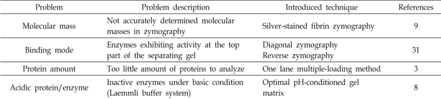
Table 1. Development of problem-based zymography techniques
Problem Problem description Introduced technique References
Molecular mass Not accurately determined molecular
masses in zymography Silver-stained fibrin zymography 9
Binding mode Enzymes exhibiting activity at the top part of the separating gel
Diagonal zymography
Reverse zymography 31
Protein amount Too little amount of proteins to analyze One lane multiple-loading method 3 Acidic protein/enzyme Inactive enzymes under basic condition
(Laemmli buffer system)
Optimal pH-conditioned gel
matrix 8
Fig. 2. Isolation of microbes showing proteolytic activity from soil and performing zymography with culture supernatants of isolated microbes. *Brevundimonas naejangsanensis sp. Nov. was identified based on 16S rRNA gene sequences and fatty acids compo- sions [19].
zymography 포함) 및 효소 활성 분석을 통해 AJ 효소의 경우 homodimer 임이 밝혀졌다[7].
네 번째, 효소 활성인자(activator) 또는 저해인자(inhibitor) 연구; zymography 기술을 이용하여 인체 혈전용해효소 플라 스민(plasmin)의 전구체인 플라스미노겐(plasminogen)의 전 환인자인 플라스미노겐 활성인자(plasminogen activator) [6]
탐색 및 효소 활성 저해 인자(inhibitor) [21] 발굴, 그리고 베타 -글루칸(β-glucan)과 같은 고분자 소재를 전기영동 겔에 직접 중합체를 만들어 저분자로 생물 전환(bioconversion)하는 효 소의 탐색 및 구조 연구도 가능하다[32].
Zymography 기술의 한계와 극복
Zymography 기술의 장점은 상기에서도 언급하였듯이 활 성을 지닌 효소 단백질의 분자량을 겔 상에 시각화하여 측정 이 가능할 뿐만 아니라 측정 가능한 단백질 효소 농도가 나노 그램(nanogram) 수준으로 민감도가 아주 높아 미생물 배양액 을 아무런 전처리 없이 적용이 가능하다는 것이다. 하지만, zymography의 이러한 장점이 오히려 효소 연구에 단점으로 작용할 경우가 있는데, 예를 들어 기술의 높은 민감도로 인해
실제 활성을 지닌 단백질을 측정하기가 곤란할 경우가 있다 (Table 1) [9]. 또한, 겔 상의 기질이 단백질 시료의 전기영동 시 이동상에 영향을 주어 정확한 분자량의 측정이 어렵다[20].
이러한 문제점뿐만 아니라 zymography를 이용하여 효소 단 백질을 연구하는 과정에서 수반되는 zymography 기술의 문 제점과 해결 사례를 서술하고자 한다(Table 1).
첫 번째, 분자량 측정의 문제점; Zymography 기술은 겔 상에서 직접적으로 그리고 아주 미량의 시료로도 효소 활성을 확인 할 수 있는 훌륭한 기술이라 할 수 있지만, Katrina 등이 보고 한 바와 같이 겔 상에 첨가된 기질이 시료 단백질의 이동 상에 영향을 주어 실제적으로 SDS-전기영동 겔과 zymog- raphy 겔 상의 분자량의 차이가 있어 비교가 어려울 수 있다 [20]. 일반적으로 효소 활성을 위해 SDS-전기영동 겔과 zy- mography 겔을 동시에 같은 조건(시료 조건 및 전기영동 조 건)하에서 수행하게 된다. 전기영동 후 SDS-전기영동 겔은 은 염색(silver staining)을 하고 zymography 겔은 효소 단백질의 복원 및 활성화(step 2) 단계를 거쳐 Coomassie blue 염색을 통해 효소 활성을 시각화 한다. Zymography 겔을 SDS-전기 영동 겔로 오인하여 zymography 겔을 은 염색을 수행하게
되었고, 이 과정에서 “silver-stained fibrin zymography (은 염색 피브린 zymography)” 기술이 개발되었다[9]. 그 결과, 하나의 zymography 겔 상에서 효소 활성과 활성을 지닌 단백 질을 동시에 확인할 수 있게 되어 기존의 zymography의 단점 인 겔 상에 첨가된 기질에 의한 단백질의 이동상의 변화를 극복할 수 있게 되었다.
두 번째, binding mode의 문제점; Binding mode 란 zy- mography 겔 상에서 이동되지 않고 겔의 상단에 머물러 활성 을 나타내는 현상을 말한다. 이 현상을 Brown [1]과 Lantz [27]
등은 시료 내 단백질 효소가 겔 상에 중합된 기질과 결합하여 분리 겔(resolving gel) 상으로 더 이상 이동하지 못하고 겔 상단부에 머물러 기질을 분해하여 활성을 나타낸다고 정의하 였다. 이 현상을 silver-stained fibrin zymography 겔 상에서 도 겔의 상단부에서 활성과 활성을 지닌 단백질의 확인이 가 능하였다[9]. Binding mode 현상을 규명하기 위해 세 종류의 zymography 기술(direct zymography, reverse zymography 그리고 diagonal zymography)을 접목시켜 이 현상이 규명되 었다[31]. Binding mode 현상은 2가지 요인에 의한 것으로 하 나는 Brown과 Lantz가 주장한 효소와 기질의 결합에 의한 현상(Brown & Lantz binding mode), 그리고 다른 하나는 효 소 단백질의 높은 pI 값(8.8 이상)으로 인해 일반 전기영동 조 건(Laemmli buffer system, pH 8.8) [26]에서는 분리 겔 상으로 이동하지 못하고 겔 상단부에 머물러 기질을 분해하는 현상 (Choi & Kim binding mode) 이다.
세 번째, 전기영동 적용 시료 단백질의 농도; 일반적으로 단백질을 분석하기 위해서는 많은 양의 단백질 농도를 필요로 한다. 이를 위해서 다양한 단백질 농축 방법들을 선택적으로 사용하지만 시료의 변성 및 손실이 불가피하다. 특히, 효소 단백질을 분석하기 위해서는 비 변성 상태로 활성 분석 및 구조 분석을 수행하여야 한다. 단백질 분석 기술의 발달로 소 량의 단백질 시료로도 프로테오믹스/MALDI-TOF (Matrix- assisted laser desorption/ionization time-of-flight) 기술로 데 이터베이스 기반 효소 단백질의 분석이 가능하다[34]. 하지만, 미량 함유 단백질의 경우 활성 및 구조 분석을 위해서는 농축 을 위한 전 처리가 요구되는데, 이 경우 비 변성의 단백질 상태 로 많은 양의 시료 단백질을 적용하기 위해 “one-lane multi- ple-loading 법”을 2차원 전기영동에 접목하였다[3]. 이 방법은 IEF (isoelectric focusing)의 특성(단백질은 자신의 등전점에 도달하게 되면 머무르게 된다는 점)을 바탕으로 하여 2차원 전기영동을 전개하는 방법이다. 이 기술을 이용하여 프로테오 믹스/MALDI-TOF 기술로는 분석이 어려운 Bacillus sp. DJ 유 래의 쉽게 자가 분해(autolysis)되는 효소를 분석이 가능하였 다[3].
네 번째, 산성 조건의 활성 단백질; SDS-전기영동 겔을 이용 한 단백질의 활성 측정은 전기영동 수행하는 조건(Laemmli buffer system, pH 8.8)에서 수행되어지기 때문에 산성 조건에
서 활성을 지닌 단백질의 활성을 측정하기에는 부적합하다.
대표적인 항균 단백질인 니신(nisin)과 같은 산성 조건에서만 활성을 지닌 단백질의 활성을 측정하기 위해 최적 pH 조건의 겔 기질(optimal pH-conditional gel matrix, OP-CGM, pH 4.0)의 제조 및 전기영동 겔의 OP-CGM으로의 전기적 이동법 (electrotransfer)이 도입되었다[8].
Zymography 기술을 이용한 다종 효소 동시 검출 첫 번째, 다종 기질 샌드위치 zymography법; 다종 효소(단 백질 분해효소, 지질 분해효소 그리고 섬유소 분해효소)를 동 시 검출을 위한 새로운 방법의 zymography 기술이 도입되었 는데 그 원리는 단백질 표면의 음전하를 이용한 전기적 이동 법(electrotransfer)을 이용하여 겔 상의 단백질을 서로 다른 3종류의 기질로 이동시키는 것이다[4](Fig. 3). 이 기술의 원리 는 농축된 단백질 시료를 SDS-전기영동 겔을 통하여 단백질을 분자량에 따라 분리시킨다. SDS에 의해 불 활성화된 단백질은 2.5%(v/v) Triton X-100을 처리하여 겔 속에 포함되어 있는 SDS를 제거를 한다. 활성화된 단백질을 포함한 겔을 서로 다 른 3 종류의 기질에 전기적 이동(electrotransfer) 방법으로 단 백질들을 기질로 이동시킨다(Fig. 3A). 이 기술을 이용하여 3 종류의 서로 다른 기질을 사용하여 토양에서 분리한 4종의 균주를 분석한 결과, Paenibacillus sp. 균주와 두 종류의 Bacillus sp. 균주로부터 3종류의 cellulase를 검출하였고, Staphylococcus sp. 균주로부터 한 종류의 lipase를 그리고 2종류의 Bacillus sp. 균주로부터 2종류의 protease를 검출되었다[4].
두 번째, 혼합 기질 zymography법; 혼합기질 zymography 기술은 하나의 기질 겔에 서로 다른 효소에 대한 기질을 동시 에 혼합하여 중합체를 만든 후 효소 활성을 측정하는 방법이 다. 서로 다른 기질로서 단백질 분해효소(protease) 검출을 위 한 단백질 기질(fibrin gel)과 지질 분해효소(esterase 및 lipase) 검출을 위한 glycerol tributyrate가 사용된다. 상기의 다종 기 질 샌드위치 zymography법과 같이 SDS-전기영동에 의해 분 리된 단백질을 전기적 이동법(electrotransfer)을 이용하여 혼 합 기질 겔로 이동시키는 것이다[2]. 효소활성 측정방법은 Fig.
3(B)의 모식도에서 보는 바와 같이, 먼저 lipase 활성을 육안으 로 측정 한 후 동일 기질 gel을 Coomassie 염색약을 이용하여 protease 활성을 확인한다. 토양 및 된장으로부터 분리한 3 종류의 미생물을 이용하여 혼합기질 zymography를 바탕으로 protease 및 lipase를 확인한 결과, Staphylococcus sp. 균주로부 터 6 개의 lipase 효소 활성의 확인이 가능하고, 그리고 두 종류 의 Bacillus spp. 균주로부터 4 개의 protease band의 확인이 가능하다[2].
결 론
Zymography 기술은 효소에 의한 중합된 기질을 분해하는
A
B
37oC for 24 hr 37oC for 6 hr
Fig. 3. Schematic diagram of electrotransfer method for multiple enzymes detection. A, Simultaneous detection of multiple enzymes using multiple substrate gels. B, Simultaneous detection of protease and lipase using mixed substrate (glycerol tributyrate and fibrin).
것을 기반으로 가수 분해 효소를 탐색 및 효소 특성을 연구하 는 탁월한 기술이다. 전기적으로 분리된 단백질들 중 효소 활 성을 지닌 단백질의 분자량을 직접 측정이 가능할 뿐만 아니 라 효소 활성의 민감도(sensitivity)가 아주 높아 종종 잘못 해 석되는 경우가 있다. 따라서, 본 리뷰에서는 zymography기술 의 장점 및 응용을 상세히 설명하였다. 또한, zymography 기 술의 장점이 오히려 단점 및 한계임을 알았고 이를 해결하기 위한 새로운 zymography 기술과 단백질 기술 등을 접목하여 이를 극복할 수 있었다. 앞으로도 새로운 zymography 기술이 지속적으로 소개될 것이며 이와 함께 zymogrphy의 문제점 또한 노출될 것이다. 현재, 국내·외 효소 연구와 관련된 zy- mography 기술은 활성 단백질의 분자량을 측정가능 하다는 점을 활용하여 암 전이 관련 효소인 metrix metalloproteinases (MMPs) 및 가수분해효소(protease, cellulase, amylase, 및 li- pase 등)의 분자량을 확인을 위한 기술로 국한되어 연구에 적 용되고 있다. 그래서, zymography 기술의 장단점을 최적화하 여 모든 연구자들이 다양한 연구 분야에서 쉽게 적용 가능한 보편화된 기술이 되었으면 한다.
Zymography 기술을 통해 다양한 기능성 효소를 탐색, 개 량 및 확보는 미래 산업에 꼭 필요한 효소를 포함한 단백질
뱅크 구축 및 바이오 융합형 유용 효소를 대량 확보하고 활용 하는 선도 기술 개발에 크게 기여할 것으로 기대된다. 또한, 기존의 효소 발굴 시스템으로는 불가능했던 난 배양성 또는 배양이 불가능한 미생물 유래의 효소를 메타게놈(metage- nome) 환경 유전체를 통해 유용 효소의 발굴이 가능하며 zy- mography 기술의 확립은 다양한 효소 자원의 확보에 유용하 게 활용될 수 있을 것이다.
References
1. Brown, T. L., Yet, M. G. and Wold, F. 1982. Substrate-con- taining gel electrophoresis: sensitive detection of amylolytic, nucleolytic, and proteolytic enzymes. Anal. Biochem. 122, 164-172
2. Choi, N. S., Choi, J. H., Kim, B. H., Han, Y. J., Kim, J. S., Lee, S. G. and Song, J. J. 2009. Mixed-substrate (glycerol trib- utyrate and fibrin) zymography for simultaneous detection of lipolytic and proteolytic enzymes on a single gel.
Electrophoresis 30, 2232-2237.
3. Choi, N. S., Choi, J. H., Yoon, J. H., Lee, S. G. and Song, J. J. 2009. Identification of a serine protease from a Bacillus sp. using multiple loading of O’Farrell-type isoelectric fo-
cusing slab two-dimensional gel. Biotechnol. Lett. 31, 975-978.
4. Choi, N. S., Kim, B. H., Park, C. S., Han, Y. J., Lee, H. W., Choi, J. H., Lee, S. G. and Song, J. J. 2009. Multiple-layer substrate zymography for detection of several enzymes in a single sodium dodecyl sulfate gel. Anal. Biochem. 386, 121- 122.
5. Choi, N. S. and Kim, S. H. 1999. Application of fibrin zy- mography for determining the optimum culture time for protease activity. Biotechnol. Techniq. 13, 899-901.
6. Choi, N. S. and Kim, S. H. 2000. Two fibrin zymography methods for analysis of plasminogen activators on gels.
Anal. Biochem. 281, 236-238.
7. Choi, N. S., Song, J. J., Chung, D. M., Kim, Y. J., Maeng, P. J. and Kim, S. H. 2009. Purification and characterization of a thermo acid-stable fibrinolytic enzyme from Staphylo- coccus sp. strain AJ isolated from Korean salted-fermented Anchovy-joet. J. Ind. Microbiol. Biotechnol. 36, 417-426.
8. Choi, N. S., Jeong, S. Y., Yang, H. J., Ahn, K. H., Park, C.
S., Kim, C. Y., Kim, J. S., Yoon, B. D. and Kim, M. S. 2010.
Activity assay for nisin-like acidic bacteriocins using an op- timal pH-conditioned gel matrix. Anal. Biochem. 397, 259- 261.
9. Chung, D. M., Kim, K. E., Ahn, K. H., Park, C. S., Kim, D. H., Koh, H. B., Chun, H. K., Yoon, B. D., Kim, H. J., Kim, M. S. and Choi, N. S. 2011. Silver-stained fibrin zymog- raphy: separation of proteases and activity detection using a single substrate-containing gel. Biotechnol. Lett. 33, 1663- 1666.
10. Duarte, J. G., Leone-lgnacio, K., da Silva, J. A. C., Fernandez- Lafuente, R. and Freire, D. M. G. 2016. Rapid determination of the synthetic activity of lipases/esterases via transester- ification and esterification zymography. Fuel 177, 123-129.
11. Granelli-Piperno, A. and Reich, E. 1978. A study of proteases and protease-inhibitor complexs in biological fluids. J. Exp.
Med. 148, 223-234.
12. Gross, J. and Lapiere, C. M. 1962. Collagenolytic activity in amphibian tissues: a tissue culture assay. Proc. Natl. Acad.
Sci. USA. 48, 1014-1022.
13. Haddar, A., Agrebi, R., Bougatef, A., Hmidet, N., Sellami- Kamoun, A. and Nasri, M. 2009. Two detergent stable alka- line serine-proteases from Bacillus mojavensis A21: Purifica- tion, characterization and potential application as a laundry detergent additive. Bioresour. Technol. 100, 3366-3373.
14. Hammami, A., Hamdi, M., Abdelhedi, O., Jridi, M., Nasri, M. and Bayoudh, A. 2017. Surfactant- and oxidant-stable al- kaline proteases from Bacillus invictae: Characterization and potential applications in chitin extraction and as a detergent additive. Int. J. Biol. Macromol. 96, 272-281.
15. Heussen, C. and Dowdle, E. B. 1980. Electrophoretic analysis of plasminogen activators in polyacrylamide gels containing sodium dodecyl sulfate and copolymerized substrates. Anal.
Biochem. 102, 196-202.
16. Hibbs, M. S., Hasty, K. A., Seyer, J. M., Kang, A. H. and Mainardi, C. L. 1985. Biochemical and immunological char- acterization of the secreted forms of human neutrophil gelatinase. J. Biol. Chem. 260, 2493-2500.
17. Hmidet, N., Jemil, N. and Masri, M. 2019. Simultaneous pro- duction of alkaline amylase and biosurfactant by Bacillus methylotrophicus DCS1: application as detergent additive.
Biodegradation 30, 247-258.
18. Houde, M., De Bruyne, G., Bracke, M., Ingelman-sundberg, M., Skoglund, G., Masure, S., Van Damme, J. and Opdenak- ker, G. 1993. Differential regulation of gelatinase B and tis- sue-type plasminogen activator expression in human Bowes melanoma cells. Int. J. Cancer 53, 395-400.
19. Kang, S. J., Choi, N. S., Choi, J. H., Lee, J. S., Yoon, J. H.
and Song, J. J. 2009. Brevundimonas naejangsanensis sp. nov., a proteolytic bacterium isolated from soil, and reclassifica- tion of Mycoplana bullata into the genus Brevundimonas as Brevundimonas bullata comb. nov. Int. J. Syst. Evol. Microbiol.
59, 3155-3160.
20. Katrina, M. H., Penheiter, A. R., Gathman, A. C. and Lilly, W. W. 1996. Anomalous Estimation of Protease Molecular Weights Using Gelatin-Containing SDS–PAGE. Anal. Bio- chem. 233, 140-142.
21. Kim, S. H. and Choi, N. S. 1999. Electrophoretic analysis of protease inhibitors in fibrin zymography. Anal. Biochem.
270, 179-181.
22. Kim, S. H., Choi, N. S. and Lee, W. Y. 1998. Fibrin zymog- raphy: a direct analysis of fibrinolytic enzymes on gels. Anal.
Biochem. 263, 115-116.
23. Kobayashi, T., Kakizaki, I. and Nakamura, T. 2019. Proteo- glycan-substrate gel zymography for the detection of chon- droitin sulfate-degrading enzymes. Anal. Biochem. 568, 51-52.
24. Kocabay, S., Cetinkaya, S., Akkaya, B. and Yenidünya, A.
F. 2016. Characterization of thermostable β-amylase iso- zymes from Lactobacillus fermentum. Int. J. Biol. Macromol. 93, 195-202.
25. Kurz, L., Hernández, Z., Contreras, L. M. and Wilkesman, J. 2017. Sequential detection of thermophilic lipase and pro- tease zymography. In Zymography: Methods in Mol. Biol. (pp.
271-277): Springer.
26. Laemmli, U. K. 1970. Cleavage of structural protein during the assembly of the head of bacteriophage T4. Nature 227, 680-685.
27. Lantz, M. S. and Ciborowski, P. 1994. Zymographic techni- ques for detection and characterization of microbial pro- teases. Methods Enzymol. 235, 563-594.
28. Masure, S., Billiau, A., Van Damme, J. and Opdenakker, G.
1990. Human hepatoma cells produce an 85 kDa gelatinase regulated by phorbol 12-myristate 13-acetate. Biochim.
Biophys. Acta 1054, 317-325.
29. Masure, S., Proost, P., Van Damme, J. and Opdenakker, G.
1991. Purification and identification of 91-kDa neutrophil gelatinase. Release by the activating peptide interleukin-8.
Eur. J. Biochem. 198, 391-398.
30. Paemen, L., Martens, E., Narga, K., Masure, S., Roets, E., Hoogmartens, J. and Opdenakker, G. 1996. The gelatinase inhibitory activity of tetracyclines and chemically modified tetracycline analogues as measured by a novel microtiter assay for inhibitors. Biochem. Pharmacol. 52, 105-111.
31. Park, C. S., Kang, D. O., Lee, W. Y., Chun, S. S., Lim, S.
초록:자이모그라피 기술의 문제점과 해결
강대욱1․최낙식1,2*
(1창원대학교 생명보건학부, 2㈜케이프로텍 기업부설연구소)
효소는 세제, 식품, 사료, 의약품 및 의료용 분야 등 산업 전반적인 응용 분야에서 널리 사용되고 있으며, 산업 제품 및 공정에서 주요 요인이다. 효소를 선별, 확인, 및 특성 분석을 위해 zymography 기술이 일상적으로 사용됩 니다. Zymography 기술은 SDS-전기영동을 통해 단백질을 분리한 후 포함된 기질을 겔 상에서 분해하는 기능성 효소를 검출하는 데 널리 사용되는 단순하고 민감하며 정량화가 가능한 기술이다. 이 방법은 비 환원 조건하에서 SDS-전기영동 겔에서 전기영동에 의한 단백질의 분리와 겔 상에서 효소 활성을 검출하는 다목적 2 단계의 기술이 다. 이는 SDS-전기영동 겔에 기질을 중합시키고 전기영동 분리 후 효소 반응 완충용액에서 복원된 가수분해 효소 에 의해 분해되는 것을 기반으로 하는 기술이다. 미생물 배양액, 식물 추출물, 혈액, 조직 배양액, 식품 속 효소 및 메타 프로테옴을 포함한 어떤 종류의 생물학적 시료들을 zymography에 적용하고 분석이 가능하다.
Zymography의 장점은 전처리 없이 혼합된 시료를 적용하여 SDS-전기영동 겔 상에서 활성을 지닌 단백질을 직접 육안으로 검출이 가능할 뿐만 아니라 나노그람(nanogram) 수준에서 활성을 확인이 가능하다. 그래서 이 zymog- raphy 기술은 다양한 분야에 응용이 가능하다. 하지만, 이러한 장점이 오히려 단점으로 작용하여 실험적 오류를 범할 수 있는 경우가 많다. 본 총설에서 zymography 기술의 장점, 단점, 및 문제점 해결에 관해서 서술하였다.
Y., Moon, J. Y., Kim, D. H. and Choi, N. S. 2015. Identifica- tion of two types binding modes using reverse or diagonal electrophoretic zymography. Aca. J. Biotechnol. 3, 52-55.
32. Park, C. S., Yang, H. J., Kim, D. H., Kang, D. O., Kim, M.
S. and Choi, N. S. 2012. A screening method for β-glucan hydrolase employing trypan blue-coupled β-glucan agar plate and β-glucan zymography. Biotechnol. Lett. 34, 1073- 1077.
33. Park, J. W., Cho, S. Y. and Choi, S. J. 2008. Purification and characterization of hepatic lipase from Todarodes pacificus.
BMB Reports 41, 254-258.
34. Park, S. G., Kho, C. W., Cho, S., Lee, D. H., Kim, S. H. and Park, B. C. 2002. A functional proteomic analysis of secreted fibrinolytic enzymes from Bacillus subtilis 168 using a com- bined method of two-dimensional gel electrophoresis and zymography. Protomics 2, 206-211.
35. Phitsuwan, P., Tachaapaikoon, C., Kosugi, A., Mori, Y., Kyu, K. L. and Ratanakhanokchai, K. 2010. A Cellulolytic and Xylanolytic Enzyme Complex from an Alkalothermoanaer- obacterium, Tepidimicrobium xylanilyticum BT14. J. Microbiol.
Biotechnol. 20, 893-903.
36. Picart, P., Diaz, P. and Pastor, F. I. J. 2007. Cellulases from two Penicillium sp. strains isolated from subtropical forest
soil: production and characterization. Lett. Appl. Microbiol.
45, 108-113.
37. Pillai, P., Mandge, S. and Archana, G. 2011. Statistical opti- mization of production and tannery applications of a kerati- nolytic serine protease from Bacillus subtilis P13. Proc. Bio- chem. 46, 1110-1117.
38. Schwarz, W. H., Bronnenmeier, K., Gräbnitz, F. and Stau- denbauer, W. L. 1987. Activity staining of cellulases in poly- acrylamide gels containing mixed linkage β-glucans. Anal.
Biochem. 164, 72-77.
39. Sookkheo, B., Sinchaikul, S., Phutrakul, S. and Chen, S. T.
2000. Purification and Characterization of the Highly Ther- mostable Proteases from Bacillus stearothermophilus TLS33.
Prot. Exp. Purif. 20, 142-151.
40. Su, L. J., Liu, H., Li, Y., Zhang, H. F., Chen, M., Gao, X.
H., Wang, F. Q. and Song, A. D. 2014. Cellulolytic activity and structure of symbiotic bacteria in locust guts. Gen. Mol.
Res. 13, 7926-7936.
41. Tapizquent, M., Fernández, M., Barreto, G., Hernández, Z., Contreras, L. M., Kurz, L. and Wilkesman, J. 2017. Zymogra- phy detection of a bacterial extracellular thermoalkaline es- terase/lipase activity. In Zymography: Methods in Mol. Biol.
(pp. 295-300): Springer.