Determination of Monoglycoceramides in Biological Samples using Enzymatic Deacylation and Reverse-phase HPLC
Mi-Hwa Choi, Kyeong-Mi Choi, Youn-Sun Lee, So-Young Ji, Ju-Hyun Cho, Yong-Moon Lee, Yeo-Pyo Yun and Hwan-Soo Yoo#
College of Pharmacy and CBITRC, Chungbuk National University, Cheongju 361-763, Korea
Abstract — Glycosphingolipids are structural components of mammalian cell membranes and are involved in essential cel- lular physiology such as cell-cell interaction, recognition, transmembrane signaling, proliferation and cell death. In this study, the simple quantitative method of monoglycoceramides-containing glucosylceramide and galactosylceramide was developed.
The glycosylceramides extracted from culture cells and rat plasma were resolved by TLC, deacylated by SCDase and ana- lyzed by HPLC-fluorescence detector at an excitation wavelength of 340 nm and an emission wavelength of 455 nm. Limit of detection was approximately 0.1 pmol and limit of quantification was about 1 pmol for both monoglycoceramide standards.
The recoveries of standard glucosylceramides from intra- and inter-day assays were 113.8 and 88.8% and those of galac- tosylceramides were 110.7 and 123.9%, respectively. The monoglycoceramide contents of SW-620 cells and rat plasma were 141.5±5 pmol/1×106 cells and 3.9±0.3
µM, respectively. The present analytical method provides a reproducible quan-
tification and total content of monoglycoceramide which may be as a potential biomarker for lipid imbalance-related human diseases.Keywords □ monoglycoceramide, glucosylceramide, galactosylceramide, SCDase, HPLC, SW-620 cells
스핑고지질은 동물, 식물, 효모 및 Sphingomonas를 포함하는 세균에서 광범위하게 생성된다.1)생체 내 스핑고지질 대사는 신 생합성, 전환 및 분해 과정으로 구성되며, 세포의 소포체에서 발 생하는 스핑고지질 신생합성(de novo biosynthesis)은 아미노산 인 세린(serine)과 지방산인 팔미테이트(palmitate)를 전구체로 하여 serine palmitoyltransferase의 작용에 의해 스핑고지질 모 핵인 3-ketodihydrosphingosine이 생성된다.2,3)이 물질은 환원되 어 dihydrosphingosine이 되며, 지방산이 축합된 dihydroceramide 가 생성되고, 이어서 이중결합이 포함된 sphingosine 골격구조를 가지고 있는ceramide로 전환된다.4,5)스핑고지질 대사 중간체인 ceramide는 ceramide kinase에 의하여 인산화되어 ceramide-1- phosphate가 형성되거나, sphingomyelin synthase에 의하여
sphingomyelin으로 전환되며, galactosyl ceramide synthase에 의하여 당스핑고지질의 생합성이 시작된다.6-8)
당스핑고지질은 동물세포의 세포막 구성성분으로서 필수적이 며, 뇌와 말초신경의 수초(myelin sheath)에 많이 포함되어있다.
Glucose나 galactose가 β-glycoside 결합을 통해 ceramide의 기 본골격인 sphingoid base의 첫 번째 탄소에 위치하고 있는 수산 기에 연결되어 있으며, 당이 결합되어있는 이들 물질을 각각 글 루코실세라마이드(Glucosylceramide) 또는 갈락토실세라마이드 (galactosylceramide)라고 불리운다(Fig. 1). 세포에서 당스핑고지 질의 주요 기능은 세포와 세포간 상호 인식(cell-cell recongnition) 및 작용(cell-cell interaction), 세포막 신호전이(transmembrane signaling), 세포증식 및 사멸을 포함한다.9-12)세포성장이나 개체 발생과 같은 발생학적인 상태뿐만 아니라 바이러스에 의한 변형 이나 발암과 같은 병리학적인 상태에 의해서 당스핑고지질의 발 현은 다양하게 변한다.13-15)당스핑고지질에서 생리활성이 강한 단당세라마이드를 구성하고 있는 글루코실세라마이드와 갈락토
#본 논문에 관한 문의는 저자에게로 (전화) 043-261-3215 (팩스) 043-268-2732 (E-mail) [email protected]
실세라마이드는 신경세포에서 액손 성장 및 마이엘린 기능손 상에 관련있는 것으로 보고되었다.16,17)따라서, 질병이나 세포 의 상태에 따른 당스핑고지질의 발현 형태를 분석하는 것은 질 병 진단이나 예후를 평가하는 측면에서 임상적인 중요성을 나 타낸다.
당스핑고지질 분석방법은 가스크로마토그라피, 액상크로마 토그라피, mass spectrometry 및 NMR(nuclear magnetic resonance) 분광법이 알려져 있다.18-20) 최근에는 NMR 분광법을 보조로 하여 mass spectrometry를 사용한 당스핑고지질 구조분 석법이 보고되었다.21)일반적으로 널리 알려진 당스핑고지질 분 석은 TLC를 사용하여 당스핑고지질을 분리한 후 MALDI-MS 분 석으로 구조를 상세하게 동정하는 방법이 있다.22)이 방법은 LC- MS 방법과 비교 시 민감도는 훨씬 높지만 분석하고자 하는 당스 핑고지질을 PVDF membrane으로 부착시키는 과정이 포함되어 야 하는 문제점을 안고 있다.22)단당세라마이드를 탈아실화한 후 액상크로마토그라피에 의한 분석방법이 보고되어있으나 내부표준 물질로 사용된 C6-NBD-GlcCer이 분석하고자 하는 단당세라마이 드와 동일하게 검출되지 않는 단점이 있다.23)기존에 알려진 당 스핑고지질 분석법은 Mass spectrometry나 NMR spectroscopy 등을 사용하므로 감도나 상세 세부구조를 분석할 수 있는 장점이 있으나, 분석기기의 구매 및 유지비용의 부담이 있으므로 일반 실 험실에서 쉽게 사용이 어려우므로 생체 내 당스핑고지질을 용이 하게 분석할 수 있는 새로운 분석법의 개발이 필요하다.
따라서 본 연구에서 세포 또는 생체 내에서 단당스핑고지질 함량변화와 세포증식, 세포사 및 세포변이와의 상관관계 규명을 통한 사람에게서 발생하는 질병 예방 및 치료 예후에 대한 예측 에 응용하고자 단당세라마이드의 HPLC에 의한 새로운 분석 방 법을 개발 하였다.
실험방법
재료 및 시약
글루코실세라마이드(glucosylceramide)와 갈락토실세라마이드 (galactosylceramide)는 Avanti polar lipids, Inc.(Alabaster, AL, USA)에서 구입하였다. High-performance thin-layer chromato- graphy silica-gel plate(HPTLC)와 메탄올 및 에탄올은 Merck사 (Darmstadt, Germany)에서 구입하였으며, 클로르포름과 암모니 아수는 Sigma Chemical Company(St. Louis, MO, USA)로부터 구입하였다. o-phthalaldehyde(OPA)는 Molecular Probes, Inc.
(Eugene, OR, USA)에서 구입하였고, sphingolipid ceramide N- deacylase(SCDase)는 Takara Bio, Inc.(Shiga, Japan)로부터 구입 하였으며, 그 외 시약은 최상급 순도를 가진 것을 사용하였다.
세포배양
직장암에서 유래된 SW-620세포는 ATCC로부터 구매하였으며 (Manassas, VA, USA), 100 units/ml penicillin-streptomycin과 10% 우태혈청 (fetal bovine serum, FBS)이 함유된 RPMI-1640 배지를 사용하여 37oC, 5% CO2항온기에서 배양하였다.
실험동물
본 연구는 충북대학교 동물윤리위원회에서 실험계획서를 승인 받은 후 수행하였다. 5주령의 Sprague Dawley 흰쥐를 중앙실험 동물(Seoul, Korea)에서 공급받아 충북대학교 약학대학 동물실 에서 약 1주일간 적응시켜 사용하였으며, 물과 사료는 자유롭게 섭취하도록 하였다. 동물실의 온도(23±2oC), 습도(50±10%) 및 명암주기(12시간)는 자동 조절되도록 하였다. 채혈 후 4oC에서 3,000×g로 10분간 원심분리 후 상등액을 취하여 혈장을 당스핑 Fig. 1 − Chemical structures of sphingolipids. Structures of sphingolipids include glucosylcermide, galactosylceramide, sphingosine,
dihydrosphingosine, ceramide-1-phosphate and ceramide.
HP-TLC에 의한 단당세라마이드의 분리
건조 지질 추출잔사에 30 µl 메탄올을 첨가하여 용해한 후 HP- TLC silica-gel plate에 점적하고, 클로르포름 : 메탄올 : 2.5N 암모 니아수(60 : 35 : 8, v/v/v)의 용매조건에서 전개하였다. 10% 황산 용액에 HP-TLC silica-gel plate를 침적한 후 150oC 건조오븐에서 단당세라마이드 밴드를 발색시켰다. Sample lane 상의 단당세라 마이드 표준물질과 동일한 위치의 Rf 값을 갖는 밴드를 분리하여 단당세라마이드를 메탄올로 용출하였다. 당스핑고지질 용출액을 polypropylene tube로 옮긴 후 진공농축기를 사용하여 건조시켰다.
SCDase 효소에 의한 단당세라마이드의 탈아실화 반응 효소반응 완충액(25 mM Tris-HCl buffer와 1% sodium cholate를 혼합한 용액, pH 7.5)에 15% fatty-acid-free BSA 및 150µU sphingolipid ceramide N-deacylase(SCDase)를 첨가한 후 37oC에서 12시간 동안 반응시켰다. 탈아실화 반응이 종료된 후 에탄올 첨가하여 효소반응액 중 BSA를 침전시키고, 원심분 리에 의하여 상층액을 분리시킨 후 건조시켰다. 이때 생성된 단 당스핑고신(글루코실스핑고신, 갈락토실스핑고신)은 OPA 시약 [50 mg OPA, 1 ml ethanol, 200µl β-mercaptoethanol, 50 ml 3%(w/v) boric acid buffer(pH 5.5)]과 반응함으로써 형광유도체 로 전환되었다.
HPLC 분석
당스핑고지질 분석에 사용한 HPLC 시스템은 Shimadzu model LC-10AT pump, SIL-10AL auto sampler system과 Nova-pak C18 역상컬럼(4 µm, 100 mm×8 mm)으로 충진 된 Radial-pak cartridge으로 구성되었다. 형광검출기(Shimadzu RF-10XL)의 조 건은 여기파장 340 nm와 방출파장 455nm 이며, 메탄올/ 증류수/
triethylamine(92 : 8 : 0.1, v/v/v)로 조성된 이동상을 사용하여 1 ml/min 속도 조건에서 단당세라마이드를 분리하였다. HPLC 크로마토그램은 Borwin system manager software(JMBS, France)를 사용하여 분석하였다.
분석방법의 유효화
유효성 검증을 위한 항목으로서 검출한계, 정량한계, 정확도,
회수율을 결정하였다.
실험결과 및 고찰
HP-TLC를 활용한 단당세라마이드 정제 및 HPLC 분리 조건 확립
최근에 SCDase를 사용한 당스핑고지질 정량분석법 개발이 보 고되었으며, 이때 효소반응에 의해 단당세라마이드에서 지방산 을 방출시킴으로써 생성되는 단당스핑고신을 순상 HPLC로 분 석하는 방법이 발표되었다.23)본 연구에서 생체 중 단당세라마 이드 함량은 HP-TLC에 의한 물질 분리, SCDase에 의한 단당스 핑고신 생성 및 HPLC 분석에 의해 결정되었다. 단당세라마이드 인 글루코실세라마이드와 갈락토실세라마이드 표준물질은 클로 르포름 : 메탄올 : 2.5N 암모니아수(60 : 35 : 8, v/v/v)로 구성된 전 개용매를 사용한 HP-TLC 분석에서 Rf값이 0.79임을 확인할 수 있었다(Fig. 2A). 글루코실세라마이드와 갈락토실세라마이드는
Fig. 2 − Separation of sphingolipids by TLC and HPLC. (A) Sphingolipids were resolved by TLC. (B) Monoglycoceramide was separated from sphingosine and dihydrosphingosine by HPLC.
동일한 Rf치를 가지므로 HP-TLC를 사용하여 두 이성질체의 분 리가 불가능함을 알 수 있었다. 특히, TLC 조건에서 생체 내에 서 생성되는 생리활성 스핑고지질인 스핑고신, 디하이드로스핑 고신, 세라마이드 및 세라마이드-1-인산의 Rf값은 TLC상에서 각각 0.80, 0.76, 0.21 및 0.97을 나타내었다(Fig. 2A). 따라서, TLC 크로마토그램에서 단당세라마이드는 세라마이드 및 세라마 이드-1-인산과 분리되었으나 스핑고신 및 디하이드로스핑고신은 단당세라마이드와 거의 동일한 Rf값을 나타냈다. 그러나 역상컬 럼을 사용한 HPLC분석 시 단당세라마이드는 스핑고신 및 디하 이드로스핑고신과 뚜렷이 분리되었으며, 각각 머무름 시간은 9.4, 11.7 및 15.4분으로 나타내었다(Fig. 2B). 생리활성을 나타내는
세라마이드, 세라마이드-1-인산, 스핑고신 및 디하이드로스핑고 신은 단당세라마이드 함량분석에 영향을 미치지 않으며, 동일 시 료로부터 세라마이드를 포함한 다른 종류의 스핑고지질의 함량 도 분석이 가능할 것으로 사료된다. 기존의 추출물로부터 직접 HPLC를 사용하는 방법과 비교 시 지질추출물로부터 TLC 정제 를 통하여 비특이적 피크가 없는 단당세라마이드의 HPLC 크로 마토그램을 생성함으로써 정량의 정밀성을 증가시켰다.
탈인산화 및 HPLC에 의한 단당세라마이드 검출
글루코실세라마이드와 갈락토실세라마이드 표준물질은 SCDase 에 의한 탈아실화 반응에 의해 글루코실스핑고신과 갈락토실스
Fig. 3 − HPLC analysis of monoglycoceramide by enzymatic deacylation. (A) Scheme for the conversion of glucosylceramide and galactosylceramide into their sphingosine counterparts by sphingolipid ceramide-N-deacylase (SCDase), and HPLC analysis following OPA derivatization. (B) HPLC chromatograms for glucosylceramide, galactosylceramide and their mixture.
의한 분리가 이루어지지 않음을 확인하였다(Fig 3B). 글루코실세 라마이드와 갈락토실세라마이드의 최소검출농도는 두 물질 모두 약 0.1 pmol(S/N=3)이고, 최소정량농도는 1 pmol(S/N=10)을 나 타내었다.
단당세라마이드 분석의 최적화
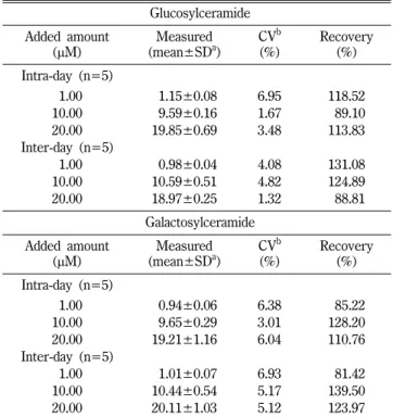
단당세라마이드인 글루코실세라마이드와 갈락토실세라마이드 의 표준물질을 각각 1, 10, 20 µM의 농도로 에탄올에 담아 실험 일내 분석의 회수율을 측정한 결과 20 µM 농도의 글루코실세라 마이드 시료에서 90.5~131.8%의 범위를 가졌으며, 평균 값은 113.8%였다. 또한 갈락토실세라마이드 시료에서는 89.4~125.8%
의 범위의 회수율이 측정되었으며, 평균 값은 110.8%로 나타났 다. 실험일내 분석의 정밀도에 대한 표준편차(SD)와 변동계수 (CV) 값은 20 µM 의 글루코실세라마이드에서 각각 0.7%와 3.5%
였으며, 20 µM 농도의 갈락토실세라마이드에서는 각각 1.2%와 6.0%로 나타났다. 실험일간 분석의 회수율을 측정한 결과는 20 µM 의 글루코실세라마이드 시료에서 평균 값은 88.8%로서 79.1~
112.9%의 범위를 가졌으며 20 µM 농도의 갈락토실세라마이드 시료에서는 82.8~134.8%의 범위의 회수율이 측정되었으며, 평 균 값은 124.0%로 나타났다. 실험일간 분석의 표준편차와 변동 계수 값은 20 µM 의 글루코실세라마이드에서 각각 0.3%와 1.3%
였으며, 20 µM 농도의 갈락토실세라마이드에서는 각각 1.0%와 5.1%로 나타났다(Table I).
단당세라마이드 추출 효율의 최적조건을 확립하기 위하여 Folch
방법(CHCl3 : MeOH=2 : 1) 및 Bligh and Dyer 방법(CHCl3 : MeOH=2 : 1)에서 사용된 추출용매 와 에탄올을 사용하여 배양 세포 및 흰쥐 혈장으로부터 단당세라마이드의 추출 효율을 비교
하였다.24,25)지질추출 및 TLC로부터 단당세라마이드 용출과정
에서 사용된 용매조건에서 단당세라마이드의 HPLC 피크면적은 각 용매조성에 따라 유의성있는 차이를 보이지 않았다(Table II).
따라서, 본 연구에서는 실험과정의 간편성을 위하여 지질 추출 에서 에탄올을 사용하였고, TLC 점적 및 분리된 단당세라마이 드의 실리카겔로 부터의 용출용매로서 메탄올을 사용하였다.
생체시료 중 단당세라마이드 정량
SW-620 세포와 흰쥐 혈장으로부터 글루코실세라마이드와 갈
20.00 18.97±0.25 1.32
088.81
Galactosylceramide Added amount
(µM) Measured
(mean±SDa)
CVb (%)
Recovery (%) Intra-day (n=5)
01.00 00.94±0.06
6.38085.22
10.00
09.65±0.29
3.01 128.2020.00 19.21±1.16 6.04 110.76
Inter-day (n=5)
01.00 01.01±0.07
6.93081.42
10.00 10.44±0.54 5.17 139.50
20.00 20.11±1.03 5.12 123.97
aStandard deviation. bCoefficient of variation.
Table II - Monoglycoeramide extraction efficiencies of solvent systems in SW-620 cells and rat plasma Solvent systems for
lipid extraction (v/v)
Solvent systems for TLC elution (v/v)
HPLC peak area of monoglycoceramide
SW-620 Cells (n=5)
CHCl3: MeOH=2 : 1 CHCl3: MeOH=2 : 1 16710702±90587
CHCl3: MeOH=2 : 1 MeOH 16250983±87112
CHCl3: MeOH=1 : 2 CHCl3: MeOH=1 : 2 15271920±79713
CHCl3: MeOH=1 : 2 MeOH 15743392±98456
EtOH MeOH 16734756±91934
Rat Plasma
(n=5)
CHCl3: MeOH=2 : 1 CHCl3: MeOH=2 : 1 22710572±84551
CHCl3: MeOH=2 : 1 MeOH 19737271±99871
CHCl3: MeOH=1 : 2 CHCl3: MeOH=1 : 2 17946883±93938
CHCl3: MeOH=1 : 2 MeOH 19968295±81556
EtOH MeOH 22270510±88664
락토실세라마이드의 함량분석을 수행하였다. SW-620 세포와 사 람의 혈청시료에 대한 HPLC 크로마토그램에서 단당세라마이드 (글루코실세라마이드 및 갈락토실세라마이드) 피크가 머무름 시 간 9.7분에서 관찰되었다(Fig. 4). 최근에 보고된 SCDase를 사용 한 단당세라마이드의 분석법에서 내부표준물질로 사용된 C6- NBD-GlcCer가 시료로부터 추출된 단당세라마이드와 동일한 크 로마토크램에서 피크를 검출할 수 없는 단점이 있다.23)본 분석 법은 내부표준물질로 사용된 디하이드로스핑고신은 단당세라마 이드로부터 탈아실화된 단당스핑고신과 같은 HPLC 크로마토그 램에서 피크가 검출되므로 생체 내 함량분석에 대한 신뢰성이 높 은 것으로 사료된다. 생체 내 단당세라마이드 함량은 내부표준 물질로 생체시료에 첨가한 디하이드로스핑고신(C18-base)의 피 크면적으로부터 산출하였다. 내부표준물질을 사용하지 않은SW- 620세포와 흰쥐 혈장을 분석한 HPLC크로마토그램에서 디하이 드로스핑고신이 전혀 검출되지 않았다(Fig. 4). 내부표준물질은 천연에서 생성되지 않는 화학합성 단당세라마이드를 사용하는 것이 이상적이지만 현재 사용할 수 있는 내부표준물질이 없으므 로 디하이드로스핑고신을 내부표준물질로 사용하였다. SW-620
세포 및 흰쥐혈장의 단당세라마이드 함량은 141.4±6.8 pmol/
1×10⁶cells 및 3.9±0.2 µM을 나타낸다. 따라서, 본 분석방법은 배양세포와 혈장은 물론 동물 및 사람의 조직으로 시료의 분석 범위를 확대할 수 있으리라 생각되며, 단당세라마이드 함량분석 을 통하여 생체 내 생리활성 기전 규명과 지질관련 질환의 중요 한 생체 표지물질로 작용할 수 있는 가능성을 제시할 수 있다.
결 론
생체 시료의 지질추출물에 함유되어있는 단당세라마이드는 세 라마이드 및 세라마이드-1-인산으로부터 HP-TLC에 의하여 분리 될 수 있었으며, 이때 TLC에서 단당세라마이드와 유사한 Rf값 을 갖는 스핑고신은 HPLC분석에 의해서 머무름 시간이 다른 피 크로 분리되었다. TLC 정제과정을 통하여 background가 깨끗한 HPLC크로마토그램이 얻어지며, 글루코실세라마이드와 갈락토실 세라마이드 피이크는 동일한 머무름 시간에서 용출되는 것으로 밝혀졌다. 따라서 두 종류의 단당세라마이드는 분리되지 않고 단 일피크로 나타나며, 이 피크는 측정된 생체시료의 글루코실세라 Fig. 4 − Monoglycoceramide contents of SW-620 cells and rat plasma. (A) SW-620 cells were harvested, and (B) blood was withdrawed from Sprague-Dawley rats and plasma was prepared by centrifugation at 3,000×g for 10 min. The biological samples were analyzed with or without 200 pmol dihydrosphingosine as an internal standard.
으로 응용 할 수 있을 것으로 사료된다.
감사의 말씀
이 논문은 2008년도 충북대학교 학술연구지원사업의 연구비 지원에 의하여 연구되었음(This work was supported by the research grant of the Chungbuk National University in 2009).
참고문헌
1) Hannun, Y. A. and Obeid, L. M. : Principles of bioactive lipid signalling: lessons from sphingolipids. Nat. Rev. Mol. Cell. Biol.
9, 139 (2008).
2) Hannun, Y. A. and Obeid, L. M. : The Ceramide-centric universe of lipid-mediated cell regulation: stress encounters of the lipid kind. J. Biol. Chem. 277, 25847 (2002).
3) Linn, S. C., Kim, H. S., Keane, E. M., Andras, L. M., Wang, E.
and Merrill, A. H., Jr. : Regulation of de novo sphingolipid biosynthesis and the toxic consequences of its disruption.
Biochem. Soc. Trans. 29, 831 (2001).
4) Pewzner-Jung, Y., Ben-Dor, S. and Futerman, A. H. : When do Lasses (longevity assurance genes) become CerS (ceramide synthases)?: Insights into the regulation of ceramide synthesis. J. Biol. Chem. 281, 25001 (2006).
5) Causeret, C., Geeraert, L., Van der Hoeven, G., Mannaerts, G. P. and Van Veldhoven, P. P. : Further characterization of rat dihydroceramide desaturase: tissue distribution, subcellular localization, and substrate specificity. Lipids 35, 1117 (2000).
6) Wijesinghe, D. S., Massiello, A., Subramanian, P., Szulc, Z., Bielawska, A. and Chalfant, C. E. : Substrate specificity of human ceramide kinase. J. Lipid Res. 46, 2706 (2005).
7) Tafesse, F. G., Ternes, P. and Holthuis, J. C. : The multigenic sphingomyelin synthase family. J. Biol. Chem. 281, 29421 (2006).
8) Raas-Rothschild, A., Pankova-Kholmyansky, I., Kacher, Y.
and Futerman, A. H. : Glycosphingolipidoses: beyond the enzymatic defect. Glycoconj. J. 21, 295 (2004).
receptors in lymphoid and myeloid cells determined by nanoelectrospray ionization tandem mass spectrometry. Rapid.
Commun. Mass. Spectrom. 24, 2295 (2010).
12) Chakrabandhu, K., Huault, S., Garmy, N., Fantini, J., Stebe, E., Mailfert, S., Marguet, D. and Hueber, A. O. : The extracellular glycosphingolipid-binding motif of Fas defines its internalization route, mode and outcome of signals upon activation by ligand.
Cell. Death. Differ. 15, 1824 (2008).
13) Hakomori, S., Nudelman, E., Levery, S., Solter, D. and Knowles, B. B. : The hapten structure of a developmentally regulated glycolipid antigen (SSEA-1) isolated from human erythrocytes and adenocarcinoma: a preliminary note.
Biochem. Biophys. Res. Commun. 100, 1578 (1981).
14) Hakomori, S. I. : Structure and function of glycosphingolipids and sphingolipids: recollections and future trends. Biochim.
Biophys. Acta. 1780, 325 (2008).
15) Kannagi, R., Yin, J., Miyazaki, K. and Izawa, M. : Current relevance of incomplete synthesis and neo-synthesis for cancer-associated alteration of carbohydrate determinants-- Hakomori's concepts revisited. Biochim. Biophys. Acta. 1780, 525 (2008).
16) Schwarz, A. and Futerman, A. H. : Distinct roles for ceramide and glucosylceramide at different stages of neuronal growth. J.
Neurosci. 17, 2929 (1997).
17) Coetzee, T., Fujita, N., Dupree, J., Shi, R., Blight, A., Suzuki, K. and Popko, B. : Myelination in the absence of galactocerebroside and sulfatide: normal structure with abnormal function and regional instability. Cell. 86, 209 (1996).
18) Ledeen, R. W. and Yu, R. K. : Gangliosides: structure, isolation, and analysis. Methods Enzymol. 83, 139 (1982).
19) Vance, D. E. and Sweeley, C. C. : Quantitative determination of the neutral glycosyl ceramides in human blood. J. Lipid Res. 8, 621 (1967).
20) Levery, S. B., Toledo, M. S., Straus, A. H. and Takahashi, H. K. : Structure elucidation of sphingolipids from the mycopathogen Paracoccidioides brasiliensis: an immunodominant beta- galactofuranose residue is carried by a novel glycosylinositol phosphorylceramide antigen. Biochemistry 37, 8764 (1998).
21) Zhang, X. and Kiechle, F. L. : Review: Glycosphingolipids in health and disease. Ann. Clin. Lab. Sci. 34, 3 (2004).
22) Wheeler, S. F. and Harvey, D. J. : Negative ion mass spectrometry of sialylated carbohydrates: discrimination of N- acetylneuraminic acid linkages by MALDI-TOF and ESI-TOF mass spectrometry. Anal. Chem. 72, 5027 (2000).
23) Zama, K., Hayashi, Y., Ito, S., Hirabayashi, Y., Inoue, T., Ohno, K., Okino, N. and Ito, M. : Simultaneous quantification of glucosylceramide and galactosylceramide by normal-phase
HPLC using O-phtalaldehyde derivatives prepared with sphingolipid ceramide N-deacylase. Glycobiology 19, 767 (2009).
24) Folch, J., Lees, M. and Sloane Stanley, G. H. : A simple method for the isolation and purification of total lipides from animal tissues. J. Biol. Chem. 226, 497 (1957).
25) Bligh, E. G. and Dyer, W. J. : A rapid method of total lipid extraction and purification. Can. J. Biochem. Physiol. 37, 911 (1959).