https://doi.org/10.5468/ogs.2020.63.3.270 pISSN 2287-8572 · eISSN 2287-8580
Introduction
Preeclampsia is defined by new-onset hypertension and either proteinuria or end-organ dysfunction after 20 weeks of ges- tation in previously normotensive women [1]. It involves mul- tiple organ systems and is unique to pregnancy. Preeclampsia complicates 2–10% of pregnancies [2] and represents one of the most common causes of maternal mortality and se- vere maternal morbidity. The offspring of mothers with pre- eclampsia are also at increased risk of neonatal morbidity and mortality [3-6]. Hence, the early detection and management of the disease are necessary to decrease its global burden.
According to new terminology (ACOG 2014), preeclamp- sia without evidence of end-organ damage is termed pre-
A study to compare maternal and perinatal outcome in early vs. late onset preeclampsia
Pooja Wadhwani, MD
1, Pradip Kumar Saha, MD, MAMS, FIMSA
1, Jaswinder Kaur Kalra, MD
1, Shalini Gainder, MD
1, Venkataseshan Sundaram, MD, DM
2Departments of 1Obstetrics and Gynecology, 2Neonatology, Postgraduate Institute of Medical Education and Research (PGIMER), Chandigarh, India
Objective
The risk factors, clinical trends, and maternal and fetal health of early- and late-onset preeclampsia have not been adequately studied. We examined the effects of early- and late-onset preeclampsia on maternal and perinatal outcomes as well as the known risk factors of preeclampsia.
Methods
One hundred and fifty women with preeclampsia were consecutively enrolled in each group. Those who developed preeclampsia before 34 weeks of gestation were identified as having early-onset preeclampsia, while those who developed at 34 weeks or later were identified as having late-onset preeclampsia. Maternal and perinatal outcomes were compared between groups.
Results
Compared with the late-onset group, the early-onset group had higher rates of abruptio placentae (16% vs. 7.3%;
P=0.019), but there was no intergroup difference in the composite maternal outcomes. A significantly higher number
of women with early-onset preeclampsia developed severe features during the disease course, and most required treatment with antihypertensive drugs. Late-onset preeclampsia was more prevalent among primigravid mothers.
Babies born to mothers with early-onset preeclampsia had a significantly higher rate of adverse outcomes.
Conclusion
These study findings indicate that women with early-onset preeclampsia had more adverse outcome than those with late-onset preeclampsia, but the difference was not statistically significant. There were more babies with adverse perinatal outcomes in the early-than late-onset group.
Keywords: Early-onset preeclampsia; Late onset preeclampsia; Maternal outcome; Perinatal outcome
Received: 2019.08.19. Revised: 2019.10.21. Accepted: 2019.11.07.
Corresponding author: Pradip Kumar Saha, MD, MAMS, FIMSA Department of Obstetrics and Gynecology, Postgraduate Institute of Medical Education and Research (PGIMER), Sector 12, Chandigarh 160012, India
E-mail: [email protected] https://orcid.org/0000-0002-3200-4124
Articles published in Obstet Gynecol Sci are open-access, distributed under the terms of the Creative Commons Attribution Non-Commercial License (http://creativecommons.
org/licenses/by-nc/3.0/) which permits unrestricted non-commercial use, distribution, and reproduction in any medium, provided the original work is properly cited.
Copyright © 2020 Korean Society of Obstetrics and Gynecology
eclampsia without severe features [1]. However, the presence of end-organ damage defines severe preeclampsia. In recent years, a new disease classification based on onset timing consisting of early-onset preeclampsia (EO-PE) occurring be- fore 34 weeks of gestation and late-onset preeclampsia (LO- PE) occurring at or after 34 weeks of gestation has gained attention [7-9]. The diagnostic criteria are the same for EO-PE and LO-PE; in fact, this simple division has better prognostic implications than mild vs. severe terminology [10].
Few retrospective studies have defined the outcomes of EO-PE and LO-PE and compared them with the outcomes of pregnancies without preeclampsia, whereas studies compar- ing the risk factors, clinical trends, laboratory parameters, and composite outcomes of maternal and fetal health in the 2 groups are limited. The clinical parameters and associated prognostic implications of the two groups may reflect the different mechanisms of the disease.
This prospective study examined the effects of EO-PE and LO-PE on composite maternal and perinatal outcomes and identified the similarities and differences in laboratory param- eters and clinical presentations of the 2 groups.
Materials and methods
This study was performed in the Department of Obstetrics and Gynecology of the Post Graduate Institute of Medical Education and Research, Chandigarh, India, from July 2016 to October 2017. The total of 300 women with preeclampsia who attended the outpatient department and labor room were recruited. Of these 300 preeclamptic women, 150 women were prospectively recruited in each subgroup and written consent was taken from all participants.
The study was approved by our institute’s Ethics Committee (INT/IEC/2017/774) and Research Committee. The study sub- jects diagnosed with preeclampsia who were supervised and delivered at the Department of Obstetrics and Gynecology were included in this study. Preeclampsia was defined accord- ing to American College of Obstetrics and Gynecology crite- ria [1]. We recruited patients with preeclampsia with or with- out severe features. Only subjects with confirmed gestation and disease onset timing (preeclampsia) were included. One hundred and fifty eligible patients were consecutively recruit- ed in each group (Fig. 1). Those who developed preeclampsia before 34 weeks of gestation were identified as having EO-
PE, while those who developed preeclampsia at or beyond 34 weeks of gestation were identified as having LO-PE.
Patients with chronic kidney disease, heart disease, severe anemia (hemoglobin <7 g/dL), anti-phospholipid antibody syndrome, or a history of drug abuse were excluded from the study.
Maternal and fetal monitoring was performed according to standard guidelines. Labetalol was given as an antihyper- tensive. All patients with preeclampsia and severe features were admitted to the hospital. Maternal clinical, hematologi- cal, and biochemical monitoring was performed at regular intervals. Fetal surveillance in the form of a biophysical profile, non-stress test, and Doppler ultrasonography was performed. If severe features developed, the pregnancy was terminated at 34 weeks and magnesium sulfate was given during delivery. Continuous intrapartum fetal monitoring was performed.
1. Comparison of maternal and perinatal outcomes The following maternal and perinatal outcomes were com- pared between groups:
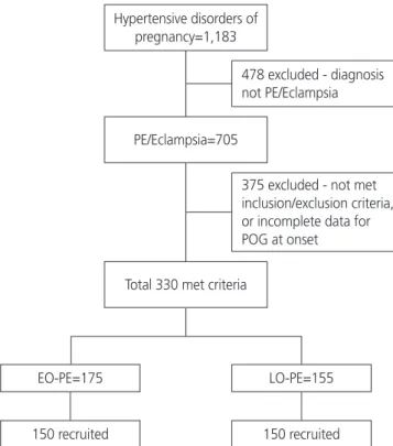
Fig. 1. Consort flow chart showing sample size selection. PE, preeclampsia; POG, period of gestation; EO-PE, early-onset pre- eclampsia; LO-PE, late-onset preeclampsia.
Hypertensive disorders of pregnancy=1,183
478 excluded - diagnosis not PE/Eclampsia
375 excluded - not met inclusion/exclusion criteria, or incomplete data for POG at onset
PE/Eclampsia=705
Total 330 met criteria
150 recruited
EO-PE=175 LO-PE=155
150 recruited
i. Adverse maternal outcomes such as hemolysis, elevated liver enzymes, low platelet count (HELLP) [1] syndrome, ec- lampsia [1], abruptio placentae, postpartum hemorrhage, acute kidney injury [11], venous thromboembolism, cerebro- vascular accident, pulmonary edema, and disseminated intra- vascular coagulopathy were compared between groups;
ii. Adverse perinatal outcomes such as small for gesta- tional age (SGA), stillbirth (intrauterine fetal death after 24 completed weeks of pregnancy) low birth weight, 5-minute Apgar score <7, acidosis at birth (pH <7.1), neonatal in- tensive care unit (ICU) admission, and the development of complications such as intraventricular hemorrhage, hypoxic- ischemic injury, respiratory distress syndrome, and necrotizing enterocolitis were compared between the groups. Abnormal umbilical artery Doppler findings (increased systolic/diastolic ratio for that period of gestation, absent end-diastolic flow, and reverse end-diastolic flow) were also compared between groups;
iii. Differences in laboratory parameters in preeclamptic women in the 2 groups;
iv. Difference in the development of imminent and severe features in the 2 groups;
v. Rate of labor induction;
vi. Cesarean section rate. Neonates were classified as SGA when they had a birth weight of less than the 10th percentile for gestational age. Lubchenko charts were used to convert
this classification into small for gestational dates [12]; and vii. Total number of neonates with complications (composite neonatal outcome).
2. Statistical analysis
Discrete categorical data are presented as number or percentage (%), continuous data assumed to be normally distributed are shown as mean and standard deviation, and skewed data are shown as median and interquartile range.
The normality of the quantitative data was checked by the Kolmogorov-Smirnov test of normality. Student’s t-test or the Mann-Whitney U test was used to compare the 2 groups depending upon the normality of the data. Proportions were compared using the χ
2or Fisher’s exact test depending on their applicability for the 2 groups. The statistical tests were 2-sided and performed at a significance level of α=0.05. The analyses were conducted using SPSS Statistics (version 22.0;
IBM Corp., Armonk, NY, USA).
Results
A total of 300 patients (150 patients in each group) were followed until discharge after delivery. Table 1 summarizes the baseline demographic details of women with EO-PE and those with LO-PE.
Table 1. Maternal demographic characteristics in women with early- and late-onset preeclampsia (PE) group
Characteristics EO-PE LO-PE P-value
Age (years) 27.21±4.4 26.43±4.04 0.111
BMI (kg/m2) 22.52±1.95 23.26±1.46 <0.001
Primigravida 64 (42.7) 83 (55.3) 0.028
Diabetes in pregnancy 10 (6.7) 7 (4.7) 0.454
Chronic hypertension 36 (24.0) 24 (16.0) 0.083
History of PE in previous pregnancies 30 (20.0) 14 (9.3) 0.009
Family history of hypertension 16 (10.7) 13 (8.7) 0.558
Systolic blood pressure on admission (mmHg) 0.900
Mean±SD 148.91±16.07 148.67±16.93
Minimum–maximum 118–200 110–200
Diastolic blood pressure on admission (mmHg) 0.445
Mean±SD 96.87±10.89 95.88±11.44
Minimum–maximum 60–120 68–140
Values are expressed as mean±SD or number (%). P-value <0.05 – statistically significant.
EO, early-onset; LO, late-onset; BMI, body mass index; SD, standard deviation.
In this study, there were 144 singleton and 6 twin deliveries in the EO-PE group and 143 singleton and 7 twin deliveries in the LO-PE group. A total of 277 live births and 36 intra- uterine fetal deaths were recorded.
Mean gestational age at diagnosis was 30.0 weeks and 36.5 weeks in EO-PE and LO-PE groups, respectively. In the EO-PE group, the mean gestational age at admission was 32.1 weeks, mean gestational age at delivery was 32.6 weeks, and mean duration of hypertension from onset to delivery was 17.83 days. In the LO-PE group, the mean ges- tational age at admission was 37.1 weeks, mean gestational age at delivery was 37.3 weeks, and mean duration of hy- pertension from onset to delivery was 5.47 days.
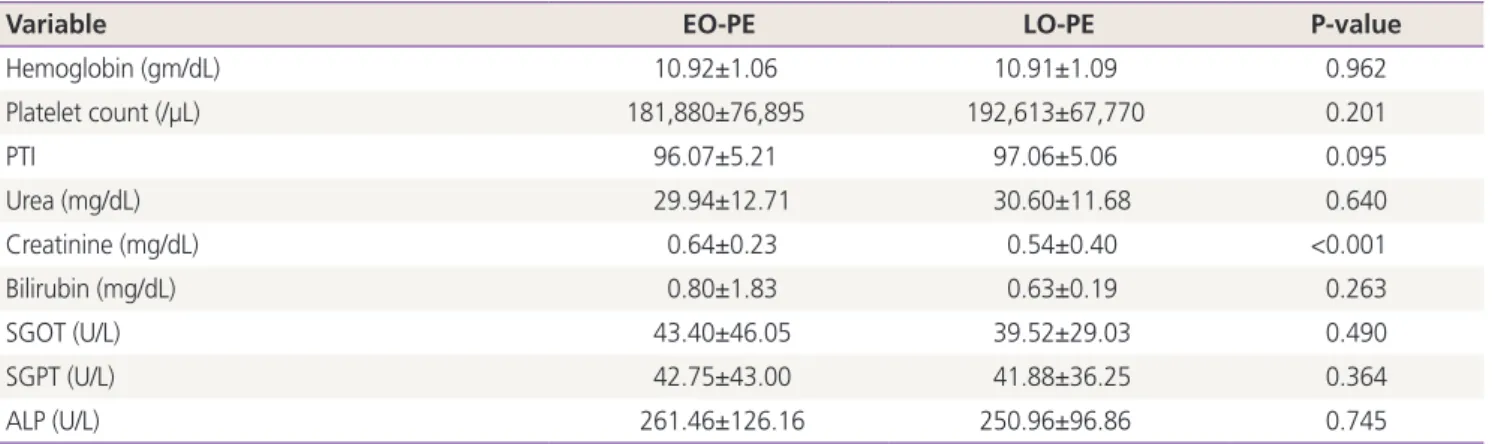
The hematological and biochemical parameters are sum- marized in Table 2. The mean serum creatinine value in EO-PE and LO-PE groups were 0.64±0.23 mg/dL and 0.54±0.40 mg/dL, respectively; the difference was statistically significant.
A significantly higher number of women with EO-PE than those with LO-PE developed severe features during their disease course and required treatment with antihypertensive drugs (P=0.01). The rate of induction was significantly higher in women with EO-PE than in those with LO-PE (P=0.045).
However, the cesarean section rate was similar between groups (P=0.354) (Table 3).
A total of 78 women (52%) with EO-PE developed adverse outcomes versus 66 women (44%) with LO-PE. The dif- ference was not statistically significant. Comparison of the maternal and perinatal outcomes of the twin gestations re-
vealed no significant differences.
Maternal complications are summarized in Table 4. Abrup- tio placentae was significantly more common in women with EO-PE than in those with LO-PE. Women with EO-PE had a significantly higher number of growth-restricted fetuses than those with LO-PE (54.6% vs. 40.0%, respectively) and a higher number of fetuses with umbilical artery Doppler abnormalities (32.6% vs. 9.3%). The rate of stillbirths was significantly higher in the EO-PE group than the LO-PE group (21.0% vs. 1.9%). Also, babies born to mothers with EO-PE had significantly higher adverse outcomes (Table 5).
Discussion
Although the causes of preeclampsia are unclear, multiple authors have suggested that the pathophysiology of EO-
Table 2. Hematological and biochemical parameters in women with early- and late-onset preeclampsia (PE) group
Variable EO-PE LO-PE P-value
Hemoglobin (gm/dL) 10.92±1.06 10.91±1.09 0.962
Platelet count (/µL) 181,880±76,895 192,613±67,770 0.201
PTI 96.07±5.21 97.06±5.06 0.095
Urea (mg/dL) 29.94±12.71 30.60±11.68 0.640
Creatinine (mg/dL) 0.64±0.23 0.54±0.40 <0.001
Bilirubin (mg/dL) 0.80±1.83 0.63±0.19 0.263
SGOT (U/L) 43.40±46.05 39.52±29.03 0.490
SGPT (U/L) 42.75±43.00 41.88±36.25 0.364
ALP (U/L) 261.46±126.16 250.96±96.86 0.745
Values are expressed as mean±standard deviation. P-value <0.05 – statistically significant.
EO, early-onset; LO, late-onset; PTI, prothrombin time index; SGOT, serum glutamic oxaloacetic transaminase; SGPT, serum glutamate-pyruvate transaminase; ALP, alkaline phosphatase.
Table 3. Disease severity correlation, labor and delivery details in early- and late-onset preeclampsia (PE) group
Treatment EO-PE LO-PE P-value
Antihypertensive therapy 129 (86.0) 111 (74.5) 0.012 PE with severe features 112 (74.7) 91 (60.7) 0.01 MgSO4 therapy 101 (67.3) 68 (45.3) <0.001 Induction of labor 104 (69.3) 96 (64.0) 0.045 Cesarean section 83 (55.3) 75 (50.0) 0.354 Values are expressed as number (%). P-value <0.05 – statistically sig- nificant.
PE differs from that of LO-PE. Early-onset disease appears to be mediated by the placenta. LO-PE, on the other hand, is mediated by maternal factors and a maternal overreaction to pregnancy [13-15]. Our data show no intergroup difference in mean maternal age, and there were significantly more multiparous women in EO-PE group. This observation is simi- lar to that of a study by Stubert et al. [16]. The frequency of
risk factors predisposing an expectant mother to developing diabetes and chronic hypertension did not differ between groups; however, patients with LO-PE had a higher mean body mass index than those with EO-PE as reported by the Valensise et al.’s study [13] of patients with two different he- modynamic states.
A history of preeclampsia in a previous pregnancy is a
Table 4. Maternal complications in women with early- and late-onset preeclampsia (PE) group
Complications EO-PE (n=150) LO-PE (n=150) Total (n=300) P-value
Complete HELLP 8 (5.3) 4 (2.6) 12 (4.0)
Partial HELLP
Deranged LFT 9 13 22 (7.3)
Thrombocytopenia 12 19 31 (10.3)
Total HELLP 29 (19.3) 36 (24.0) 65 (21.6) 0.327
Eclampsia 26 (17.3) 20 (13.3) 46 (15.3) 0.337
Abruption 24 (16.0) 11 (7.3) 35 (11.6) 0.019
Pulmonary edema 4 (2.7) 2 (1.3) 6 (2.0) 0.684
AKI 7 (4.6) 4 (2.7) 11 (3.7) 0.356
PPH 2 (1.3) 6 (4.0) 8 (2.7) 0.282
CVA 0 (0.0) 1 (0.7) 1 (0.3) 1.000
Retinal detachment 2 (1.3) 1 (0.7) 3 (1.0) 0.624
Values are expressed as number (%). P-value <0.05 – statistically significant.
EO, early-onset; LO, late-onset; HELLP, hemolysis, elevated liver enzymes, low platelet count; LFT, liver function test; AKI, acute kidney injury;
PPH, post-partum haemorrhage; CVA, cerebrovascular accident.
Table 5. Perinatal outcome parameters in women with early- and late-onset preeclampsia (PE) group
Parameters EO-PE (n=156) LO-PE (n=157) P-value
Small for gestation age neonates 83 (53.2) 60 (38.2) 0.008
Umbilical artery Doppler abnormality 49 (31.4) 14 (8.9) <0.001
Still births 33 (21.2) 3 (1.9) <0.001
Live births 123 (78.8) 154 (98.1) <0.001
Birth weight (kg) 1.502±0.58 2.416±0.59 <0.001
<2.5 148 (94.9) 87 (55.4)
≥2.5 8 (5.1) 70 (44.6)
5-min Apgar score <7 10 (6.4) 5 (3.2) <0.074
Respiratory support 44 (28.2) 5 (3.2) <0.001
Neonatal acidosis (PH <7.1) 5 (3.2) 2 (1.3) 0.144
NICU stay 34 (21.8) 4 (2.5) <0.001
Days of NICU stay (days) 10.98±11.7 6.95±4.36 0.019
Composite Neonatal outcome 63 (40.4) 20 (12.7) <0.001
Neonatal death 8 (5.1) 4 (2.5) 0.112
Values are expressed as mean±SD or number (%). P-value <0.05 – statistically significant.
EO, early-onset; LO, late-onset.