A Case of Multiple Metastatic Renal Cell Carcinoma in an Adult Patient Presenting with Ventricular Tachycardia
4
0
0
전체 글
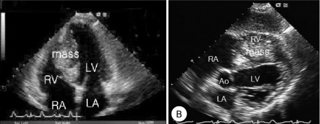
(2) 342·Korean Circulation J 2005;35:341-344. A. B. Fig. 1. Electrocardiography shows reverse of ventricular tachycardia (A) to sinus rhythm (B) by DC cardioversion (50J).. defect of the entire inferior and inferoseptal wall, with left ventricular dysfunction. However, the coronary angiography was normal, and the cardiac enzyme level was within normal limits. On esophagogastroduodenoscopy, performed due to indigestion, a gastric polyp was revealed with clear cell infiltration in the deep mucosal layer(Fig. 5B). Although a biopsy of right ventricular mass was not performed, as the patient refused, a secondary metastatic cardiac tumor of renal cell carcinoma was suspected considering the multiple metastases in both lungs and the stomach. A left nephrectomy and immunotherapy was recommended, but the patient refused. He expired one month after discharge due to cardiac arrest.. RPA. Discussion Fig. 2. Chest roentgenogram shows multiple metastatic masses of the. lung.. A. Although primary cardiac tumors are rare, cardiac metastases are not infrequent, with autopsy series ha-. B. Fig. 3. A right ventricular mass is noted attached to the interventricular septum on the apical four-chamber (A) and subcostal (B) view of the 2D-. echocardiography. RA: right atrium, LA: left atrium, RV: right ventricle, LV: left ventricle, Ao: aorta..
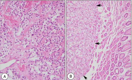
(3) Min Jeong Kwon, et al: Ventricular Tachycardia Caused by Multiple Metastatic Renal Cell Carcinoma·343. A. B. Fig. 4. A: chest CT demonstrates a cardiac mass attached to the interventricular septum protruding into the right ventricle, but without contiguous. vena caval involvement, and multiple metastases in both lungs. B: abdominal CT shows a large mass in the upper pole of the left kidney.. A. B. Fig. 5. A: the renal biopsy shows a clear type renal cell carcinoma (H & E stain, ×100). B: the gastric mucosal biopsy of the polyploid mass reveals clear cell infiltration (arrow) in the deep mucosal layer (H & E stain, ×40).. ving reported a 5 to 20% incidence of metastatic carcinomas to the heart and pericardium in patients dying of malignancies. The most common tumors associated with cardiac metastases are those of the lung and breast, as well as melanomas, leukemia, lymphomas and sarcomas.1)2) Cardiac metastases from renal cell carcinomas are rare, with incidences ranging from 1.3 to 4.2%; however, endocardial invasion by metastatic renal cell carcinomas is virtually unknown. The extension of a tumor column to the vena cava as a luminal mass, with the growth along the caval wall into the right heart chambers, has been well documented as the most common mechanism of cardiac metastasis.3)4) Additionally, tumor cells from the kidney may disseminate to the right ventricle by retrograde lymphatic or lymphohematogenous spread through the thoracic duct into the superior vena cava, or by hematogenous spread of embolic cells.5) A cardiac tumor is suspected when symptoms or signs that could be related to a lesion of a particular. size, extension or location appear. The location and size of a tumor may be such that the cardiac function is altered due to obstruction of the blood flow through a cardiac chamber, such as with a left atrial myxoma, or by interfering with the valve function, as with a rhabdomyoma, or by causing cardiac failure like with a rhabdomyosarcoma. A strategically located tumor, which occludes a coronary artery, may alter the electrocardiography and produce patterns of a current injury, such as a myocardial infarction, arrhythmia or abnormal conduction.6-9) Tachy-arrhythmia may result from valvular interference of the tumor mass or re-entry about its border with the myocardium. Accessory bundles resulting in ventricular pre-excitation have been seen in certain patients.10)11) Our patient had palpitation during ventricular tachycardia, which was supposed may have resulted from re-entry about the right ventricular mass. Renal cell carcinomas account for 2% of all cancers and 80 to 85% of malignant kidney tumors. They occur in men nearly twice as often as in women. Patients are.
(4) 344·Korean Circulation J 2005;35:341-344. generally older than 40 years on diagnosis, with the disease predominantly occurring in the seventh and eighth decades of life. Five types of carcinoma have been distinguished: clear-cell, chromophilic, chromophobic, oncocytic and collecting-duct tumors. Clear cell carcinomas make up 75 to 85% of these tumors, and a higher nuclear grade or the presence of a sarcomatoid pattern correlate with a poorer prognosis. Chromophilic carcinomas comprise approximately 14% of renal cancers and chromophobic carcinomas approximately 4%, but renal oncocytomas and collecting-duct carcinomas are rare.12)13) In our patient, the pathology was that of a conventional clear cell renal cell carcinoma. mixed with sarcomatoid. The signs and symptoms of renal cell carcinomas are usually nonspecific. The most common presentations are a hematuria in 50 to 60% of patients, abdominal pain in 40% and a palpable mass in the flank or abdomen in 30 to 40%. These three symptoms occur in combination(classic triad) in less than 10% of patients.14) One to 3% of tumors are bilateral and 25 to 30% of patients have overt metastases on initial presentation. Frequent sites include: the lung parenchyme in 50 to 60% of patients with metastases, bone in 30 to 40% and the brain in 5%. A broad range of paraneoplastic syndromes have been in less than 5% of patients, including erythrocytosis, hypercalcemia, hepatic dysfunction and amyloidosis.12) Finally, if no definite cause can be found for the onset of cardiovascular symptoms in patients with a known malignancy, the symptoms may be due to cardiac metastasis.. REFERENCES 1) Carroll JC, Quinn CC, Weitzel J, Sant GR. Metastatic renal cell. carcinoma to the right cardiac ventricle without contiguous vena caval involvement. J Urol 1994;151:133-4. 2) Toshikazu S, Iwase M, Iwase M, et al. Solitary left ventricular metastasis of renal cell carcinoma. Am Heart J 1993;125:1801-2. 3) Hunsaker RP, Stone JR. Renal cell carcinoma extending into the vena cava and right side of the heart. N Engl J Med 2001;345: 1676. 4) Masaki M, Kuroda T, Hosen N, et al. Solitary right ventricle metastasis by renal cell carcinoma. J Am Soc Echocardiogr 2004; 17:397-8. 5) Santo-Tomas M, Mahr NC, Robinson MJ, Agatston AS. Metastatic renal cell carcinoma invading right ventricular myocardium without caval involvement. J Cardiovasc Surg 1998;39: 811-2. 6) Engle MA, Ebert PA, Redo SF. Recurrent ventricular tachycardia due to resectable cardiac tumor. Circulation 1974;50:1052-7. 7) Goodwin JF. The spectrum of cardiac tumors. Am J Cardiol 1968; 21:307-14. 8) Heath D. Pathology of cardiac tumors. Am J Cardiol 1968;21: 315-27. 9) Harvey WP. Clinical aspects of cardiac tumors. Am J Cardiol 1968;21:328-43. 10) Krasuski RA, Hesselson AB, Landolfo KP, Ellington KJ, Bashore TM. Cardiac rhabdomyoma in an adult patient presenting with ventricular arrhythmia. Chest 2000;118:1217-21. 11) Enbergs A, Borggrefe M, Kurlemann G, et al. Ventricular tachycardia caused by cardiac rhabdomyoma in a young adult with tubeous sclerosis. Am Heart J 1996;132:1263-5. 12) Motzer RJ, Bander NH, Nanus DM. Renal-cell carcinoma. N Engl J Med 1996;335:865-75. 13) Weiss LM, Gelb AB, Medeiros LJ. Adult renal epithelial neoplasms. Am J Clin Pathol 1995;103:624-35. 14) Cheng A. Cardiac metastasis from a renal cell carcinoma. Int J Clin Pract 2003;57:437-8..
(5)
수치
관련 문서
The index is calculated with the latest 5-year auction data of 400 selected Classic, Modern, and Contemporary Chinese painting artists from major auction houses..
The key issue is whether HTS can be defined as the 6th generation of violent extremism. That is, whether it will first safely settle as a locally embedded group
SigmaNEST 펀칭 파워팩 제품 관리, 자동 다이나믹™ 배 열 및 펀칭 툴 관리를 갖춘 터렛 펀칭 프로그래밍을 위한 가장 완벽하고 최적화된
The “Asset Allocation” portfolio assumes the following weights: 25% in the S&P 500, 10% in the Russell 2000, 15% in the MSCI EAFE, 5% in the MSCI EME, 25% in the
1 John Owen, Justification by Faith Alone, in The Works of John Owen, ed. John Bolt, trans. Scott Clark, "Do This and Live: Christ's Active Obedience as the
The innovation of the mass media not only has changed the delivery process of a product but also has expanded into the image industry such as
In gi ngi va,LCs are found i n oralepi thel i um ofnormalgi ngi va and i n smal l er amountsi nthesul cul arepi thel i um,buttheyareprobabl yabsentfrom thejuncti onal epi thel
웹 표준을 지원하는 플랫폼에서 큰 수정없이 실행 가능함 패키징을 통해 다양한 기기를 위한 앱을 작성할 수 있음 네이티브 앱과
