https://doi.org/10.5763/kjsm.2017.35.2.77 pISSN 1226-3729 eISSN 2288-6028
운동선수의 족관절 골연골 손상에서의 치료
순천향대학교 부천병원 정형외과
1, 중앙보훈병원 정형외과
2유정우1ㆍ여의동2ㆍ이영구1
Treatment of Osteochondral Lesions of the Talus in Athletes
Jungwoo Yoo
1, Eui Dong Yeo
2, Young Koo Lee
11
Department of Orthopedics, Soonchunhyang University Bucheon Hospital, Bucheon,
2
Department of Orthopedics, Veterans Health Service Medical Center, Seoul, Korea
The definition of osteochondral lesion of the talus (OLT) is any defect involving both the articular surface and the subchondral bone of the talus. Many of these lesions are associated with acute ankle injury. Although many classification schemes for OLT have been proposed, Berndt and Harty’s 4-staging classification is most commonly used. Stage 4 lesions and symptomatic lesions under grade 3 are usually recommended to surgical treatment. The treatment approach for athletes should be more elaborate due to the need for an early return to play. Several different types of treatment are described for OLTs in athletes, including bone marrow stimulation, osteochondral autograft transfer system, and autogenous chondrocyte implantation. Osteochondral autograft transfer system shows good clinical outcome and has the advantages that could be applied to large defect and recurred lesions, however, it has some disadvantages in terms of the complications related with the donor site and the difficult approach to the medial lesions. Although autogenous chondrocyte implantation has been extensively applied for treating OLTs with successful clinical outcomes, it has some limitations that apply to athletes in terms of the 2-stage and complicated procedure and the insurance issues. Bone marrow stimulation being a simple and cost-effective procedure associated with a low complication rate and low postoperative pain has faster return to play and is recommended the first-line treatment for the OLTs of athletes.
Keywords: Athletes, Microfracture, Autografts, Talus
Received: July 14, 2017 Revised: August 16, 2017 Accepted: August 18, 2017
Correspondence: Young Koo Lee
Department of Orthopedics, Soonchunhyang University Bucheon Hospital, 170 Jomaru-ro, Wonmi-gu, Bucheon 14584, Korea
Tel: +82-32-621-6702, Fax: +82-32-621-5018 E-mail: [email protected]
Copyright ©2017 The Korean Society of Sports Medicine
CC
This is an Open Access article distributed under the terms of the Creative Commons Attribution Non-Commercial License (http://creativecommons.org/
licenses/by-nc/4.0) which permits unrestricted non-commercial use, distribution, and reproduction in any medium, provided the original work is properly cited.
서 론
거골 골연골 병변(osteochondral lesion of the talus)은 1737년 Monro
1)에 의해 처음 소개된 이래로 박리성 골연골염(osteo- chondritis dissecans), 거골원개 골절(talar dome fracture), 골연골 골절(osteochondral fracture) 등 다양한 용어로 불리며 그 발생 기전이나 치료 방법에 대해 지속적으로 연구되어 왔다 . 그리고 1959년 Berndt와 Harty
2)에 의하여 이 질환에 대한 분류가 처음 발표되었다.
거골 골연골 병변은 거골의 관절 연골과 연골하골에 발생하
는 다양한 형태의 손상 및 결손을 모두 포함하여 정의하고 있다 . 이러한 손상을 일으키는 다양한 원인이 있을 수 있으며 대부분 유사한 형태의 병변으로 나타난다. 드문 원인으로는 퇴행성 관절 질환, 관절의 부정 정렬, 무혈성 골괴사, 말초 혈관 질환이나 내분비 대사 이상 등이 있다
3). 하지만 주된 원인은 급성 발목 손상이며 많은 연구를 통하여 거골의 골연골 병변은 발목 골절이나 염좌가 발생하였을 경우 50% 이상에서 동반된다고 보고되고 있다
2,4,5). 또한, 거골의 연골 손상의 23%
는 외측 만성 발목 불안정과 연관이 있어 인대 재건술을 시행한 후에도 지속적인 통증의 원인이 될 수 있다
6). 연골의 경우 치유 능력이 떨어지는 조직이므로
7)한번 손상을 입게 되면 불가피하게 만성적인 통증이나 골 관절염으로 진행되는 경우 가 많다
8,9). 이는 운동선수에게 치명적이며 심한 경우 운동을 그만두게 되는 하나의 원인이 될 수 있다.
또한, 발목 손상은 가장 흔한 운동 손상 중 하나이며 모든 운동 손상의 15%를 차지한다. 따라서 발목 손상이 발생하였을 경우 충분한 검사를 통하여 거골 골연골 병변의 동반 여부를 확인하는 것이 중요하다.
골연골 병변에 대한 다양한 치료법이 고안되었지만 아직까 지 확립된 치료 가이드라인은 없다
10,11). 특히 운동선수의 경우 운동으로의 빠른 복귀가 매우 중요하기 때문에 치료 방법 선택에 있어 제약이 있으며 어떠한 치료가 가장 좋은지에 대해서는 많은 논란의 여지가 있다. 본 논문의 목적은 현재 임상에서 많이 사용하고 있는 거골 골연골 병변 치료에 대해 소개하고 그중 운동선수에게 가장 효과적인 치료법에 대해 알아보고자 하는 것이다.
원 인
다양한 원인 중에서도 외상은 족관절의 골연골 질환의 가장 중요한 원인으로 알려져 있다. 거골체 외측의 병변의 경우 93%–98%, 내측의 병변은 61%–70% 정도가 외상으로 인해 발생한다고 알려져 있다
11,12). 그리고 비외상성 원인으로는 허혈성 손상과 그로 인한 무혈성 골괴사, 부 전하방 경비인대 (accessory anterior-inferior tibiofibular ligament, Bassett’s liga- ment)에 의한 연부조직 충돌(soft-tissue impingement)이나 만성 발목 불안정 등이 있다
13,14).
외상성 연골 손상은 미세손상(microdamage), 둔상(blunt trauma), 연골 골절과 골연골 골절(chondral and osteochondral fracture) 세 가지 분류로 나누어 생각해 볼 수 있다
15). 발목의 염좌는 외상성 골연골 병변의 주된 원인으로 발목 관절 내에서
거골체가 회전하면서 연골 손상이 발생한다. 이 손상으로 연골 에 타박상이 생기고 연골은 점차 물러지게 되며 심한 경우 금이 가면서 작은 조각으로 나누어지게 된다
14).
거골체에 가해진 전단력(shearing force)은 연골 상층부나 연골하골에 전해져 골조직의 분리를 일으킨다. 파편은 발목 관절 내에 유리체로 떨어져 나가거나, 혹은 부분적으로 연결되 어 남아 있게 된다. 이러한 병변은 점차 치유되거나 무증상으 로 남을 수 있고 또는 더 진행하여 연골하골의 낭종을 형성하게 된다.
Elias 등
16)은 거골체 관절면을 9개의 구역으로 나누어 각각 구역의 골연골 병변의 특징을 연구하였고, 4구역(중내측)과 6구역(중외측)에서 가장 흔하게 발생한다고 하였다. 또한 Flick 과 Gould
12)는 500명의 환자를 대상으로 한 연구를 통하여 98%의 외측 병변이 발목 염좌와 같은 외상에 의해 발생한다고 발표하였다 . 손상 기전은 발목이 외상에 의하여 강제로 배굴 (dorsiflexion)되고 내번(inversion)될 때 거골원개에 전단력이 전해지며 발생한다. 이러한 외측 병변은 내측 병변에 비해 보통 얕은 경향이 있으나 일반적으로 더 심하게 나타나며 골편이 떨어져 나가는 경우가 더 흔하다. 반면에 내측 병변의 70%는 발목이 강제로 족저굴곡(plantar flexion) 및 내전될 때 축 하중(axial load)이 전해지며 발생한다
14).
환자 초기 진찰 시에 증상의 경향을 보면, 보행 시 불안정성 이 있거나, 특별한 자세에서 갑작스러운 통증이 발생한 경우에 는 발목 불안정성이 동반된 것을 예상할 수 있고, 장기간 보행 이나 발목을 과하게 사용한 뒤에 발생한 관절 통증이 있는 경우는 퇴행성 변화가 동반되어 있음을 알 수 있다.
통증의 기전
모든 족관절의 연골 손상이 통증을 유발하지는 않는다.
일부는 무증상으로 남아있기도 하고 또 일부는 연골하골 낭종을 형성하며 체중 부하 시에 깊은 발목 통증을 유발하기 도 한다. 골연골 병변에서 통증을 유발하는 몇 가지 요인이 있다 . 첫째로 골내압의 증가는 여러 연구에서 통증을 일으키 는 요인의 하나로 언급되었으며 관절의 퇴행성 변형과 연관 이 있다
13,17,18).
관절 내 압력의 증가도 퇴행성 관절 질환에서 통증의 원인이
될 수 있다. Goddard와 Gosling
19)은 관절 내 활액의 압력과
골관절염에서의 통증 사이의 상관관계를 발견하였다. 하지만
국소적인 골연골 병변의 경우 관절 내 삼출이 적어 실제로
관절 내 압력을 높이는 것으로 보이지는 않는다.
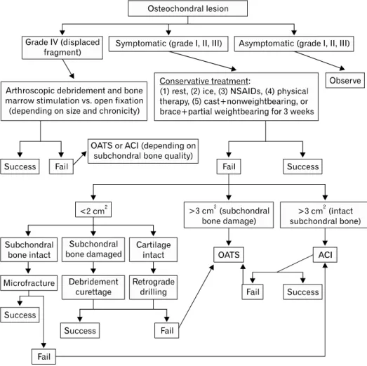
Fig. 1. The treatment algorithm for osteochondral lesion of the talus. NSAIDs: non-steroidal an- ti-inflammatory drugs, OATS:
osteochondral autograft transfer system, ACI: autogenous chon- drocyte implantation. Reproduc- ed from Dragoni et al. Foot Ankle Int 2011;32:910-6, with permission of SAGE Publica-
26)
활액막과 관절막에는 신경 말단들이 분포하고 있으며 관절 막과 관절 주변의 연부조직은 통각 유발에 중요한 역할을 한다. 관절의 염증성 변화에 따른 substance P와 calcitonin gene-related peptide 양성 뉴런의 상향 조절은 퇴행성 관절염에 서 신경펩티드와 관련된 통증 메커니즘으로 생각된다
20).
분 류
Berndt와 Harty
2)가 처음 발표한 4단계의 법은 아직까지도 가장 널리 사용되는 분류법으로 이후 많은 변형된 형태의 분류법이 발표되었다. 이는 단순 방사선 사진의 형태에 따라 4가지 단계로 구분한 분류로 주로 연골 골절에 대한 것이었다.
하지만 단순 방사선 사진 소견 만으로는 정확한 진단이 어려우 며 관절경 소견과도 상관관계가 크지 않았다
21). 최근에는 다양 한 영상 기술의 발달에 따라 컴퓨터단층촬영 , 자기공명영상과 관절경 소견을 바탕으로 한 새로운 분류들이 발표되었다
22-25). 일부는 기존의 4단계 분류에 낭종 아류형을 추가하였고 또는 관절 연골의 변화를 분류에 추가하였다.
영상 검사 소견과 실제 관절경 소견 사이의 연관성에 대한 여러 연구가 진행되었고 Pritsch 등
21)은 단순 방사선 사진과 관절경 소견 사이에 연관성은 떨어진다고 발표하였다. 반면에 자기공명영상 소견은 관절경 소견과 높은 상관관계를 보였으 며 병변의 중증도는 실제에 비해 낮게 보이는 경향이 있었다
24).
치 료
지금까지 다양한 치료법이 개발되었고 각각의 치료법은 장단점이 있으므로 환자 상태에 맞는 치료법을 선택하는 것이 중요하다 . Dragoni 등
26)은 기본적인 4단계 분류체계를 기반으 로 하여 증상의 유무와 병변의 크기에 따른 치료 알고리즘을 발표하였다(Fig. 1).
먼저 4등급에서는 수술적 치료를 고려하고, 1, 2, 3등급의
경우에는 증상의 유무에 따라 수술을 결정하게 된다. 증상이
없는 경우는 경과 관찰을 하고 증상이 있는 경우라도 우선적으
로 보존적 치료를 시행해 보고 이후에도 증상이 지속되는
경우에는 수술적 치료를 시행한다
26).
보존적 치료에는 생활 습관의 변경, 단계적 체중 부하, 재활 치료, 보조기, non-steroidal anti-inflammatory drugs (NSAIDs) 사용 등이 포함된다
27,28). 증상이 적고 자기공명영상에서 관절 연골의 일부만 침범한 거골 골연골 병변 환자에 대한 보존적 치료로 임상적이나 방사선학적으로 만족스러운 결과를 보였 다
29). 하지만 대부분 증상이 있는 환자들에게서는 만족스러운 결과를 얻기 어려웠다 . Lee 등
30)이 시행하였던 연구에 의하면 보존적 치료를 통해 39%에서 증상의 호전을 보였으나 61%의 환자에 대해서는 수술적 치료가 필요하였다. Verhagen 등
11)은 14개 연구의 메타 분석을 통하여 201명의 보존적 치료를 시행 받은 환자 중 45%인 91명에서만 만족스러운 결과를 보였다고 발표하였다.
수술적 치료로 현재까지 다양한 치료 방법이 개발되었다.
소파술(curettage)과 절편 제거술(fragment excision), 미세 골절 술(microfracture)과 전향적 및 후향적 천공술(antergrade or retrograde drilling) 등의 골수 자극술(bone marrow stimula- tion)
31), 골연골 자가 이식술(osteochondral autograft transfer system)과 골연골 동종 이식술(osteochondral allograft transplan- tation)
27), 그리고 자가 연골세포 이식술(autologous chondrocyte implantation)과 혈소판 풍부 혈장 치료(platelet-rich plasma therapy) 등의 세포 기반 치료술(cell-based repair technique) 등이 현재 사용되고 있는 대표적인 치료법이다
32).
각각의 치료법은 연구마다 어느 정도의 편차는 있지만 대부 분 만족스러운 결과를 보였다. Zengerink 등
33)의 메타 분석 연구에서는 보존적 치료를 포함한 많은 수술적 치료법 중에서 절제술 및 소파술은 77%, 절제술 및 소파술과 골수 자극술은 85%, 골연골 자가 이식술은 87%, 자가 연골세포 이식술은 76%의 치료 성공률을 보였다.
하지만 아직까지 거골 골연골 병변 치료에 대한 충분한 장기 추시 결과나 정립된 치료방법은 없다
10,11). 또한, 각각의 치료법은 수술 전 준비 기간과 수술 후 재활 기간, 술기의 난이도 , 수술 비용 등 많은 부분에서 차이가 있기 때문에 단순 한 치료 성공률만으로 수술 방법의 우위를 결정할 수는 없다.
따라서 수술적 치료 방법을 결정하기 전 환자의 전신적 상태, 급성 또는 만성의 경과 여부 , 치료 시기, 재발 여부, 빠른 복귀의 필요성, 환자의 경제적 상태 등 다양한 인자들을 고려하여 치료법을 선택해야 한다.
운동선수의 족관절 골연골 병변은 일반인들과는 다르게 접근해야 한다. 치열한 경쟁이 필요한 스포츠 선수들에게 초기 치료의 기간이 길어진다면 운동으로의 복귀가 늦어지고 그로 인해 꾸준한 경기력의 유지가 어려워지게 되며 결국에는 조기
은퇴를 해야 할 상황에 처할 수 있다. 그렇기 때문에 운동선수 에게 치료의 성공은 일반인들의 기준보다 더 엄격하게 판단되 어야 하고 초기 치료법의 선택에 있어서 단순히 성공률이 가장 높은 치료보다는 빠른 운동으로의 복귀가 가능한 최소 침습 수술(minimal invasive surgery)을 선택해야 할 것이다
34).
골수 자극술
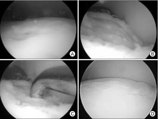
미세골절술로 대표되는 골수 자극술은 관절경 시술 시 간단 하게 시행할 수 있어 골연골 병변 치료 시 널리 시행되는 치료법이다. 골수 자극술은 천공술이나 미세골절술을 통하여 연골하골에 작은 구멍을 만들어 줌으로써 중간엽 줄기세포 (mesenchymal stem cell)를 연골 결손 부위로 이동시켜 치유하 는 방법으로 연골하골에 구멍이 만들어지면 섬유성 응괴가 결손 부위를 채우게 되며 염증 반응이 시작되고 이어서 사이토 카인(cytokine)과 성장 인자들이 분비되어 치유를 촉진하게 된다
31). 이때 중간엽 줄기세포가 섬유성 응괴 속으로 이동하여 분화 및 증식하여 주로 제1형 콜라겐을 함유한 섬유 연골성 치유조직으로 변화하게 된다(Fig. 2)
35).
골수 자극술은 관절경 수술과 함께 빠르고 간단하게 시행할 수 있다는 장점이 있다 . 또한, 최소 침습적이며 수술 후 합병증 이나 통증도 적어서 초기치료로 선호되고 있다
31). 무엇보다도 골수 자극술은 다른 치료법에 비하여 빠른 운동으로의 복귀가 가능하다는 점에서 운동선수의 치료에 있어서 큰 장점이 있다.
슬관절의 골연골 병변에 대한 한 연구
36)에서 미세골절술은 골연골 자가이식술에 비해 1개월 정도 운동 복귀가 늦은 반면 에 발목관절의 골연골 병변 환자를 대상으로 한 Saxena와 Eakin
6)의 연구에서 미세골절술은 골연골 자가이식술과 유사 한 임상적 증상 호전을 보였으나 운동으로의 복귀는 미세골절 술이 약 4개월, 골연골 자가 이식술이 약 5개월로 미세골절술 이 1개월 정도 더 빨랐다. 또한, 미세골절술을 시행한 환자를 대상으로 체중 부하 시기를 달리하여 비교한 연구에서 수술 후 2주째부터 부분 체중 부하를 시작한 환자군은 수술 후 6주 동안 체중 부하를 하지 않은 환자군과 임상적, 방사선학적 결과에서 차이를 보이지 않았다. 이는 미세골절술 후 조기 체중 부하 및 재활치료를 시작할 수 있다는 점에서 미세골절술 후 빠른 운동으로의 복귀를 지지해 주고 있다
37).
다만 미세골절술은 병변의 크기가 1.5 cm
2이상이거나 장경
이 15 mm 이상인 경우 치료 성공률이 높지 않았다
38).
Chuckpaiwong 등
39)은 105명의 환자를 대상으로 한 연구에서
병변의 크기가 15 mm 이하의 경우 모든 환자에서 좋은 결과를
Fig. 2. Arthroscopic microfrac- ture technique. (A) Arthroscopic view of the medial talar dome lesion. (B) The same lesion after curettage and (C) microfracture.
(D) The healed lesion filling with fibrous cartilage after 2 years.
얻었지만 15 mm 이상의 병변의 가진 32명의 환자에서는 단 1명만이 성공적인 치료를 하였다고 보고하였다.
골연골 이식술
일반적으로 골연골 이식술은 큰 골연골 결손 부위에 사용되 는 수술방법이다 . 이식되는 골연골의 종류에 따라 골연골 자가 이식술과 골연골 동종 이식술 등으로 불리며 동종골 이식의 경우 과거에는 동결과 신선 동결 동종골(frozen and fresh-frozen allograft)을 사용한 이식도 시행하였지만, 연골세포의 활성도 저하에 따른 높은 실패율 때문에 현재는 신선 동종골을 이용한 수술법이 주로 사용되고 있다
40-42). 골연골 이식술의 큰 장점 중의 하나는 섬유연골로 치유되는 골수 자극술과 달리 원래의 관절연골 성분인 초자연골의 복원이 가능하다는 점이다
28). 골연골 자가이식술은 많은 연구에서 좋은 결과를 얻었다.
Imhoff 등
43)은 골연골 자가이식술을 시행한 26명의 환자를 장기 추시하여 22명에서 American Orthopedic Foot and Ankle Score (AOFAS), visual analogue scale 점수 면에서 만족스러운 결과를 얻었다 . Scranton 등
44)도 50명의 낭종성 거골 결손 환자 에게 골연골 자가이식술을 시행 후 36개월 이상 추시하여 45명에서 Karlsson-Peter 점수 등을 평가하여 만족스러운 결과 를 얻었다 . 또한 골연골 자가이식술은 이전 치료에 실패하였을 경우 구제치료로써 시행할 수 있다 . Kreuz 등
45)은 이전 치료에
실패하였던 35명의 거골 골연골 병변 환자에게 골연골 자가이 식술을 시행하여 평균 4년간 추시하였고 35명 모두에서 의미 있는 AOFAS의 향상을 얻었다.
이처럼 골연골 자가이식술은 많은 장점을 가진 치료법이지 만 운동선수에게 적용하는 데에는 몇 가지 단점을 가지고 있다. 우선 골연골 공여부와 연관된 합병증을 들 수 있다.
자가 골연골 이식술의 경우 공여 골연골은 주로 동측 슬관절의 대퇴외과(lateral femoral condyle)나 대퇴과간 절흔(intercon- dylar notch) 그리고 동측 거골 골연골 등에서 채취하게 된다
32,46). 그러나 채취 후 공여부 슬관절에 지속적인 통증이나 내반슬 변형과 같은 합병증이 보고되기도 하였다
47).
또 다른 단점으로는 수술 시 거골의 골연골 결손부위로의 접근이 어렵다는 점이다. Peters 등
48)이 시행한 연구에서 제한 적 경골 천장 성형술을 이용한 연부조직 접근 방법으로 절골술 없이 거골의 많은 부분을 노출 시킬 수 있었지만 거골의 중간 후방부의 10% 정도는 접근이 불가능하다고 하였으며, Young 등
49)이 시행한 연구에서 거골의 내측에 발생한 골연골 병변의 경우 대부분 시상면에서 가운데 또는 후측에 있기 때문에 관절경과 관절 절개술(arthrotomy) 등만으로는 접근이 쉽지 않았다. 이런 경우 족관절 내과 골절제술을 시행해야 하고 골절제술을 시행한 경우 수술 후 수술 부위로 장기간 통증이 남을 수 있고 오랜 기간의 관절 고정이 필요하다(Fig. 3).
그리고 골연골 이식술을 시행한 환자들은 수술 후에 수술
Fig. 3. The fluoroscopic images of osteochondral autograft trans- fer system. (A) The resection of the lateral malleolus was done during the approach to the lat- eral talar dome lesion. (B) The image shows plating of the lat- eral malleolus after the pro- cedure.
Fig. 4. These pictures show second look arthroscopy after autologous chondrocyte im- plantation of osteochondral le- sion of the talus. (A, B) Com- plete degree of defect repair and filling of the defect, (C, D)
<50% incomplete degree of de- fect repair and filling of the defect. Reproduced from Lee et al. Scand J Med Sci Sports 2012;22:510-5
50).
전과 같은 정도의 운동량으로 회복되지 않는 경향이 있다는 점이다. Paul 등
51)이 시행한 연구에서 골연골 이식술이 임상적 으로 좋은 결과를 보이더라도 환자들은 수술 후 스포츠 활동에 덜 적극적이며 컨택트 스포츠(contact sport)의 참여 빈도가 감소하였다. 또 Tegner와 Lysholm
52)과 Marx 등
53)이 시행한 연구에서도 골연골 이식술을 시행 받은 환자의 평균 활동 수준은 유의하게 감소하였다.
세포 기반 치료술
가장 최근에 연구되고 있는 치료 방법으로는 세포 기반
치료술인 자가 연골세포 이식술이 대표적이다
32,54). 자가 연골
세포 이식술은 자신의 연골세포를 골연골 결손부에 이식함으
로써 골연골 자가이식술과 같이 원래의 관절연골과 생화학적,
생체역학적으로 유사한 초자연골 조직으로 골연골의 결손을
치유하는 치료법이다(Fig. 4). 자가 연골세포 이식술은 2가지
단계로 진행되는데 첫 단계에서는 본인의 연골세포를 슬관절 이나 족관절 등에서 채취한 뒤 배양, 증식시키는 과정이며 두 번째 단계는 결손 부위에 증식된 세포를 이식하는 과정이다 .
임상적으로 여러 연구가 이 치료법을 이용하여 좋은 결과를 얻었다. Giannini 등
55)은 8명의 비교적 큰 결손 부위(평균 3.3 cm
2) 환자를 대상으로 한 26개월간의 추시에서 AOFAS의 의미 있는 증가를 얻었다 . 그리고 육안검사에서도 이식부위가 연골 로 모두 덮여 있었으며 조직검사에서도 제 2형 콜라겐과 프로 테오글리칸(proteoglycan)을 모든 조직에서 확인할 수 있었다.
Lee 등
50,56,57)은 족관절 골연골 병변에 대한 자가 연골세포 이식에 대해 세 차례 연구를 시행하였다. 먼저 종골의 전방 돌기에서 연골세포를 채취하여 6주간 배양을 한 뒤 수술적 치료를 진행하였으며 분석은 변형된 magnetic resonance observation of cartilage repair tissue 체계를 이용하여 치유연골 형성에 영향을 줄 수 있는 여러 요인을 분석하였다. 결과적으 로 나이와 병변의 크기가 자가 연골세포 이식술 후 치유 연골 형성에 영향을 주는 요인으로 생각되며 병변의 위치나 깊이, 수술 전 AOFAS 등은 영향을 주지 못했다.
세포 기반 치료술은 많은 연구를 통하여 임상적 유용성이 입증되고 있으나 아직까지 충분한 수의 장기 추시 연구가 진행되지 않았으며 현재 국내에서 보험적용과 관련한 비용문 제로 실제로 임상에서 사용하기는 어렵다 . 또한, 치료를 위하 여 두 번의 수술이 필요하고 수술 후 운동으로의 복귀 기간도 길기 때문에 운동선수의 초기 치료로는 적절하지 않을 것으로 생각된다.
결 론