* Corresponding author
Phone: +82-51-620-6337, Fax: +82-51-620-6330 E-mail: [email protected]
Cloning and Characterization of cDNA for Korean Rockfish ( Sebastes schlegeli ) Insulin-like Growth Factor- I
Mi-Jin Kwon1, Jae-Yoon Jo2 and Taek-Jeong Nam1*
1Faculty of Food Science and Biotecnology, 2Department of Aquaculture, College of Fisheries Science, Pukyong National University, Busan 608-737, Korea
Abstract To understand the comprehensive mechanisms of biological function for insulin-like growth factor-I (IGF-I) in vertebrates, we have investigated the cDNA sequence of this gene in the korean rockfish (Sebastes schlegeli). The mature form of korean rockfish IGF-I was found to be comprised of 67 amino acid residues, showing about a 7 kDa molecular weight.
In this study, we used the polymerase chain reaction (PCR) to obtain a korean rockfish IGF-I (KR IGF-I) cDNA fragment, and methods of rapid amplification of cDNA ends (RACE) to obtain a full length of the KR IGF-I sequence.
The KR IGF-I encoded for a predicted amino acid sequence showed identities of 93.6 %, 90.7 %, and 85.4 % in comparison with flounder, chinook salmon, and human IGF-I, respectively. To obtain recombinant biologically active polypeptides, korean rockfish B-C-A-D domains were amplified using the PCR, then the isolated cDNA was expressed in the E. coli BL21(DE3). The recombinant KR IGF-I protein biological function was measured by stim- ulation of [3H] thymidine incorporation, suggesting the cDNA codes for the korean rockfish proIGF-I.
Key words : Insulin-like growth factor-I (IGF-I), korean rockfish (Sebastes schlegeli), rapid amplification of cDNA ends (RACE)
Introduction
The insulin-like growth factors (IGF-I and IGF-II) are single chain polypeptides, structurally related to proinsulin, and play a major role in the regulation of normal growth and development. The biological actions of IGFs are mediated by a family of cell surface re- ceptors that induces the type I and type II IGF receptors [4,12]. Furthermore, IGF binding proteins are also im- portant modulators of the biological actions of IGFs [1].
It is generally accepted that IGF-I is produced pre- dominantly in the liver in response to the binding of growth hormone to its specific receptor and mediates the growth-promoting effect of growth hormone [13, 17]. IGF-I contains the NH2-B-C-A-D-COOH domain;
the single peptide and E domain are removed during proteolytic processing to generate the mature form of
IGF-I [4,5].
IGF-I cDNA sequences are highly conserved in dif- ferent vertebrates, including humans [18], chicken [9], rats [19], salmonids [8,10,11], eel [6], and tilapia [2].
In vertebrates, the effects of IGF-I on growth, differ- entiation, and proliferation are mediated by growth hor- mone and nutrition conditions [5,7,13]. In spite of the importance of IGF-I for growth and development in vertebrate, there is not a clear understanding of the mechanisms of the IGF-I system in fish since the most diverse group of vertebrates is fish.
Hence, the present study aimed to establish the cDNA sequence of prepro-IGF-I and to investigate the bio- logical role in korean rockfish. As well, we found that the expression of the korean rockfish IGF-I matured peptide by the Escherichia coli gene expression system.
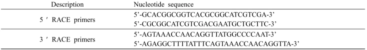
Table 1. Primers and used in RACE
Description Nucleotide sequence
5 ¢ RACE primers 5’-GCACGGCGGTCACGCGGCATCGTCGA-3’
5’-CGCGGCATCGTCGACGAATGCTGCTTC-3’
3 ¢ RACE primers 5’-AGTAAACCAACAGGTTATGGCCCCAAT-3’
5’-AGAGGCTTTTATTTCAGTAAACCAACAGGTTA-3’
Materials and Methods Materials
All chemicals were purchased from the Sigma Chemical Company (St. Louis, Mo). Polyacrylamide was purchased from National Diagnostics (Atlanta, USA). Prestained molecular weights standards and [methyl-3H]thymidine (1 μCi /㎍) were purchased from Amersham Pharmacia biotech (Little Chalfont, UK).
EDTA and Tween 20 were obtained from USB (Little Chalfont, UK). A Mighty Small Ⅱ Apparatus (Hoefer Scientific Instruments, San Francisco, CA) was used for protein electrophoresis. Tissue culture plates were ob- tained from Falcon Labware Division, Becton Dickinson (Oxnard, CA).
RT-PCR cloning and sequence analysis Two degenerate primers were designed from the alignment of IGF-I amino acid sequences from human, salmonide, carp. Liver of korean rockfish (Sebastes Schlegeli) was cracked with syringe in TRI reagent (Sigma) and descending size of needles. The RNA sam- ple was suspended in DEPC-treated water after ethanol precipitation, spectrophotometrically quantified at A260
and A280 nm, and then analyzed for their integrities on agarose gel (1%) with formaldehyde.
RT-PCR was performed (denaturation at 94oC for 2 min, followed by 35 cycles of 1 min at 94oC, 30 s at 52oC, 4 min at 72oC, and finally an elongation step at 72oC for 10 min) with one step RNA PCR kit (TAKARA) and the PCR products were cloned into pT7 Blue T vector (Novagen) for nucleotide sequence determination.
To obtain the full-length sequence of korean rockfish IGF-I (KR IGF-I), rapid amplification of cDNA ends (RACE) methods were performed using gene specific primers (Table 1). 3’ and 5’ RACE were performed according to the menufacturer’s instructions (Clontech).
cDNA clones appeared on the LB-ampicillin plate and next the QIAGEN plasmid extraction miniprep kit was used to extract DNA. The entire cDNA, digested with
Pst I and Eco RI restriction enzyme, was subcloned into pT7 Blue T vector and transformed into JM109, and then sequenced by ABI autosequencer. The nucleic acid sequence from the clones was used to search the GenBank database.
Southern blot analysis
The products of RACE were separated by agarose gel electrophoresis, and southern blotting was carried out on amplification products after transfer on nylon membrane (ICN BIOTRANSTM). The blot was prehy- bridized (1h, 42oC) in a solution of 5X SSC (1X SSC is 0.8% NaCl and 0.4% sodium citrate), 0.02% SDS, 0.1% N-laurysarcosine and 1% blocking reagent. Then, the blot was hybridized (18 h, 42oC) by adding 5 μl of a DIG labeled DNA probe which has been sequenced and identified as part of KR IGF-I. Using PCR and degenerate oligonucleotide primers, which are specific for two highly conserved sequences found in several vertebrates, a resultant cDNA fragment was used as a probe.
The 108 bp DNA probe was synthesized by PCR DIG probe synthesis kit (Roche). The blot was washed at room temperature for 15 min in 2X SSC, 0.1% SDS and for 15 min in 2X SSC, 0.1% SDS at 68oC for 30 min in 0.1X SSC, 0.5% SDS and then at 68oC for 30 min in 0.1X SSC, 0.5% SDS. The hybridized probe was detected with the DIG DNA Detection kit (Roche) and visualized by chemiluminescence reaction.
Expression of recombinant KR IGF-I pro- tein (rKR IGF-I)
rKR IGF-I, from B domain to D domain, was ampli- fied by PCR and two oligonucleotide primers; KR5 pri- mer (sense primer : 5’-GGCCATGGGACCGGAGAC CCTGTGCGGG-3’) and KR6 primer (antisense primer : 5’-CCGGATCCAGCTGCCTTGCTAGTCTTGGC-3’).
The PCR products were constructed with the histidin (His) gene fusion system (Novagen) of expression vec- tor pET 22b(+) with an N-terminal histidine tag for KR IGF-I. To overexproduce the fusion protein the ex-
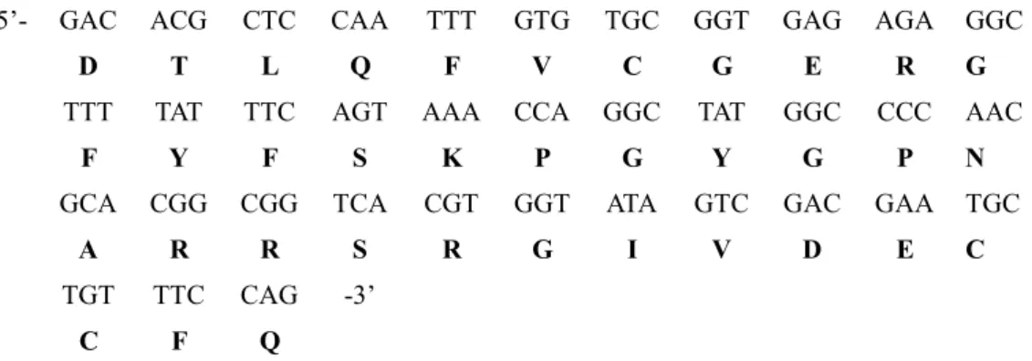
5’- GAC ACG CTC CAA TTT GTG TGC GGT GAG AGA GGC
D T L Q F V C G E R G
TTT TAT TTC AGT AAA CCA GGC TAT GGC CCC AAC
F Y F S K P G Y G P N
GCA CGG CGG TCA CGT GGT ATA GTC GAC GAA TGC
A R R S R G I V D E C
TGT TTC CAG -3’
C F Q
Fig. 1. Nucleotide and deduced amino acid sequence of the partial cDNA obtained by PCR from total RNA of korean rockfish.
pression vector was transformed into E. coli BL21(DE3) cells and selected by ampicillin. Colony hybridization and sequencing were used to identify the correct direction for insertion.
A single colony of BL21(DE3) E. coli cells contain- ing a recombinant pET-IGF-I plasmid was incubated in 100 ml of LB broth and incubated at 37oC for 18 h with shaking. The culture was then transferred into 500 ml of LB broth and incubated at 37oC with shaking until the absorbance at 600 nm was 0.6. Then 0.1 mM isopropyl-thio-D-galactoside (Sigma) was added to the culture medium and the culture was incubated at 37oC for 3 h with shaking.
The cells were subsequently pelleted and stored at -80oC. After thawing, the cells were resuspended in 10 ml of binding buffer containing 160 mM Tris-HCl (pH 7.5), 4 M NaCl, and 40 mM imidazole and then lyzed by sonication. The pellet containing insoluble rKR IGF-I was resuspended in 15 ml of the binding buffer supplemented with 6 M guanidine-HCl and incubated on a rotating platform for 24 h at 4oC to denature the protein. rKR IGF-I in the denaturated state was recov- ered in the supernatant fraction after centrifugation at 10,000g for 30 min. This supernatant was loading on a Ni2+ chelation resin column (Novagen) and the elution was carried out with 80 mM Tris-HCl (pH 7.5) contain- ing 2 mM NaCl, 4 M imidazole, and 6 M guani- dine-HCl. Proteins were analyzed by 15% SDS-PAGE and stained with Coomassie brilliant blue R-250.
Bioassay
The bioactivity of rKR IGF-I protein was measured with an in vitro assay. [3H]Thymidine incorporation in- to DNA in a CHES-214 embryonic cell was studied.
CHES-214 (3X 10⁴ cells/well) were seeded in 24-well
plates in DMEM medium supplemented with 10% FBS for 24 h. After 24 h of serum free incubation, the cells were incubated with or without various amounts of IGF-I (0–100 nM) for 18 h. The cells were then pulse-labeled with [3H]Thymidine (1 μCi/ml) for 2 h at 25oC. Then 0.5 ml of 3 N NaOH was added, and after 5 min the mixture was transferred to scintillation vials. After addition of 10 ml of aquasol, the solution was counted in a scintillation counter(Phamacia). Every parameter in this experiment was repeted three times.
Results
Isolation and sequence analysis of KR IGF-I cDNA
To identify and isolate the KR IGF-I cDNA, firstly two degenerate primers withen the conserved regions of human, salmonid, and carp IGF-I cDNAs. RT-PCR was performed using the first strand cDNA templates prepared from liver of korean rockfish. Cloning and se- quence analysis of the PCR product revealed a 108 bp partial sequence of an unknown IGF-I cDNA (Fig. 1).
To obtain the full-length sequence of KR IGF-I, 5’
RACE and 3’ RACE were used (Table 1) with two sets of gene-specific primers. The amplification and se- quencing of the 3’ and 5’ RACE revealed a 500 bp sequence of the translated IGF-I mRNA of the korean rockfish liver (Fig. 2). Nucleotide sequence determi- nation of this fragment showed that the full-length se- quence described in Fig. 4 is a single mRNA transcript with the same cDNA sequence carrying an ORF of the preproIGF-I sequence.
By southern blotting of the RACE products and hy- bridization with a KR IGF-I partial DNA probe which had been sequenced before, the RACE products were
verified (Fig. 3).
¬ 0.5kb
M 1 2 3 4 5
Fig. 2. Amplification of korean rockfish IGF-I cDNA by RACE.
5’ and 3’ RACE amplification of korean rockfish liver IGF-I.
Double strand IGF-I cDNA was synthesized from korean rockfish liver mRNA and ligated to cDNA adaptor AP1. M : molecular size marker (1 Kb ladder), lane 1, 2 : 5’ RACE products, lane 3, 4 : 3’ RACE products.
(A)
(B)
Fig. 3. Southern blot analysis of RACE products.
RACE products digested with EcoR I and Pst I from clones with insert DNA. RACE products were electrophoresed in a 1% agarose gel, transferred onto nitrocellulose membrane and hybridized with 108 bp korean rockfish IGF-I cDNA.
Panel (A) : 1% agarose gel electrophoresis patterns. Panel (B) : Southern hybridization.
Structure of KR IGF-I
The cDNA sequence corresponding to the coding re- gion of the KR IGF-I preprohormone, as indicated in Fig. 4, is 558 bp long. The deduced IGF-I protein con-
Fig. 4. Nucleotide and deduced amino acid sequence of the Korean rockfish IGF-I cDNA obtained from RACE. Double lined letters indicate amino residues of the domain expressed in E. coli host vector system. Underlined and bolded se- quences show polyadenylation signal and poly(A) tail, respectively.
sists of 185 amino acids spanning the signal peptide, domains B, C, A, D and E. The B domain consisted of 29 amino acids (aa), the C domain of 9 aa, the A domain of 21 aa, the D domain of 8 aa, and E domain of 67 aa. The E domain of this IGF from korean rock- fish is shorter than the other teleost IGF-I is. Amino acid sequence comparison also shows that this KR IGF-I has sequence identities of 93.6%, 90.7% and 85.4% in comparison with flounder, chinook salmon and human, respectively. It is concluded that the KR IGF-I cDNA encodes for a preproIGF-I sequence (Fig.
5).
Recombinant KR IGF-I (rKR IGF-I) protein expression and biological function
To investigate the biological properties of rKR IGF-I, the cDNA encoding, the KR IGF-I was subcloned downstream of the bacteriophage T7 promoter in an ex- pression vector pET 22b(+). E. coli BL21 (DE3) was transformed with the resulting plasmid, pET 22b(+) / KR IGF-I, and the transformed bacteria were induced to produce the recombinant protein with a polyhistidine tag at its N-terminus. Extracts were prepared from E.
coli and analyzed by the SDS-PAGE. As shown in Fig.
6, the whole-cell extracts contain the recombinant pro- tein of about 7 kDa as one major protein band (Lane 2), which is not present in the bacteria carrying the con- trol plasmid (Lane 1). This rKR IGF-I was found to localize in the insoluble fraction of cell extracts and was successfully purified under denaturing conditions with a Ni2+ column (Lane 3).
After purification of the rKR IGF-I, the rKR IGF-I revealed a single band of 7 kDa on the SDS-PAGE.
To test biological function of rKR IGF-I polypeptides,
Fig. 5. Comparison of deduced amino acid sequence between human and several fishes. Dot line(-) indicates identical ami- no acid sequence.
Fig. 6. Expression of korean rockfish IGF-I mature peptide in E. coli BL21(DE3).
Cells were cultured in LB medium with 100 μg/ml ampicillin at 37oC until OD600 reached 0.6. Then, 0.1 mM IPTG was added for induction of Korean rockfish mature protein synthesis. The cells were harvested after 180 min induction.
The fusion proteins were purified by Ni2+ chelation resin col- umn (Novagen). Lane 1 : the expression protein with the pET 22b(+) vector alone, lane 2 : the expression protein with the pET 22b(+) ligated IGF-I insert, lane 3: the expression pro- tein after purification as a single band.
0 200 400 600 800 1000 1200 1400
0 5 50 100
IGF-I (nM)
[3H]-Thymidine Incorporation (CPM)
**
*
Fig. 7. Effects of recombinant korean rockfish IGF-I protein on stimulated CHES-214 cell proliferation. The following ef- fects were measured for different concentrations of recombi- nant korean rockfish IGF-I. Asterisks indicate that the value is significantly different (*p < 0.01, **p < 0.001) from the control group (0 mM). The [3H]-thymidine into DNA syn- thesis experiment was repected three times.
they were analyzed by in vitro assay of incorporation of [3H]thymidine into DNA differentiation. The test quantities were between 0 and 100 nM and in- corporation ability significantly increased over 50 ng (Fig. 7, *p<0.01 and **p<0.001).
Discussion
To isolate cDNA encoding for IGF-I in korean rock-
fish, a strategy based on regions of nucleotide con- servation was used to generate the fragment of cDNA encoding the B-, C-, A-, D domains. A sequence nu- cleotide alignment was carried out on IGF-I sequences of several fish species to design primers for the RT-PCR and RACE. PCR products generated by 3’ and 5’ RACE procedures of about 500 bp were combined to generate the composite full cDNA sequence. A com- parison of the nucleotide and deduced amino acid se- quences of the korean rockfish IGF-I with that from other species indicated that the cDNA sequence ob- tained encompassed the entire mature peptide. The ri- gion of cDNA corresponding to the mature peptide was 558 bp in length. The conservation of the deduced ami- no acid sequence in the B and A domains clearly in- dicates the sequence obtained is a number of the IGFs gene family. At the amino acid level, the IGF-I exhibits highly identity to IGF-I of different vertebrates. These results suggest that IGF-I is well conserved among vertebrates.
Previously, we have shown that in korean rockfish in vivo administration of GH increases endocrine pro- duction of IGF-I in the liver (data not shown). It has been well established that in the teleost, hepatic IGF-I expression is regulated by growth hormone [4,7,13].
Recombinant bovine IGF-I resulted in hypoglycemia while mammalian IGF-I can stimulate growth rate of coho salmon [14]. Perez-Sanchez el al [16] found that the recombinant human IGF-I inhibited GH release after IGF-I stimulation, and GH release was stimulated in a dose dependent manner. In order to understand the biological function of IGF-I in fish, it is essential to obtain pure IGF-I. Many studies have used bovine and human proteins to examine the physiological role in fish [3,14-16]. Although these IGF-I proteins should pro- duce highly active stimulation and regulation effects, it is not sufficient for a function to be associated with fish. In the present study, to characterize the IGF-I pro- tein function of fish, we have developed the IGF-I / His tag fusion protein expression system. The rKR IGF-I protein was expressed in E. coli and appeared to significantly stimulate thymidine incorporation in dose-dependent manners. These data suggest that the rKR IGF-I protein may have a similar function in fish as compared with those in mammals.
Thus, it is reasonable that the rKR IGF-I protein pro- duced by the E. coli can be used for large scale purifica- tion of biological functional protein through fusion pro-
tein expression. In turn, this is an important application potential in analyzing the function of intact fish IGF-I protein.
요 약
어류의 insulin-like growth factor-I (IGF-I)의 생화학 적 작용기작을 연구하기 위하여 한국산 조피볼락의 IGF-I cDNA 유전자 cloning을 행하였다. 완전한 cDNA 유전자 염기서열은 PCR과 RACE 방법을 통 하여 얻어진 DNA로부터 결과를 얻을수 있었다. 결 정된 IGF-I의 염기서열은 flounder, chinook salmon, human IGF-I의 염기서열과 비교한 결과 각각 93.6%, 90.7%, 85.4%의 높은 상동성을 보였다. 생화학적으 로 활성이 있는 재조합 IGF-I을 얻기 위하여 IGF-I의 B-C-A-D domain 부분을 PCR로 얻은 뒤 E. coli BL21(DE3)에 넣어 overexpression 시켰다. Ni-NTA colummn을 사용하여 순수한 재조합 단백질을 정제 할수 있었다. 정제된 단백질은 SDS-PAGE 상에서 7 kDa의 단일 band를 보여 주었으며 [3H]-thymidine 결 합정도를 측정하는 방법으로 활성을 가지고 있음을 확인할수 있었다.
Acknowledgements
This work was supported by Korea Sea Grant Program of the Ministry of Maritime Affairs &
Fisheries(MOMAF-2001).
References
1. Baxter, R. C. and Martin, J. L. 1989. Binding proteins for the insulin-like growth factors : structure, regulation and function. Prog. Growth Factor Rev. 1, 49-68.
2. Chen, J. Y., Chen, J. C., Chang, C. Y., Shen, S. C., Chen, M. S. and Wu, J. L. 2000. Expression of recombinant tilapia insulin-like growth factor-I and stimulation of ju- venile tilapia growth by injection of recombinant IGFs polypeptides. Aquaculture 181, 347-360.
3. Chen, J. Y., Tsai, H. L., Chang, C. Y., Wang, J. I., Shen, S. C. and Wu, J. L. 1998. Isolation and characterization of tilapia (Oreochromis mossambicus) insulin-like growth factors gene and proximal promoter region. DNA Cell.
Biol. 17, 359-376.
4. Cohick, W. S. and Clemmons, D. R. 1993. The insulin- like growth factors. Annu. Rev. Physiol. 55, 131-153.
5. Daughaday, W. H. and Rotwein, P. 1989. Insulin- like growth factor-I and II : peptide, messenger ribonucleic acid and gene structure, serum and tissue concentration.
Endocrine Rev. 10, 68-91.
6. Duan, C. and Inui, Y. 1990. Evidence for the presence
of a somatomedin-like plasma factors in the Japanese eel, Anguilla japonica. Gen. Comp. Endocrinol. 79, 326-331.
7. Duan, C. and Plisetskaya, E. M. 1993. Nutrition regu- lation of insulin-like growth factor-I mRNA expression in salmon tissue. J. Endocrinol. 139, 243-252.
8. Duguay, S. J., Park, L. K., Samadpour, M. and Dickhoff, W. W. 1992. Nucleotide sequence and tissue distribution of three insulin-like growth factor-I prohormones in salmon. Mol. Endocrinol. 6, 1202-1210.
9. Kajimoto, Y. and Rotwein, P. 1991. Structure of the chicken insulin-like growth factor-I gene reveals con- served promoter element. J. Biol. Chem. 266, 9724-9731.
10. Kavsan, V. M., Grenbenjuk, V. A., Koval, A. P., Skorokhod, A. S., Roberts, C. T. Jr. and Leroith, D. 1994.
Isolation of a second nonalleic insulin-like growth fac- tor-I gene from the salmon genome. DNA Cell. Biol. 13, 555-559.
11. Kavsan, V. M., Koval, A. P., Grenbenjuk, V. A., Chan, S. J., Steiner, D. E. and Roberts, C. T. Jr. 1993. Structure of the chum salmon insulin-like growth factor-I gene.
DNA Cell. Biol. 12, 729-737.
12. LeRoith, D., Werner, H., Beitner-Johnson, D. and Roberts, C. T. Jr. 1995. Molecular and cellular aspects of the insulin-like growth factor-I receptor. Endocrine Rev. 16(12), 143-164.
13. Mathews, L. S., Norstedt, G.. and Palmiter, R. D. 1986.
Regulation of insulin-like growth factor-I gene ex-
pression by growth hormone. Proc. Natl. Acad. Sci. USA.
83, 9343-9347.
14. McCormick, S. D., Kelley, K. M., Young, G.., Nishioka, R. S. and Bern, H. A. 1992. Stimulation of coho salmon growth by insulin-like growth factor-I. Gen. Comp.
Endocrinol. 86, 398-406.
15. McCormick, S. D., Sakamoto, T., Hasegawa, S. and Hirano, T. 1991. Osmoregulatory actions of insulin-like growth factor-I in rainbow trout (Oncorhynchus mykiss).
J. Endocrinol. 130, 87-92.
16. Perez-Sanchez, J., Weil, C. and Le-Bail, P. Y. 1992.
Effects of human insulin-like growth factor-I on release of growth hormone by rainbow trout (Oncorhynchus my- kiss) pituitary cells. J. Exp. Zool. 262, 287-290.
17. Roberts, C. T., Brown, A. L., Graham, D. E., Seelig, S., Berry, S., Gabbay, K. H. and Rechler, M. M. 1986.
Growth hormone regulates the abundance of insulin-like growth factor I RNA in adult rat liver. J. Biol. Chem.
261, 10025-10028.
18. Rotwein, P., Pollock, K. M., Didier, D. K. and Krivi, G. G. 1986. Organization and sequence of the human insulin-like growth factor-I gene. J. Biol. Chem. 261, 4828-4832.
19. Shimatsu, A. and Rotwein, P. 1987. Mosaic evolution of the insulin-like growth factors : organization, se- quence, and expression of the rat insulin-like growth fac- tor-I gene. J. Biol. Chem. 262, 7894-7900.