• 검색 결과가 없습니다.

Headache without Cerebrospinal Fluid Leakage Haeng-Seon Shim

N/A
N/A
Protected

Academic year: 2021

Share "Headache without Cerebrospinal Fluid Leakage Haeng-Seon Shim"

Copied!
4
0
0

로드 중.... (전체 텍스트 보기)

전체 글

(1)

ISSN: 1229-6538

Korean J Clin Geri 2013;14(2):83-86

CASE REPORT

Received: July 29, 2013 Revised: December 2, 2013 Accepted: December 14, 2013.

Corresponding author: Myoung-Keun Shin

Department of Anesthesiology and Pain Medicine, Samsung Changwon Hospital, 158, Palyong-ro, Masanhoiwon-gu, Chanwon 630-723, Korea Tel: +82-55-290-6078, Fax: +82-55-290-6578, E-mail: [email protected]

Copyright Ⓒ 2013 The Korean Academy of Clinical Geriatrics

This is an open access article distributed under the terms of the Creative Commons Attribution Non-Commercial License (http://creativecommons.org/ licenses/by-nc/3.0) which permits unrestricted non-commercial use, distribution, and reproduction in any medium, provided the original work is properly cited.

Headache without Cerebrospinal Fluid Leakage

Haeng-Seon Shim

1

, Sung-Il Bae

1

, Seung-Won Choi

2

, Chang-Jin Oh

2

, Myoung-Keun Shin

1

Department of Anesthesiology and Pain Medicine,

1

Samsung Changwon Hospital, Sungkyunkwan University School of Medicine, Changwon,

2

Samsung Medical Center, Sungkyunkwan University School of Medicine, Seoul, Korea

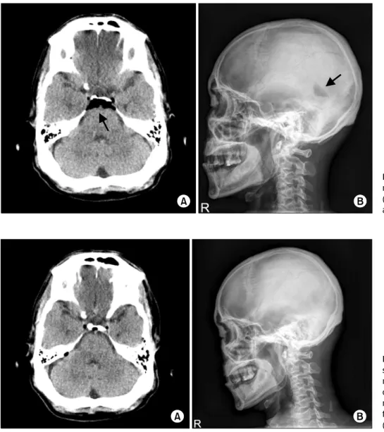
Headache can occur after a lumbar epidural block. There are several reasons for the development of headache such as hypotension, postdural puncture, vascular injection, subarachnoid hemorrhage and pneumocephalus. We report a case of a 63-year-old male patient who developed a severe headache without neurologic symptoms after a lumbar epidural block with the loss of resistance technique (LORT) using air. We excluded the possibility of postdural puncture headache because there was no cerebrospinal fluid (CSF) leakage. Brain CT was performed and it revealed pneumocephalus. After conservative treatment, the patient recovered and was discharged on the fourth day of hospitalization.

Key Words: Cerebrospinal fluid leakage, Headache, Loss of resistance technique, Lumbar epidural block, Pneumocephalus

INTRODUCTION

Headache can occur due to several reasons after a lumbar epidural block. Postdural puncture headache is the most common complication of unintentional dural puncture with an epidural needle. In most of the cases, headache after a lumbar epidural block is usually due to CSF leakage through the dural puncture site. If there is headache without CSF leakage after a lumbar epidural block with the loss of resist- ance technique (LORT) using air, we can consider pneumo- cephalus. We experienced a case of severe headache in a pa- tient after a lumbar epidural block and brain CT demon- strated pneumocephalus. We report a case of pneumocephalus with literature review.

CASE REPORT

A 63-year-old male patient was diagnosed with herniated intervertebral disc during orthopaedic examination. He weigh- ed 67 kg and was 169 cm tall. He complianed of numb- ness in both legs since the last 1 year. The pain in his legs was aggravated since the last 1 week. He was admitted to the orthopedics ward. He was then transferred to the pain clinic. He had no systemic diseases. He complained of a low back pain, numbness in both legs, and radicular pain.

Hence, we performed a lumbar epidural block. In the sitting

position, axenic disinfection of the back and local infiltration

with 2% lidocaine at the L4/5 level were performed. Lumbar

epidural block was performed at the L4/5 interspace with a

22 G Tuohy needle via the median approach with the LORT

using air. After the LORT using 3 ml air, an extra 3 ml air

(2)

84 Korean J Clin Geri 2013;14(2):83-86

Figure 1. Brain CT shows pneu- mocephalus in suprasellar cistern (A). Skull lateral shows ventricular air density (B).

Figure 2. After 4 days, brain CT shows a partially resolved pneu- mocephalus, unremarkable ventri- cular system (A). Skull lateral does not show any significant abnormal findings and ventricular air density (B).

was injected because a false negative reaction was suspected.

Immediately, the patient winced and complained of severe headache. We immediately stopped the epidural procedure, and with the use of a glass syringe we confirmed that there was no CSF leakage. We did not inject 0.125% bupivacaine 10 ml with triamcinolone 40 mg. We repositioned the pa- tient into the supine position and administered an infusion of fluids with 100% O

2

administration via a mask. Fortuna- tely, he had no other neurologic symptoms. After rest for 1 hour, his headache improved but mild headache persisted.

At that time, his blood pressure (BP), heart rate (HR), and pulse oximetry (SpO

2

) were 120/70 mmHg, 58 bpm, 100%, respectively. There were no other symptoms, and we asked the patient to visit the hospital if any other symptoms developed. He was discharged from the hospital. Because of

a continuous headache for 2 hours, he revisited the neuro- surgery ward of the hospital. We performed brain CT and skull series to rule out the possibility of pneumocephalus or any other findings. The brain CT showed a large pneumo- cephalus in the suprasellar cistern and a small amount air in the ambient cistern, quadrigeminal cistern, and superior cer- ebellar cistern. There was no evidence of hemorrhage (Fig. 1A).

The skull series showed ventricular air density (Fig. 1B). He was admitted to the neurosurgery ward. We reperformed brain CT and skull series after 4 days. Brain CT showed a partially resolved pneumocephalus, unremarkable ventricular system, and no evidence of intracranial hemorrhage (Fig.

2A). The skull series did not show any significant abnormal

findings and ventricular air density (Fig. 2B). He was dis-

charged on the fourth day of hospitalization.

(3)

Haeng-Seon Shim, et al: Pneumocephalus 85

DISCUSSION

Lumbar epidural steroid injections have been used as a routine therapeutic procedure for pain management. Despite a low morbidity, possible complications that can occur as a result of an epidural injection include dural puncture, vas- cular injection, and subarachnoid hemorrhage. After a lum- bar epidural block, headache is generally caused due to dural puncture. But, headache can occur due to pneumocephalus, subarachnoid hemorrhage, or migraine. If air is used in the LORT, pneumocephalus can be considered. Pneumocephalus, known as intracerebral aerocele or pneumatocele is defined as the presence of gas within any of the intracranial com- partments of the cranial vault.

1)

Several symptoms such as headache, nausea, vomiting, seizure, dizziness, lethargy, con- fusion, slow arousal, and obtundation may develop. Air em- bolism and cardiac arrest are associated with pneumoce- phalus in rare circumstances. Tension pneumocephalus, in whichthe volume of intracranial air places pressure on the brain, may require a neurosurgical evacuation.

As in this case, the LORT with air can be considered as a cause of pneumocephalus.

2)

This procedure results in pneu- mocephalus, nerve root compression, subcutaneous emphyse- ma, venous air embolism, and insufficient analgesia resulting from unblocked segments caused by epidural air.

3,4)

After postdural puncture, generally a positional headache occurs at 24∼48 hours. Pneumocephalus headaches are typically not position dependent.

An anatomical study of the lumbar epidural space in hu- man autopsy subjects was performed in 1986 after develop- ment of the method called epiduroscopy.

5)

In this case, we can assume that pneumocephalus developed due to air en- tering the subdural space. Although the subdural space is not directly connected to the subarachnoid space, it is con- nected to the intracranial space and the floor of the third ventricle in the cranial cavity through the lower border of the second sacral vertebra.

6,7)

Reina et al. described that in the subdural space there is a cellular junction called the du- ra-arachnoid interface between the laminar arachnoid portion and inner surface of the dura.

8)

In this case, we can assume that the air used in LORT entered the subdural space and

then entered into the suprasellar cistern, ambient cistern, quadrigeminal plate cistern, and superior cerebellar cistern.

During routine epidural catheter placement, the bevel of a Tuohy needle may be unintentionally advanced through the dura mater and the epidural catheter placed into the sub- dural compartment may not return CSF.

9)

A subsequent small test dose of local anesthetic may not produce dis- cernible sensory or motor block, while a full epidural dose of local anesthetic results in extensive sensory and motor block.

10)

Factors that may increase the incidence of subdural catheter placement include the following: previous back sur- gery, dural puncture at the same or an adjacent interspace, and rotation of the needle by 180 degrees after locating the epidural space.

11)

The duration and severity of symptoms are related to the amount of air inside the cranium. Therefore, we can reduce the incidence of pneumocephalus by using saline instead of air for the LORT.

12)

Treatment of pneumocephalus includes conservative meas- ures including oxygen therapy, supine position, aggressive hydration, caffeine, analgesics.

3,13)

Supplemental high oxygen concentration can hasten absorption of an intracranial air collection. Oxygen therapy accelerates the absorption of in- tracranial air by increasing the diffusion gradient for nitro- gen between the air collection and the surrounding cerebral tissue.

14,15)

These patients are usually monitored by skilled nursing staff who look for the signs of emergent neurological complications. Surgery is also an option for some patients if their condition does not improve in a timely manner.

In conclusion, in this case, there was headache without

CSF leakage due to pneumocephalus that is assumed to

have developed because of the air used for LORT that en-

tered the subdural space and then entered into the supra-

sellar cistern through the weak region of the dura. When

the LORT with air is chosen, we should use a small

amount of air or saline. When there is a headache without

CSF leakage after a lumbar epidural block, we should con-

sider pneumocephalus and prompt treatment should be

initiated. Also, we must minimize the amount of air used in

this procedure and we should be aware of the diagnosis and

treatment of pneumocephalus.

(4)

86 Korean J Clin Geri 2013;14(2):83-86

▪ 국문요약 ▪

요부 경막외 차단후 두통이 발생 할 수 있다. 두통의 원인으로는 저혈압, 경막 천자후 두통, 혈관 주입, 지주막하 출혈 과 기뇌증 등이 있다. 본 증례는 63세 된 남자 환자에게 공기를 이용한 저항소실법으로 경막외 차단을 시행하는 도중 에 신경학적 증상없이 극심한 두통을 호소한 경우를 보고하고자 한다. 뇌척수액 누출이 없었기 때문에 경막 차단후 두통은 배제하였다. 뇌단층 전산화촬영을 시행하여 기뇌증으로 진단 되어졌다. 적절한 치료후 환자는 회복되었고 입 원 4일 만에 퇴원하였다.

중심단어: 뇌척수액 누출, 두통, 저항소실법, 요부 경막외 차단, 기뇌증

REFERENCES

1. Schirmer CM, Heilman CB, Bhardwaj A. Pneumocephalus:

case illustrations and review. Neurocrit Care 2010;13:152-8.

2. Mateo E, López-Alarcón MD, Moliner S, Calabuig E, Vivó M, De Andrés J, et al. Epidural and subarachnoidal pneumo- cephalus after epidural technique. Eur J Anaesthesiol 1999;

16:413-7.

3. Shenouda PE, Cunningham BJ. Assessing the superiority of saline versus air for use in the epidural loss of resistance tech- nique: a literature review. Reg Anesth Pain Med 2003;28:

48-53.

4. Saberski LR, Kondamuri S, Osinubi OY. Identification of the epidural space: is loss of resistance to air a safe technique? A review of the complications related to the use of air. Reg Anesth 1997;22:3-15.

5. Blomberg RG, Olsson SS. The lumbar epidural space in pa- tients examined with epiduroscopy. Anesth Analg 1989;68:

157-60.

6. Ajar AH, Rathmell JP, Mukherji SK. The subdural com- partment. Reg Anesth Pain Med 2002;27:72-6.

7. Collier CB. Why obstetric epidurals fail: a study of epiduro- grams. Int J Obstet Anesth 1996;5:19-31.

8. Reina MA, De Leon Casasola O, López A, De Andrés JA, Mora M, Fernández A. The origin of the spinal subdural

space: ultrastructure findings. Anesth Analg 2002;94:991-5.

9. Blomberg RG. The lumbar subdural extraarachnoid space of humans: an anatomical study using spinaloscopy in autopsy cases. Anesth Analg 1987;66:177-80.

10. Lehmann LJ, Pallares VS. Subdural injection of a local anes- thetic with steroids: complication of epidural anesthesia. South Med J 1995;88:467-9.

11. Sukhani R, Garcia CJ, Munhall RJ, Winnie AP, Rodvold KA.

Lidocaine disposition following intravenous regional anesthesia with different tourniquet deflation technics. Anesth Analg 1989;68:633-7.

12. van den Berg AA, Nguyen L, von-Maszewski M, Hoefer H.

Unexplained fitting in patients with post-dural puncture headache. Risk of iatrogenic pneumocephalus with air ration- alizes use of loss of resistance to saline. Br J Anaesth 2003;

90:810-1.

13. Laviola S, Kirvelä M, Spoto MR, Tschuor S, Alon E.

Pneumocephalus with intense headache and unilateral pupil- lary dilatation after accidental dural puncture during epidural anesthesia for cesarean section. Anesth Analg 1999;88:582-3.

14. Dexter F, Reasoner DK. Theoretical assessment of normobaric oxygen therapy to treat pneumocephalus. Anesthesiology 1996;

84:442-7.

15. Kim YJ, Baik HJ, Kim JH, Jun JH. Pneumocephalus devel-

oped during epidural anesthesia for combined spinal-epidural

anesthesia. Korean J Pain 2009;22:163-6.

수치

Figure 1. Brain CT shows pneu- pneu-mocephalus in suprasellar cistern  (A). Skull lateral shows ventricular air density (B).

참조

관련 문서