구강 내 누공이 있는 낭성 병소와 제 3대구치의 병적 이동
이은희, 김대훈, 장효원, 박광호, 허종기*
연세대학교 치과대학 강남세브란스병원 구강악안면외과
<Abstract>
Cystic Lesion with Sinus Tract and Pathologic Migration of the Third Molar
Eun Hee Lee, Dae-Hoon Kim, Hyo-Won Jang, Kwang-Ho Park, Jong-Ki Huh*
Department of Oral and Maxillofacial Surgery, Gangnam Severance Hospital, Yonsei University College of Dentistry, Seoul, Korea
There are many case reports about cysts within the bones of the jaws associated with impacted third molars. When osmotic pressure is introduced into a cyst, the cyst expands and displaces the third molar. If continuity of cyst wall is lost, cystic expansion cannot occur, and the cyst cannot displace the third molar. This study analyzed four cases of pathologic migration of the third molar in ruptured cystic lesions that had formed bone tunnels and intraoral fistulas to identify the causes and factors contributing to this migration. Authors hypothesized that closure of fistulas repeated generation of pressure, it may temporarily increase the osmotic pressure within a cyst that has lost its continuity, causing displacement of the third molar. A cyst that has lost its continuity due to fistula formation within the oral cavity can cause ectopic displacement of the adjacent impacted teeth.
Key words : Third molar, Pathologic migration, Cyst, Sinus tract
Korean Journal of Oral and Maxillofacial Pathology 2017;41(5):237-244 ISSN:1225-1577(Print); 2384-0900(Online) Available online at http://journal.kaomp.org https://doi.org/10.17779/KAOMP.2017.41.5.006
* Correspondence: Jong-Ki Huh, Department of Oral and Maxillofacial Surgery, Gangnam Severance Hospital, Yonsei University College of Dentistry. 211 Eonju-ro, Gangnam-gu, Seoul 06273, South Korea Tel: +82-2-2019-4560, Fax: +82-2-3463-4052
E-mail: omshuh@yuhs.ac ORCID : 0000-0002-7381-3972
Ⅰ. INTRODUCTION
While pathologic migration of the third molars due to cystic lesions is commonly observed, pathologic migration of the third molars by a large distance - to a location deep within the mandibular ramus, maxillary sinus, coronoid process, or condylar head - rarely occurs. Pathologic migration of a tooth is commonly believed to occur when
the expansive force arising from the growth of a lesion such as cyst or tumor displace the impacted teeth.1) However, ectopic impaction of third molars has been observed in the absence of an expansive force from a tumor or cyst,2) and large displacement of third molars can occur even when a cyst has ruptured to turn into a tunnel-like structure, and formed intraoral fistulas.
Authors analyzed four cases of pathologic migration of third molars due to ruptured cystic lesions that formed bone tunnels and intraoral fistulas. This study aims to identify the causes and factors that contribute to pathologic migration of impacted third molars.
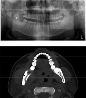
Fig. 1-A. Pre-operative panoramic view : Panoramic radiograph showing an impacted third molar associated with a radiolucent tunnel-like appearance (white arrow) in the right subcondylar region.
Fig. 1-B. Pre-operative CT view(Axial view) : migrated third molar is located in the right subcondylar region.
Fig. 1-C. Pre-operative CT view(Sagittal view) : the orifice of the cystic tract (black arrow) opened at the distal gingiva of the mandibular right second molar (#47).
1. CASE REPORT 1
A 33-year-old male patient visited the hospital with pain and swelling in the right cheek region and trismus.
Panoramic radiographs showed an impacted third molar that had been displaced toward the back of the right mandibular ramus by a large distance (Figure 1-A). A radiolucent lesion with a tunnel-like appearance was observed (Figure 1-B).
It surrounded the crown of the third molar, connected to the distal region of the right mandibular second molar, and possibly to the oral cavity (Figure 1-C). Incision and drainage were performed on right buccal region under local anesthesia.
Although pain and swelling were reduced after the treatment, the trismus, fistula, and abscess persisted. Therefore, extraction of the impacted tooth and enucleation of the cyst were performed under general anesthesia. The ectopic impacted third molar was extracted by lingual approach, and the cystic
lesion was diagnosed as dentigerous cyst (Figure 2-A,B).
2. CASE REPORT 2
A 56-year-old female patient visited the hospital with swelling of the left side of the palate. The patient showed signs of swelling and tenderness extending from the central incisors to the posterior molars of maxilla. A dark black serum-like fluid was observed during aspiration. Panoramic radiographs and computed tomography scans showed a radiolucent lesion surrounding the crown of the impacted third molar, extending from central incisor to first molar of the maxilla (Figure 3-A). Erosion of the buccal cortex of maxilla, and perforation of the maxilla towards the palate were observed (Figure 3-B,C). Extraction of the impacted tooth and enucleation of the cyst were performed under general anesthesia. During the surgery, a fistula opened to
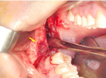
Fig. 2-A. Fig. 2-A. Intraoperative view showing fistula formation (yellow arrow) on the lingual side of the distal portion of the mandibular right second molar (#47).
Fig. 2-B. Extracted mandibular right third molar (#48) & cystic wall & fistula tract.
Fig. 3-A. Pre-operative panoramic view: Panoramic view showing an impacted third molar associated with a radiolucent tunnel-like appearance (white arrow) in the left maxillary sinus region.
Fig. 3-B. Pre-operative CT view(Axial view): Migrated third molar is located in the left maxillary sinus. Two cystic lesions are separated by a bony wall (white arrow)
Fig. 3-C. Pre-operative CT view(Sagittal view): The orifice of the cystic tract (white arrow) opened at the distal gingiva of the maxillary left second molar(#27).
the distal area of the second molar was found, and the cystic lesion was confirmed pathologically as a dentigerous
cyst (Figure 4-A,B).
Fig. 4-A. Intraoperative view showing fistula formation (yellow arrow) on the distal side of the maxillary left second molar (#27) region.
Fig. 4-B. Intraoperative view showing the maxillary left third molar (#28, yellow arrow) in the left maxillary sinus.
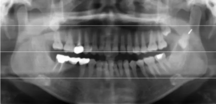
Fig. 5-A. Pre-operative panoramic view: Panoramic view showing an impacted third molar associated with a radiolucent tunnel-like appearance (white arrow) in the right ramus region.
Fig. 5-B. Pre-operative CT view: The orifice of the cystic tract (white arrow) opened at the distal gingiva of the mandibular right second molar (#47).
Fig. 5-C. Intraoperative view showing fistula (yellow arrow) formation
on the lingual side of the distal region of the mandibular right second
molar (#47).
Fig. 6-A. Pre-operative panoramic view (8 years before operation):
Panoramic view showing an impacted third molar associated with a radiolucent tunnel-like appearance (white arrow) in the left ramus region.
Fig. 6-B. Pre-operative panoramic view (6 years before operation)
Fig. 6-C. Pre-operative panoramic view (5 years before operation) Fig. 6-D. Pre-operative panoramic view (just before surgery) 3. CASE REPORT 3
A 23-year-old female patient visited the hospital with a cyst on the right side of the mandible that was discovered coincidentally on panoramic radiograph without any symptoms.
No abnormal findings were observed during the clinical examination. However, a radiolucent lesion extending from the center of the right mandibular ramus to the distal area of the mandibular right second molar was found on panoramic radiographs and computed tomography scans (Figure 5-A,B).
A cystic lesion surrounded the crown of the mandibular right third molar, which was displaced to the center of the mandibular ramus. A tunnel-like fistsula was connected to the oral cavity.
Extraction of the impacted teeth and enucleation of the cyst were performed under general anesthesia. During the surgery, a fistula opened to the distal area of the mandibular right second molar was observed (Figure 5-C). The cystic lesion was diagnosed as a lesion that had transitioned from
keratocystic odontogenic tumor to unicystic ameloblastoma.
4. CASE REPORT 4
A 52-year-old female patient visited the hospital with a discomfort around the left mandibular ramus and third molar, and swelling on left cheek. The patient felt no tenderness on palpation. The periodontal pocket depth was measured to be 9 mm on the distal area of the left mandibular second molar. Panoramic radiographs showed an impacted third molar displaced by a large distance to the posterior of the left mandibular ramus (Figure 6-D).
Computed tomography scans showed an impacted left third molar located directly below a mandibular notch and in close contact with the inferior alveolar nerve on the lingual side (Figure 6-E). It was also observed that a radiolucent lesion with a tunnel-like appearance extended from a cystic lesion to the distal area of the left mandibular second molar,
Fig. 6-E. Pre-operative CT view (just before surgery) : the orifice of the cystic tract (white arrow) opened at the distal gingiva of the mandibular left second molar (#37).
Fig. 6-F. Intraoperative view showing fistula formation (yellow arrow) on the distal side of the mandibular left second molar (#37) region.
Fig. 6-G. Extracted mandibular left third molar(#38) & cyst wall. Fig. 6-H. Cyst wall & tunnel like sinus tract
and the cystic lesion surrounded the dental crown ofimpacted third molar. Tunnel-like lesion was connected from the dental crown to the oral cavity.
The patient underwent extraction of impacted tooth and enucleation of the cyst under general anesthesia. During the surgery, a sinus tract extending from the distal area of the left mandibular second molar to the dental crown of the left mandibular third molar was found(Figure 6-F). This soft tissue and cystic lesion were carefully removed from normal tissues by approaching from lingual side of the mandibular ramus(Figure 6-G,H). A cystic lesion was confirmed as chronic nonspecific inflammation with granulation tissue.
There are panoramic radiographs 5, 6, and 8 years before
the first visit of our department, taken by the local dental clinic. We confirmed that the fistula extending from the distal area of the left mandibular second molar to the impacted mandibular third molar had existed 8 years ago.
The radiographs showed the third molar has been displaced gradually from the center of the mandibular ramus to directly below the mandibular notch, and a sinus tract with a tunnel-like appearance has extended(Figure 6-A,B,C). No signs of cystic expansion were observed during this period, and the patient did not report any unusual clinical symptoms. However, it was observed that as the impacted tooth moved directly below the mandibular notch, and a sinus tract with a tunnel-like appearance had extended.
Ⅱ. DISCUSSION
The causes of ectopic impaction of the mandibular third molars are still unknown.1) According to the literature, pathologic migration of impacted teeth can occur as a result of displacement of the tooth germs of the third molars, abnormal patterns of eruption,3,4) lack of space between the mandibular ramus and the mandibular second molars, an imbalance between the eruption direction of the third molars, or the mandibular base can contribute to displacement of impacted third molars.5,6) As explained earlier, the third molars may migrate as a result of growth of a lesion such as a cyst or a tumor.7,8) In other words, as it grows, a lesion may put pressure on the dental crown, potentially displacing the impacted tooth into the opposite direction of the eruption path.
Displacement of the third molars is known to occur as a result of cyst expansion caused by introduction of osmotic pressure into the cyst. However, when the continuity of a cystic wall is lost, cyst expansion can not occur because of the abscesce of osmotic pressure, and subsequently the cyst cannot displace impacted third molars.
In the present case report, displacement of the adjacent impacted third molars by a large distance was observed even with cysts that had lost their continuity as a result of fistula formation. As this is a rare observation, a different approach must be used to explain the causes of ectopic displacement of cysts and impacted teeth.
In the case of pathological migration of third molars due to a cyst that has lost its continuity as a result of fistulas, other factors may cause the pathological migration. First, the possibility of pathological displacement of a tooth even when the expansive force of a cyst is reduced due to cyst wall rupture must be considered. Second, it is possible that a cyst that expanded throughout the mandible displaced the impacted teeth, and then ruptured, leading to drainage or reduced osmotic pressure, and eventually to bone deposition
and repair.9-11) In the case of the second hypothesis, displacement of adjacent anatomical structures must also occur until the cyst ruptures, and the displaced anatomical structures such as the inferior alveolar canal must remain in their displaced position even after bone recovery is complete following the loss of expansive force due to cyst rupture. If the second hypothesis is true, displacement of not only impacted third molars but also adjacent anatomical structures such as the inferior alveolar canal must occur.
However, in the four cases discussed in this study, no such displacement of anatomical structures was observed on radiographs. Therefore, the possibility of tooth displacement due to other factors must be considered even after the cyst’s continuity has been lost due to cyst rupture.
Even after the cyst wall loses its continuity, osmotic pressure can temporarily increase within the cyst upon closing of the fistula, and displacement of impacted teeth may occur through repeated generation of this pressure. In other words, if a fistula repeatedly opens and closes over a long period of time, an expansive force will be created even at low osmotic pressure resulting from fistula closure.
The possibility of gradual tooth displacement through repetition of this process over a long period of time must also be considered. In general, the cyst wall is circular or oval in shape, and compresses the adjacent impacted teeth.
However, in the present cases, cyst walls formed a tunnel-like duct connecting the cyst to the oral cavity. Such changes in the morphology of cysts may have been the result of repeated opening and closing of fistulas.
In the fourth case discussed in this study, gradual displacement of an impacted third molar even in the absence of active growth and expansion of the cyst over several years was observed on panoramic radiographs. This finding confirms the possibility of pathologic migration of an impacted tooth due to a cyst that has lost its continuity as a result of fistula formation in the four cases.
In 2008, Kim et al. reported cases of impacted teeth positioned on the mandibular condyle in the absence of lesions such as cysts or tumors.2) Based on this report, pathologic migration of the third molars can be classified into three types: Type 1 – pathologic migration of third molar with cystic lesion without tunnel-like sinus tract, Type 2 – pathologic migration of third molar with cystic lesion and tunnel-like sinus tract, and Type 3 – pathologic migration of third molar without cystic lesion and tunnel-like sinus tract.
In all four cases, tooth displacement occurred in the direction opposite to that of fistula formation. This suggests that the direction of the formation of a bone tunnel, and the location of a fistula may be closely associated with the migration of an impacted tooth.
Except in one patient, the patients’ radiographs taken before they visited the hospital could not be obtained, and as a result, we could not conduct a detailed follow-up of the patterns of displacement of impacted teeth at different time periods. Because we could not analyze cyst growth, fistula formation, and displacement patterns of impacted third molars at different time periods, we could not analyze the factors that affect displacement of impacted teeth in depth. Additional research on the association between the timing of fistula closure and displacement of impacted teeth is needed.
Ⅲ. CONCLUSION
1. A cyst that has lost its continuity due to fistula formation within the oral cavity can cause ectopic displacement of the adjacent impacted teeth.
2. Displacement of tooth containing a cyst tends to occur in the direction opposite to that of fistula formation.