서 론
중피종은 중피세포에 석면 가루(asbestos fiber)가 직접 작용하여 발생한다고 알려져 있다[1]. 이 종양은 화학적, 방사선 요법에 반응하지 않은 것으로 알려져 있으나, 세 포사멸에 대한 종양 저항(tumor resistance) 기전을 알 수 있
다면 획기적인 치료를 발견할 수 있고, 또한 기존 치료의 효과를 증가시키는 방법이 될 수 있을 것으로 생각된다[2].
저자들은 Adenoviral p53에 민감하게 반응하는 중피종 세포주인 염증 및 표피세포 아유형(subtype)에 adenovirus 유전자 핵산전달감염(transfection)으로 중피세포암 치료의 새로운 방법에 대하여 평가하고자 하였다.
A Experimental Study of PTEN (Phosphatase and Tensin) Role in Mesothelioma
Seog Ki Lee, M.D.*, Kweon Cheon Kim, M.D.**
Background: Conventional treatment for mesothelioma is largely ineffective. We evaluated the novel approach of adenoviral gene transfection of PTEN gene in mesothelioma cancer cell lines, inflammatory and epithelial subtype, which are sensitive to adenoviral p53. Meterial and Method: Binary adenoviral PTEN and LacZ (Ad/GT-LacZ and Ad/GV16) vectors were used for transduction of the mesothelioma cell lines, REN (p53 sensitive). Protein levels were determined by Western blotting assay. Apoptosis was assessed by fluorescence-activated cell sorter analysis of subdiploid populations. Cell viability was determined with the XTT assay. Statistical analysis was performed with analysis of variance and the Student t test. Result: 72 hours after the treatment of adenoviral PTEN gene, cell killing were 32.9% for REN compared to control cell (2.5%) at MOI of 20. Also we observed the over-expression of proapoptotic protein, bax and decreased expression of bcl-2 protein in REN cells. But the expression of BCL-xl, Bak, Bad proteins were not altered. Conclusion: Adenovirus Pten-mediated overexpression of the Bax gene induces apoptosis and decreased cellular viability in p53-sensitive mesothelioma cells. These data suggest that the trans- fection of PTEN gene may represent a alternative gene therapy strategy to treat mesothelioma.
(Korean J Thorac Cardiovasc Surg 2003;36:852-857) ꠏ
Key words: 1. Mesothelioma 2. Gene therapy 3. Cell death
중피종에서 PTEN (Phosphatase and Tensin)의 역할에 대한 실험적 연구
이 석 기*․김 권 천**
*조선대학교 의과대학 흉부외과학교실
Department of Thoracic & Cardiovascular Surgery, Chosun University College of Medicine, Gwangju, Korea
**조선대학교 의과대학 외과학교실
Department of Surgery, Chosun University College of Medicine, Gwangju, Korea
†이 논문은 2001년 조선대학교연구비의 지원을 받아 연구되었음.
논문접수일:2003년 6월 18일, 심사통과일:2003년 9월 5일
책임저자 : 김권천 (501-717) 광주광역시 동구 서석동 588번지, 조선대학교부속병원 외과 (Tel) 062-220-3072, (FAX) 062-228-3441, E-mail: kckim@chosun.ac.kr
본 논문의 저작권 및 전자매체의 지적소유권은 대한흉부외과학회에 있다.
대상 및 방법
1) 세포주(Cell line)중피암에서 유도된(Mesothelioma-derived) 세포주에서 p53 단백질이 결여된 비정상적인 p53을 가진 REN (inflamma- tory and epithelial subtype)은 W. Roy Smythe에서 구했으며, 이 세포주은 37oC, 5% CO2을 가진 가습된 배양기에 10%
fetal bovine serum, 1% nonessential amino acids와 100μg/ml penicillin과 100μg/ml streptomycin을 함유한 RPMI 배지에 보존하였다.
2) Adenoviral 핵산전달감염(Transfection)
Adenoviral PTEN은 Introgen Therapeutics (Houston, Texas) 에서 공급을 받았으며, Cytomegalovirus promotor (Ad CML- lacZ)하에 모든 실험을 진행하였다. 세포들은 초기에 심어 진 후 수를 기록하고 밤사이 12시간 동안 배양하였다. 이 세포들은 PBS에서 씻은 후 다른 다양한 농도(multiplicity of infection: MOI)으로 핵산전달감염하였다. 20% fetal bovine 혈청이 채워진 Viral RPMI 배지에 4시간 동안 노출 후 최 종적으로 10% 혈청이 되도록 하였다.
3) 세포 생존력 분석(Cell viability assay)
XTT 분석 도구를 모든 세포 생존력 연구에 사용하였으 며, 세포는 2,000 cells/well의 농도로 96 well plate에 심어 졌다. 하룻밤 동안 배양된 후 MOI 0에서부터 MOI-120까 지 다양한 MOI에서 Ad-PTEN으로 4시간 동안 핵산전달감 염하고 72시간 동안 세포를 배양하였다. XTT 표시된 시약 과 전자와 결합된 시약(electron coupling reagent)을 50:1 비율로 XTT 반응 혼합물은 각각 well plate에 첨가하였다.
최종적으로 모든 판(plates)는 450 nm Dynatech colormetric plate reader로 결과를 판독하였다. 이 데이터의 분석은 Microsoft Excel를 이용하여 하였으며, 이 실험들은 각 3차 례씩 시행하였다.
4) Bcl-군(family) 발현(Western blot 분석)
Western blot 분석으로 Adenovirus PTEN 핵산전달감염 후 BCL군 발현를 관찰하였으며, 중피종 세포는 2,000,000 cells/plate 농도로 직경 6 cm 판(plates)에 심어졌고, 하루 밤사이 12시간 배양하고 MOI 60에서 Ad-PTEN와 함께 핵 산전달감염한 후 24시간, 48시간 및 72시간에 모든 세포 를 모았다. 전체 세포 용해질은 SDS-PAGE 견본 완충제 (buffer)와 함께 plated cell monolayers을 이용하였다. 용해
질내 단백질은 BCA 단백질 분석 시약(Piercem Rockford, IL)을 이용하여 각각 단백질 성분을 확인하고, SDS polya- crylamide gradient (12%) gel과 5μg의 세포 용해제(lysate) 를 첨가하였으며, BAK, BAX, BCL-XL, BCL-2와 caspaspase 3, β-Actin 등의 단백질을 분리하기 위하여 100oC에서 전 기영동을 하였다. 그 후 단백질은 high bond-ECL mem- branes으로 전이하고 Amersham ECL 방법으로 일차성과 이차성 항체와 함께 배양하였다. β-Actin, BCL-XL, BAK, BAX 단백질은 향광물질을 이용하여 X-ray 필름상에서 이 들 단백질 발현을 확인하였다.
5) 세포사멸을 위한 DNA 농도 분석
세포사멸 정도는 flow cytometry로 확인하였으며, flurore- scence-activated cell sorter analysis은 다음과 같은 방법으로 이용하였다. 세포들을 6 cm plate에 2,000,000 cells/plate로 24시간 동안 배양한 후 4시간 동안 MOI of 60에서 Ad- PTEN와 함께 핵산전달감염한 후 24시간, 48시간, 72시간 에 세포들을 트립신처리하여 원심분리기로 모았고, PBS 을 첨가하였다. 밤사이 -20oC 70% ethanol로 고정하였고, 세포들에게 Prodidium iodide와 RNAse (Boehringer Mann- heim Co, Indianapolis, IN)을 첨가하고 빛이 차단된 상태에 서 37oC에서 30분 동안 배양하였다.
세포사멸 정도는 세포의 sub-G1 population을 농도로 나 타내었고, 이 실험은 3번 반복하였다.
통계학적 처리는 analysis of variance와 student t test를 이용하였고, p value는 0.05 이하일 때 의미가 있는 것으로 간주하였다.
결 과
1) PTEN의 핵산전달감염(transfection)은 세포의 죽 음을 유도한다.
중피종 세포주(cell line)에서 세포사(cell death)를 유발하 는 PTEN 유전자(gene) 능력을 측정하기 위하여 대조군 (untreated REN cell)과 비교하였고, ad-PTEN는 다양한 MOI로 세포주에 핵산전달감염하였으며, 핵산전달감염 48 시간 이후에 ad-PTEN이 첨가된 세포주에서 의미 있는 세 포사멸이 되는 것을 관찰할 수 있었다(Fig. 1).
MOI of 20에서 72시간 후 대조군(2.5%)에 비해 32.9±
5.5%로 세포사멸 증가가 통계학적 의미가 있었으며(p value=0.04), MOI of 60에서 5일 이상 세포사멸 정도는 72 시간에서 가장 높은 세포사멸을 보였으며, 그 후로는 증
가되지 않았다.
2) PTEN의 핵산전달감염(transfection)는 세포사멸 (apoptosis)을 증가시킨다.
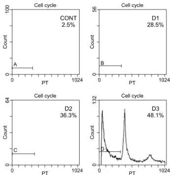
세포주에서 PTEN이 세포사멸을 증가시키는 것을 확인 한 후 세포사멸 정도를 측정하였다. 세포사멸 정도는 pro- pidium-iodide flow cytometery에서 sub-G1을 표시하는 곡선 하방에 있는 면적으로 측정하였다(Fig. 2). 대조군과 비교 하였을 때 PTEN-induced REN 세포의 세포사멸 정도가 의 미가 있을 정도로 증가하였다. MOI of 60에서 REN 세포 주의 세포사멸 양은 대조군에서 2.5%였고, PTEN 유전자 핵산전달감염 후 1일째 28.5%, 2일째 36.3%, 그리고 3일째 48.1%였다(Fig. 3). 세포사멸 증가는 3차례 반복된 실험에서 대조군 세포에 비해 통계학적으로 의미가 있었다(p=0.025).
3) Ad-PTEN 핵산전달감염(transfection)은 BAX 단백 질의 발현 증가와 BCL-XL의 발현 감소를 나타 낸다.
PTEN 핵산전달감염에 의한 전구세포사멸 단백질(pro- apoptotic proteins)인 BAX, BAK과 항세포사멸 단백질(anti- apoptotic proteins)인 BCL-2, BCL-XL 발현을 알아보고자 시행하였으며(Fig. 4), Ad-PTEN 처리된 REN 세포에서 BAX 단백질은 발현 증가(overexpression)를 보였고, BCL-2 단백질 발현 감소가 나타났고, BCL-XL, BAK 그리고 BAD는 변화가 없었다.
액틴 단백질 표현(Actin protein expression)은 대조군으로 모든 분석에서 사용되었다.
Fig. 1. Transfection of PTEN induces cellular death.
Fig. 2. DNA analysis in PTEN treated REN cell. These graphs show that 72 hours after treatment with adenoviral PTEN gene, cell killing was 32.9% for the REN cell compared to the control cell (2.5%) at MOI of 60. CONT=Untreated REN cell;
D1=24 hours after treatment of ad-PTEN; D2=48 hours after treatment of ad-PTEN; D3=72 hours after treatment of ad- PTEN.
Fig. 3. Apoptotic rate of Ad-PTEN treated REN cell on flow cytometry. This shows the amount of PTEN observed the con- trol, 2.5%; at 1 day, 28.5%; at 2 days, 36.3%; and at 3 days, 48.1%. CONT=Untreated REN; D1=24 hours after treat- ment of ad-PTEN; D2=48 hours after treatment of ad-PTEN;
D3=72 hours after treatment of ad-PTEN.
고 찰
PTEN (phosphate and tensin homolog deleted from choro- mosome 10)은 종양 억제 유전자(tumor suppressor gene)으 로 MMA1 (mutated in multiple advanced cancer) 혹은 TEPI (TGF-regulated and epithelial cell-enriched phosphatase)로 알 려져 있으며, 사람에서 발생하는 대부분의 종양에서 이종접 합성(heterozygosity (LOH))을 상실하는 지놈 부분(genomic region)인 염색체(chromosome) 10q23에 위치하고 있다[3].
PTEN은 protein tyrosine phosphatase와 dual specificity protein phosphatase signature를 가지고 있어서 in vitro에서 tyrosine-, serine-, threonine-phosphorylated peptides에서 탈인화(depho- spholation)시킬 수 있었다[4]. 여러 연구에서 PTEN은 ph- osphatidylinositol-3 phosphatase이며, phosphoinositol-3-kinase (PI3K)의 생성물이고 다양한 세포형에서 세포사멸을 방지 하는 신호 인자인 PKB/Akt를 인산화(phosphorylation)하여 활성화시키는데 필요한 phosphatidylinositol(3-5)-triphosphate (PIP3)를 탈인화(dephophoryate)한다[5,6]. PTEN은 세포 성 장과 생존을 조절하는 중요한 역할하는 PI3K 신호화(sig- nalling)을 방지하고, PTEN의 세포 성장 기능은 PI3K/Akt 경로를 차단하는 이상의 작용을 하고 있다는 사실이 점점 확실해지고 있으며, 또한 비교적 잘 알려져 있는 lipid ph- osphatase 활성과 더불어 protein phosphatase 활성화하는 역
할을 가지고 있다[4,7]. Weng 등[8]은 유방암에서 PTEN은 protein phosphatase 활성화를 통해 cyclin D를 억제(down regulating)시키고 lipid phosphatase 활성화에서 p27을 증가 (up-regulating) 시킴으로써 G1 활성화를 조정한다고 제시한 바 있다. 또 다른 최근보고는 protein과 lipid phosphatase 활 성화는 최소한 MGF-7 유방암 모델에서 PTEN을 매개로 한 성장 억제(PTEN-mediated growth suppression)과 관련이 있다고 하였다[9]. 저자들은 이러한 PTEN역할에 관심을 갖고 중피종 세포주에서 PTEN의 역할에 관해 연구하고자 하였다.
세포 죽음은 세포사멸과 관련된 복합적인 세포화 과정 (complex cellular program)에 의존하고 있다[10]. 세포 사멸 은 DNA 손상을 받은 세포를 찾아내어 없애는 기능이 있 기 때문에 세포사멸 반응 실패는 결국은 종양을 발생시키 고[11], 화학적 및 방사선 요법에 의한 종양 치료 효과를 감소시키기 때문에 세포사멸은 종양연구에서 중요성이 증가하고 있다[12].
세포사멸과 운명을 조절하는 단백질로 중요한 두 군인 BCL-2군과 Caspase군이 있다[13]. BCL-2군은 구조적으로 전구세포사멸 단백질(proapoptotic protein) 및 항세포사멸 단백질(antiapoptotic protein)로 구성되어 있으며, 항세포사 멸 단백질로 BCL-2, MCL-2 및 BCL-XL가 있고, 전구세포 사멸 단백질은 BAX, BAK, BCL-XS 및 BAD가 있다[14].
이합체화(Dimerization)은 BH1, BH2, BH3 단백질에서 three conserved domain을 통해 매개된다[13]. 다른 단백질 helical BH3 loop가 또 다른 단백질 세 가지 모두 영역 (domain)에 의해 형성된 열(cleft)에 부착될 때 이들 단백질 은 연관성을 갖게 된다[13]. 초기 보고와 반대로 이종이합 체화(heterodimerization)는 항세포사멸 기능을 위해서는 필 요하지 않았으나, BH3 영역을 포함하는 단백질에서 전구 세포사멸 기능을 위해서는 필요하다[13]. 이들 BCL-2, BCL- XL, BAX 같은 단백질은 세포막 특히 사립체(mitochodria) 에 부착하여 사립체막 전압(potential)과 투과성(permeability) 에 영향을 주는 COOH-terminal domain을 함유하고 있으 며, 이 과정은 cytochrome C, procaspase 3, 그리고 사립체 로부터 세포사멸을 유도하는 요소로 caspase을 활성화시 키는 물질을 유출한다[15,16]. BAX는 이런 물질을 방출하 는 것을 촉진하는 반면에 BCL-3와 BCL-XL은 이 과정을 억제한다[15,16]. BCL-XL은 포유동물의 CED-4 동종(homo- logue)인 apaf-1에 부착하여 세포사멸을 방지함으로써 pro- caspae 9와 cytochrome C와의 관련성을 억제하고 이에 따 른 일련의 과정에 영향을 미치는 caspases 활성화를 저해 Fig 4. PTEN treated REN cell line (Western blot analysis).
This shows an over-expression of BAX, decreased expression of BCL-2, and no change in the expression of BCL-XL, BAK and BAD protein. CONT=Control group; D1=24 hours after treatment of ad-PTEN; D2=48 hours after treatment of ad- PTEN; D3=72 hours after treatment of ad-PTEN.
한다[13]. 다른 한편으로 BAX와 BAK은 전구세포사멸 구 성성분은 BCL-XL의 apaf-1의 중성화를 억제하여 세포사 멸을 강화하는 작용을 한다[13].
BCL-2군은 단백질 발현은 종양 위치와 형태에 따라서 다양하다. 악성 중피종에서 BCL-2 발현은 소수 종양에서 만 보고되고 있고, 세포주 연구에서 악성 중피종 세포는 BCL-2가 아니라 BAX를 발현시키는 것으로 알려져 있다 [17.19]. BAX는 세포사멸을 촉진하고, BCL-2는 이종이합 체화(heterodimerization)에 의해 항세포사멸 기능 작용을 한 다고 생각된다[19]. 그러므로 직접적으로 세포사멸을 촉진 또는 저해시키는 것은 BCL-2와 BAX의 비(ratio)이다[19].
p53은 BCL-2에는 억제 조절 인자로 BAX에는 활성화 조절 인자로 작용하기 때문에 BAX에 비해 낮은 BCL-2 비는 wild-type p53이 생긴 것에 대한 결과일 수 있다[19,20].
Mohiudin 등[21]은 sodium butyrate로 처리한 adenoviral pro- apoptotic gene therapy (PAGT)를 통해 전구 세포사멸 단백 질(BAX, BAK)을 발현 증가시키는 것은 항세포사멸 단백 질(BCL-XL)의 감소로 세포 죽음과 세포사멸을 강화시킨 다고 제안했다. 또한 이 저자들은 중피종 세포에서 sodium butyrate가 BCL-XL 발현을 하향조절(down regulation)시켜 adenoviral BAX 유전자 핵산전달감염과 함께 BCL-2군 유 전자 표현을 ‘forced therapeutic imbalance’ 시킨다는 것을 밝혀냈다[21]. 이런 몇 가지 제안과 연관해서 저자들은 PAGT를 통한 종양 억제제 PTEN은 전구 세포사멸 단백질 인 BAX와 BAK을 상향조절(up-regulation)하고, 항세포사 멸 단백질 BCL-2군을 하향조절(down-regulation)함으로써 세 포성장을 억제한다는 가정하에 행하여진 본 연구는 Western blot 분석을 이용하여 REN 세포에서 Adenovirus PTEN 유 전자 핵산전달감염에 의해 전구 세포사멸 단백질 BAX가 발현 증가되고, 항세포사멸 단백질 BCL-2군 발현 감소됨 을 알 수 있었다.
위에서 언급한 것처럼 악성 흉막 중피종은 기존 종양 치료에 반응하지 않으므로 유전자 치료법(gene therapy)은 이 치명적 종양에 대한 새로운 치료법이 될 것으로 기대되 며[22], 중피종에 대한 Phase I 임상 시도에서 Herpes sim- plex virus thymidine kinase (HSV)을 흉막강 내에 주입하여 어느 정도 악성 중피종 치료에 효과가 있음을 밝혀냈다 [23]. 최근 in vitro 혹은 in vivo에서의 p53의 Adenoviral 전 이를 포함한 많은 유전자 치료법 접근이 Bax 발현 증가가 세포사멸을 유도한다고 발표되고 있다[22,24].
결 론
본 연구에서 PTEN 주입에 의한 BAX 발현을 증가시키 는 것이 중피종에서 유전자 치료 연구(gene therapeutic study)에서 새로운 목적이며, 궁극적으로 악성 흉막 중피 종 환자 치료에 있어서 새로운 유전자 치료가 될 수 있을 것으로 기대된다.
참 고 문 헌
1. Mossman BT, Kamp DW, Weitzman SA. Mechanisms of
carcinogenesis and clinical features of asbestos-associated cancers. Cancer Invest 1996;14(5):466-80.
2. Narasiman SR, Yang L, Gerwin BI, Broaddus VC. Resis-
tance of pleural mesothelioma cell lines to apoptosis: rela- tion to expression of Bcl-2 and Bax. Am J Physiol 1998;
275(1 Pt 1):L165-71.
3. Li JY, Clifford L, Danny P, et al. PTEN, a putative pro-
tein tyrosine phosphatase gene mutated in human brain, breast, and prostate cancer. Science 1997;275:1943-47.
4. Myers MP, Tonks NK. PTEN: sometimes taking it off can
be better than putting it on. Am J Hum Genet 1997; 61(6):
1234-8.
5. Stambolic V, Suzuki A, de la Pompa JL, et al. Negative
regulation of PKB/Akt-dependent cell survival by the tumor suppressor PTEN. Cell 1998;95(1):29-39.
6. Downward J. Mechanisms and consequences of activation
of protein kinase B/Akt. Curr Opin Cell Biol 1998;10(2):
262-7.
7. Tamura M, Gu J, Matsumoto K, Aota S, Parsons R, Yamada KM. Inhibition of cell migration, spreading, and
focal adhesions by tumor suppressor PTEN. Science 1998;
280(5369):1614-7.
8. Weng LP, Brown JL, Eng C. PTEN coordinates G(1)
arrest by down-regulating cyclin D1 via its protein phosph- atase activity and up-regulating p27 via its lipid phosph- atase activity in a breast cancer model. Hum Mol Genet
2001;10(6):599-604.9. Hlobilkova A, Guldberg P, Thullberg M, Zeuthen J, Lukas J, Bartek J. Cell cycle arrest by the PTEN tumor suppres-
sor is target cell specific and may require protein phosph- atase activity. Exp Cell Res 2000;256(2):571-7.
10. Oltvai ZN, Korsmeyer SJ. Checkpoints of dueling dimers
foil death wishes. Cell 1994;79(2):189-92.
11. Steller H. Mechanisms and genes of cellular suicide. Sci- ence 1995;267(5203):1445-9.
12. Lowe SW, Ruley HE, Jacks T, Housman DE. p53-depen-
dent apoptosis modulates the cytotoxicity of anticancer a-
gents. Cell 1993;74(6):957-67.
13. Adams JM, Cory S. The Bcl-2 protein family: arbiters of
cell survival. Science 1998;281(5381):1322-6.
14. Yang E, Korsmeyer SJ. Molecular thanatopsis: a discourse
on the BCL2 family and cell death. Blood 1996;88(2):386-
401.15. Kluck RM, Bossy-Wetzel E, Green DR, Newmeyer DD.
The release of cytochrome c from mitochondria: a primary site for Bcl-2 regulation of apoptosis. Science 1997;275
(5303):1132-6.16. Manon S, Chaudhuri B, Guerin M. Release of cytochrome
c and decrease of cytochrome c oxidase in Bax-expressing yeast cells, and prevention of these effects by coexpression of Bcl-xL. FEBS Lett 1997;415(1):29-32.
17. Chilosi M, Facchettti F, et al. Bcl-2 expression in pleural
and extrapleural solitary fibrous tumours. J Pathol 1997;
181(4):362-7.
18. Narasimhan SR, Yang L, Gerwin BI, Broaddus VC.
Resistance of pleural methothelima cell lines to apoptosis:
relation to expression of Bcl-2 and Bax. Am J Physiol
1998;275:L165-71.19. Oltvai ZN, Milliman CL, Korsmeyer SJ. Bcl-2 heterodime-
rizes in vivo with a conserved homolog, Bax, that accel- erates programmed cell death. Cell 1993;74(4):609-19.
20. Miyashita T, Krajewski S, Krajewska M, et al. Tumor sup-
pressor p53 is a regulator of bcl-2 and bax gene expres- sion in vitro and in vivo. Oncogene 1994;9(6):1799-805.
21. Mohiuddin I, Cao X, Fang B, Nishizaki M, Smythe WR.
Significant augmentation of pro-apoptotic gene therapy by pharmacologic bcl-xl down-regulation in methothelioma.
Cancer Gene Ther 2001;8(8):547-54.
22. Pataer A, Smythe WR, Yu R, et al. Adenovirus-mediated
Bak gene transfer induces apoptosis in mesothelioma cell lines. J Thorac Cardiovasc Surg 2001;121(1):61-7.
23. Sterman DH, Treat J, Litzky LA, et al. Adenovirus-me-
diated herpes simplex virus thymidine kinase/ganciclovir gene therapy in patients with localized malignancy: results of a phase I clinical trial in malignant mesothelioma.
Hum Gene Ther 1998;9(7):1083-92.
24. Swisher SG, Roth JA, et al. Adenovirus-mediated p53 gene
transfer in advanced non-small-cell lung cancer. J Natl
Cancer Inst 1999;91(9):763-71.=국문 초록=
배경: 중피종은 일반적 치료에 대하여 큰 효과가 없다고 알려져 있다. 저자들은 Adenoviral p53에 민 감하게 반응하는 중피종 세포주인 염증 및 표피세포 아유형(subtype)에 adenovirus 유전자 핵산전달감 염(transfection)으로 중피종 치료의 새로운 방법에 대하여 평가하고자 하였다. 대상 및 방법: 두 쌍의 adenoviral PTEN와 LacZ (Ad/GT-LacZ와 Ad/GV16) 매개체(vectors)에 REN (p53 sensitive)인 중피종 세 포주(methothelioma cell lines)의 형질을 도입(transduction)하였으며, 단백질 함량은 Western blotting 분 석을 이용하여 측정하였다. 세포사멸은 fluorescence-activated cell sorter analysis of subdiploid populations에 의하여 평가하였으며, 세포 생존력은 XTT 분석에 의하여 결정하였다. 통계 분석은 analysis of variance와 Student t test를 이용하여 하였다. 결과: Adenoviral PTEN 유전자로 처치된 세포 사는 72시간 후에 MOI of 20에서 대조군 2.5%에 비하여 REN군 32.9%로 상대적으로 높게 나타났다.
또한 REN cell에서의 전구세포사멸 단백질(proapoptotic protein)인 BAX 발현 증가를, BCL-2에서 발현 감소를 나타내었으나, BCL-XL, BAK 및 BAD 단백질은 변화가 없었다. 결론: Adenovirus PTEN을 매 개로 한 BAX 발현 증가는 세포사멸을 유도하고 p53에 민감한 중피종 세포들(p53-sensitive metho- thelioma cells)에서 세포 생존력을 감소시킨다. 이러한 결과는 PTEN 유전자 핵산전달감염하는 것은 중피종 치료의 새로운 대안적 방법이 될 수 있다는 것을 암시한다.
중심 단어:1. 중피종 2. 유전자 치료 3. 세포사멸