Therapeutic comparison of Surgery combined with chemotherapy and chemotherapy alone for Primary Gastrointestinal Lymphoma: A single center study
Je Hun Kim, Ho Sup Lee, Jun Seop Lee, Jin Young Lee,
Su Young Kim, Cheol Su Kim, Joung Wook Yang, and Ga In You
Department of Internal Medicine, College of Medicine, Kosin University, Gospel Hospital, Busan, Korea
원발성 위장관 림프종의 수술 후 복합 화학요법과 화학요법 단독과의 비교: 단일 기관 연구
김제훈, 이호섭, 이준섭, 이진영, 김수영, 김철수, 양정욱, 유가인 고신대학교 복음병원 내과학교실
Objectives: There is still no consensus on the optimal treatment for primary gastrointestinal lymphoma (PGIL). The aim of this study was to compare surgery combined with chemotherapy and chemotherapy alone in PGIL.
Methods: We retrospectively reviewed and analyzed the treatment outcomes of 107 patients with primary gastrointestinal lymphoma diagnosed between March 1999 and December 2009 at Kosin University Gospel Hospital. Patients were divided into two groups: 35 patients who underwent surgery combined with chemotherapy (group A) and 72 patients who were treated with chemotherapy alone (group B). And we analyzed prognostic factors associated with short survival.
Results: The 5-year progression free survival rates (PFS) of group A and B were 86.7% and 66.1%, respectively (P = 0.037), while the 5-year overall survival rates (OS) were 86.8% and 68.4%, respectively (P = 0.129). In multivariate analysis, Both PFS and OS were not changed by treatment strategies (surgery combined with chemotherapy or chemotherapy only).
The international prognostic index (IPI) was the only independent predictive factor for PFS.
Corresponding Author:Ho Sup Lee, Department of Internal Medicine, College of Medicine, Kosin University, Gospel Hospital, 262, Gamcheon-ro, Seo-gu, Busan, Korea
TEX: +82-51-990-5820 FAX: +82-51-990-5821 E-mail: [email protected]
Received:Oct. 13, 2013 Revised:Mar. 19, 2014 Accepted:Jun. 3, 2014
Original Article
Conclusions: In our study, surgery combined with chemotherapy and chemotherapy only make no difference of survival rate. And further randomized prospective studies are needed to confirm a treatment strategies at improving survival outcomes in PGIL patients.
Key Words: Chemotherapy, Primary gastrointestinal lymphoma, Surgery
림프종은 크게 호지킨 림프종(Hodgkin’s lymphoma)과 비호지킨 림프종(Non-Hodgkin’s lymphoma)으로 나눈 다. 호지킨 림프종이 비교적 동질성을 지닌 질환군인데 반 하여 비호지킨 림프종은 다양한 질환의 집합체이다. 발생위 치에 따라서 림프절 림프종(Nodal lymphoma)과 림프절외 림프종(Extranodal lymphoma)으로 나눈다.1
위장관은 림프절외 림프종이 침범하는 가장 흔한 장소 로, 비호지킨 림프종의 15%를, 모든 림프절외 림프종의 30~40%를 차지한다.2 가장 흔한 병변의 위치는 위 (74.7%)이며, 소장(8.6%), 회맹부(7.0%), 두 개 이상의 위 장관 병변(6.5%), 소장, 대장, 직장을 포함하는 병변 (3.2%)이 있다.3 위장관 내에서 모든 형태의 NHL이 생길 수 있지만 세포형으로서 중요한 2가지 아형으로는 점 막연관림프조직(Mucosa-associated lymphoid tissue, MALT)과 미만성 거대 B세포림프종(Diffuse large B-cell, DLBCL)이다.4
2011년 Kim 등이 보고한 국내 림프종 발생 현황을 살펴 보면 전체 림프종 중 비호지킨 림프종이 95.4%였고 림프절 외 림프종이 69.6%였다. 림프절외 림프종 중에서는 국내에 서도 위 림프종이 가장 많았으며, MALT 림프종이 56.1%, 미만성 거대 B세포 림프종이 35.1%였다.5
국소적 범위의 PGIL 표준 치료에 대해 과거부터 수많은 연구들이 있었다. 많은 연구에서 Helicobacter pylori(H.
pylori) 제균 요법이 위에 국한된 저등급의 위 MALT 림프 종의 합리적인 초치료임을 밝혔다.6,7 그러나 H.pylori 제균 요법에 반응하지 않거나, H.pylori 음성인 MALT 림프종, DLBCL, 다른 조직학형의 악성 림프종의 경우 치료는 정립 되어 있지 않다. 현재까지 몇몇 전향적, 후향적 연구에서 PGIL의 치료로 화학요법 단독, 화학요법과 방사선요법의 병합, 수술 후 복합 화학요법, 단독 방사선치료 등을 비교 하였으나 치료군 간의 생존율은 통계적으로 유의한 차이를
보이지 않았다.8-10 그래서 현재 PGIL의 치료에서 수술의 역할은 위장관 폐색이나 천공, 출혈 등의 화학요법의 부작 용이 있을 경우나 그러한 부작용이 예측되는 경우, 화학요 법을 할 수 없는 환자들의 경우에 해당되었다.11
표준치료가 정해지지 않은 PGIL에 대한 병기 설정과 질 병 관리에 있어 도움이 되는 다양한 예후인자들에 대한 분 석들도 있었다. Heriberto 등이 보고한 원발성 위림프종에 서 짧은 생존율과 연관된 예후 인자에는 진단 당시의 높은 LDH 수치와 불량한 전신상태가 있었다.12
본 연구는 후향적으로, 수술 후 복합 화학요법 또는 화 학요법 단독이 PGIL 환자의 생존에 미치는 영향을 분석하 였다.
방 법
1. 대상
1999년부터 2009년까지 고신대학교 복음병원에서 원발성 위장관 림프종으로 진단된 107명을 대상으로 후향적으로 분석하였다. 병기, 조직아형, 원발 부위, ECOG performa- nce score는 제한하지 않고 포함하였으며 수술적 치료만 시행하였던 환자와 H.pylori 제균요법만 시행하였던 환자는 제외하였다. 병기 설정에는 신체 검사, 일반 혈액 검사, 단 순 흉부 방사선 검사, 상부 및 하부 내시경 및 이를 통한 조직검사, 골수 생검, 흉부 및 복부 골반 CT, 양전자 방출 단층촬영(Positron emission tomography, PET)을 이용하 였다. 조직학적인 진단은 숙련된 병리의사가 확인하였다.
병기설정을 위하여 Ann-Arbor system을 이용하였으며 그 내용은 Table 1과 같다.
I Involvement of a single lymph node region (I) or a single extralymphatic organ or site (IE)
II Involvement of two or more lymph node regions on the same side of the diaphragm (II) or of an extralymphatic organ and its adjoining lymph node site (IIE)
III Involvement of lymph node sites o both sides of the diaphragm (III) or localized involvement of an extralymphatic site (IIIE), spleen (IIIS), or both (IIISE)
IV Diffuse or disseminated involvement of one or more extralymphatic organs with or without associated lymph node involvement
Table 1. The classification of PGIL according to Ann-Arbor staging system
2. 치료
모든 환자는 수술 후 복합 화학요법을 시행한 군과 H.pylori 제균요법을 제외한 화학요법 단독으로 시행한 군 으로 나누었다. 수술적 절제는 원발성 병변의 부분적 혹은 전절제 및 인접한 림프절을 포함하였고 이것은 숙련된 외과 의사에 의해 시행되었다. 화학요법은 cyclophosphamide, adriamycin, vincristine, prednisolone (CHOP) 또는 그 것을 기반으로 하여 6~8주기 치료하였다. 재발성 혹은 불 응성의 NHL 환자에게서는 dexamethasone, ifosfamide, methotrexate, and etoposide(IMVP), etoposide, cyta- rabine, cisplatin, and methylprednisolone(ESHAP), 고 용량 methotrexate 등으로 치료하였다.
3. 분석
두 군에서 치료반응의 평가는 Revised international workship Criteria에 기초하여 치료 전, 후 림프종 종괴의 크기 변화를 흉부 및 복부 골반 CT, 양전자 방출 단층촬영 (Positron emission tomography, PET)들을 통해 시행하 였다. 종결점(End point)은 전체생존율(Overall survival rates, OS)과 무진행생존율(Progression free survival rates, PFS)로 설정하였다. 전체생존율은 병을 진단받은 시 점으로부터 질환과 연관된 사망 혹은 마지막 추적관찰 시점 까지를 계산하였다. 무진행생존율은 치료를 시작한 시점으 로부터 진행되거나 마지막 추적관찰 시점까지로 계산하였 다.
4. 통계학적 분석
화학요법 단독군과 복합 화학요법군의 변수들간의 비교를 위하여 chi-square 검정법을 적용하였고, 예후인자에 따른 전체생존율과 무진행생존율의 단변량 분석을 위해 Kap- lan-Meier 법을 사용하였고, log rank 법을 이용하여 두 군의 차를 분석하였다. 단변량 분석에서 전체생존율 에 영 향을 미치는 예후인자(P < 0.05)를 분석대상에 포함시켜 Cox proportional hazards regression을 이용하여 다변량 분석을 시행하였다. 예후인자의 상대위험도와 95% 신뢰구 간을 산출하였으며, 유의수준은 0.05를 기준으로 판정하였 다. 통계 처리는 SPSS (version 12, SPSS Inc. Chicago, IL, USA) 프로그램을 사용하였다.
결 과
1. 대상 환자의 특성
대상 환자 107명 중 남성 53명(49.5%), 여성 54명 (50.5%)이었다. 전체 군의 평균연령은 56세였다. 추적기간 은 1.3개월에서 125.5개월로, 평균 추적기간은 48.5개월 이 었다(Table 2). 107명 중, DLBCL 69명(64.5%), 변연부 B 세포 림프종(Marginal zone B cell lymphoma, MZL) 28 명(26.2%), 말초성 T-세포 림프종 (Peripheral T cell lymphoma, PTCL) 6명(5.6%), 외투세포림프종(Mantle cell lymphoma, MCL), 여포성 림프종 (Follicular lymphoma, FL), 소림프구성 림프종 (Small lymphocytic lymphoma, SLL)과 같은 다른 종류의 B 세포 림프종이 4명(3.7%)이었
Characteristics/Category Number(%) Age, years
Median (range) 56 (21-79)
Sex Male Female
53 (49.5) 54 (50.5) Histologic type
Diffuse large B cell lymphoma Marginal zone B cell lymphoma Peripheral T cell lymphoma Other B cell lymphoma
69 (64.5) 28 (26.2) 6 (5.6) 4 (3.7) Gastrointestinal tract involved site
Stomach Small intestine Colon
68 (63.6) 26 (24.3) 13 (12.1) Ann Arbor Stage
IE IIE IIIE IV
29 (27.1) 33 (30.8) 25 (23.4) 20 (18.7) Bone marrow involvement
Present No
5 (4.7) 102 (95.3) LDH
< 450 IU/L ≥ 450 IU/L
74 (69.2) 33 (30.8) ECOG performance status
0-1 ≥2
67 (62.6) 40 (37.4) IPI risk,
Low
Low intermediate High intermediate High
48 (44.9) 20 (18.7) 12 (11.2) 27 (25.2) Treatment
Surgery+ Chemotherapy group Chemotherapy group
35 (32.7) 72 (67.3) Rituximab
Yes No
33 (30.8) 74 (69.2) Table 2 Patient characteristics (N=107)
ECOG, Eastern Cooperative Oncology Group; IPI, international prognostic index; and LDH, Lactate dehydrogenase
다. 침범한 위장관의 위치는 위가 68명(63.6%)으로 가장 많았고, 다음으로 소장(14명, 13.1%), 대장(13명, 12.1%), 말단 회장(10명, 9.3%) 십이지장(2명, 1.9%) 순이었다. 병 기는 Stage IE가 29명(27.1%), Stage IIE가 33명(30.8%), Stage IIIE가 25명(23.4%), Stage IV가 20명(18.7%) 이었 다.
수술 후 복합 화학요법을 시행한 군은 35명이었으며, 화 학요법을 단독으로 시행한 군은 72명이었다. 화학요법 단 독군에 비해 수술 후 복합 화학요법군에서 PGIL의 소장 침 범이 빈번하였다. 그리고 수술 후 복합 화학요법군에 비해 화학요법 단독군이 위에서 호발하였다. 화학요법 단독군에 비해 수술 후 복합 화학요법군에서 국소적인 병기가 많았 고, 다른 인자들은 두 군 간의 유의한 차이를 보이지 않았 다(Table 3).
2. 치료 결과와 생존율에 대한 예후 인자
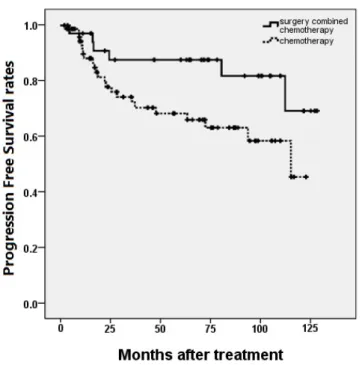
단변량 분석에서, 5년 무진행생존율은 수술 후 복합 화학 요법군이 화학요법 단독군보다 높았고(각각 86.7%, 66.1%), 국소적인 병기, 낮은 LDH 수치, 낮은 ECOG score, 낮은 IPI 위험도인 경우에 통계적으로 유의하게 높았다(각각 P
= 0.037, 0.002, 0.016, 0.016, <0.001)(Table 4, Fig.
1). 5년 전체생존율은 DLBCL, MZL, 국소적인 병기, 낮은 LDH수치, 낮은 IPI 위험도의 경우에서 유의하게 높았다(각 각 P = 0.011, 0.026, <0.001, <0.001)(Table 4). 수술 후 복합 화학요법군이 화학요법 단독군보다 5년 전체생존율이 높았으나(86.8%, 68.4%) 통계적인 유의성은 없었다(P = 0.129) (Fig. 2). Rituximab을 사용한 경우 수술 후 복합 화학요법군과 화학요법 단독군 모두 전체생존율 및 무진행 생존율이 높았지만 유의하지 않았다(P = 0.212). 다변량 분석에서, 독립적인 무진행생존율의 예측인자는 IPI 위험도 (P = 0.004)뿐이었으며, 다른 인자들은 통계학적으로 유의 하지 않았다(Table 5). 그리고 다변량 분석에서 전체생존율 의 경우 유의한 예측인자는 없었다(Table 5).
고 찰
1973년부터 2005년까지의 The Surveilance, Epide- mology, End Results (SEER) database에 의하면 PGIL의 발병율은 매년 4.67%씩 증가하고 있다. 이러한 발병율의 증가는 PGIL의 치료전략에 대해 다시 논의하게 되었다. 과 거에, 위가 주된 원발병소였던 위장관 림프종의 표준 치료 로 수술이 고려되었다. 시간이 흐르면서, 항암 화학치료와 방사선 치료가 발전하였고 이러한 치료의 효율성 및 수술 후의 환자의 삶의 질 등으로 인하여 수술을 대체하게 되었 다. 이러한 변화에 대하여 해외에서는 치료방법에 대한 많 은 비교가 이루어졌다.4 그러나 국내에서 이에 대한 연구는 매우 적다.13
본 연구의 107명 PGIL 환자는 DLBCL, MALT 림프종, PTCL, 그리고 다른 종류의 다양한 조직학적 형태들의 분포 가 다른 연구와 비슷하였다.8,9 단변량 분석에서 5년 무진행 생존율은 화학요법 단독군보다 수술 후 복합 화학요법군에 서 유의하게 높았고, 전체생존율은 두 군에서 차이가 없었 다. 하지만 다변량 분석에서 화학요법 단독군과 수술 후 복 합 화학요법군의 무진행생존율과 전체생존율의 차이가 없어 치료방법에 따른 생존율의 차이는 없는 것으로 확인되었다.
화학요법 단독군에서 병기가 높고, IPI 위험도가 높은 경우 가 많이 포함되었기 때문에 이것으로 인해 전체생존율에 영 향을 받았을 것으로 생각한다.
German multicenter study group의 연구에서 원발성 위 림프종의 치료 결과를 수술군과 비수술군과의 비교에서 5년 생존률의 유의한 차이가 없었다.8 또 Cirrocchi 등은 meta-analysis를 통하여 수술군과 비수술군의 10년 생존 율은 차이가 없으나, 수술군에서 높은 사망률, 비수술군에 서 낮은 재발율을 보였다. 또 원발성 위장관 림프종에서 광 범위 수술을 하였을 때 비 광범위 수술보다 우월한 생존율 을 보였다.14
하지만, 본 연구와 유사하게 장관의 DLBCL환자에게서 수술 후 복합 화학요법이 화학요법보다 우월한 생존율을 보 였으며,13 초기 원발성 장관 림프종에서, 외과적 절제 후 복 합 화학요법은 화학요법 단독과 비교하였을 때보다 우세한
Surgery+
Chemotherapy group (n=35)
Chemotherapy group
(n= 72) P-value Age, years
< 60
≥ 60 23 (65.7)
12 (34.3) 45 (62.5)
27 (37.5) 0.746 Sex Male
Female 16 (45.7)
19 (54.3) 37 (51.4)
35 (48.6) 0.582 Histology
DLBCL MZL Others
23 (65.7) 8 (22.9) 4 (11.4)
46 (63.9) 20 (27.8) 6 (8.3)
0.790
Gastrointestinal tract involved site Stomach
Small intestine Colon
14 (40.0) 16 (45.7) 5 (14.3)
54 (75.0) 10 (13.9) 8 (11.1)
0.685
Ann Arbor Stage IE IIE
IIIE IV
11 (31.4) 18 (51.4) 5 (14.3) 1 (2.9)
18 (25.0) 15 (20.8) 20 (27.8) 19 (26.4)
0.001
Tumor size < 10 cm
≥ 10 cm 19 (79.2)
5 (20.8) 37 (80.4)
9 (19.6) 0.900 Bone marrow involvement
Present
No 0 (0.0)
35 (100.0) 5 (6.9)
67 (93.1) 0.110 LDH < 450 IU/L
≥ 450 IU/L 26 (74.3)
9 (25.7) 48 (66.7)
24 (33.3) 0.423 ECOG performance status
0-1
≥2 22 (62.9)
13 (37.1) 45 (62.5)
27 (37.5) 0.971 IPI risk
Low
Low intermediate High intermediate High
19 (54.3) 7 (20.0) 3 (8.6) 6 (17.1)
29 (40.3) 13 (18.1) 9 (12.5) 21 (29.2)
0.432
Rituximab
YesNo 8 (22.9)
27 (77.1) 25 (34.7)
47 (65.3) 0.212 Table 3 Comparison of patients based on a treatment strategy
DLBCL, diffuse large B cell lymphoma; ECOG, Eastern Cooperative Oncology Group; IPI, international prognostic index;
LDH, lactate dehydrogenase; and MZL, marginal zone B cell lymphoma.
5-year PFS (%) p-value 5-year OS (%) p-value Age, years
< 60 ≥ 60
79.7 58.7
0.54 79.7
63.8
0.205
Sex Male Female
72.8 73.7
0.677 76.1
73.7
0.879
Histology DLBCL MZL Others
78.8 71.9 44.4
0.094 78.8
78.0 44.4
0.011
GI involve site Stomach Small intestine Colon
69.5 57.0 71.6
0.167 74.5
58.0 67.7
0.096
Ann Arbor Stage IE and IIE IIIE and IV
83.3 56.5
0.002 83.3
60.3
0.026
Tumor size < 10 cm ≥ 10 cm
73.5 55.5
0.161 77.1
55.0
0.155
Bone marrow involvement Present
No
53.3 74.3
0.522 53.3
75.8
0.408
LDH
< 450 IU/L ≥ 450 IU/L
80.7 52.6
0.016 82.6
52.1
< 0.001 ECOG performance status
0-1 ≥2
78.9 62.7
0.016 79.0
67.0
0.158
IPI risk
Low and Low intermediate High intermediate and High
84.3 50.3
< 0.001 84.4 54.6
< 0.001
Treatment
Surgery + Chemotherapy group Chemotherapy group
86.7 66.1
0.037 86.8
68.4
0.129
Rituximab Yes No
81.2 62.9
0.118 76.7
68.8
0.475 Table 4 Univariate analysis of prognostic factors
BM, bone marrow; DLBCL, diffuse large B cell lymphoma; ECOG, Eastern Cooperative Oncology Group; GI, gastrointestinal; IPI, international prognostic index; LDH, lactate dehydrogenase; MZL, marginal zone B cell lymphoma;
OS, Overall survival; and PFS, progression free survival
PFS OS
RR 95% C.I. p-value RR 95% C.I. p-value Histology
DLBCL MZL
Others 0.516 0.140-1.907 0.321
Ann Arbor Stage (%) IE and IIE
IIIE and IV 0.918 0.321-2.631 0.874 0.576 0.214-1.549 0.274 LDH
< 450 IU/L
≥ 450 IU/L 0.828 0.364-1.881 0.652 0.447 0.181-1.104 0.081 ECOG performance status
0-1
≥2 1.339 0.519-3.449 0.546
IPI risk
Low and Low intermediate
High intermediate and High 0.184 0.058-0.583 0.004 0.503 0.168-1.504 0.219 Treatment
Surgery + Chemotherapy group
Chemotherapy group 0.446 0.149-1.332 0.148 Table 5. Multivariate analysis of prognostic factors
ECOG, Eastern Cooperative Oncology Group; IPI, international prognostic index; LDH, lactate dehydrogenase; OS, overall survival; PFS, progression free survival; RR, relative risk; and 95% C.I., 95% confidence interval.
생존율을 보였다는 연구도 있다.15,16
위와 장관을 모두 포함하였던 연구에서는 PGIL의 치료에 있어서 수술은 생존율의 이득과 연관이 없지만, 사망위험도 와 전체 사망률을 증가시키지 않아 생존에 있어서 나쁜 영 향을 미치지 않고 있다고 밝혔다.17 현재는 임상적 상태에 따라 수술적 치료를 포함한 복합 화학치료를 추천하고 있으 나 소장과 대장의 림프종 치료 전략에 대한 연구가 아직 부 족하다.18
본 연구에서 이전의 연구들과 동일하게 LDH 수치, Ann arbor stage, ECOG performance status와 IPI 위험 도가 무진행생존율에 대한 예후인자임을 보였다.12 또, 수술 후 복합 화학요법이 화학요법 단독과 비교하여 나쁘지 않은 무진행생존율을 보였기에 이것이 의미 있는 치료 방법임을 보였다. 그러나, 수술 후 복합 화학요법은 다변량 분석에서 무진행생존율에 대한 유의한 예후인자가 아니었다. 그것은
본 연구에서 국소적인 병기와 소장을 침범하였던 경우가 화 학요법 단독군 보다 수술 후 복합 화학요법군에 더 많이 포 함되었고 이것은 수술이 진단과 치료를 동시에 할 수 있기 때문이며 이로 인한 결과로 생각한다.
생존율과 관련한 예후인자에 대한 다른 연구에서는 파종 성 장관 림프종에서 조기 병기보다 훨씬 낮은 생존율을 보
였다.15,16,19,20 높은 ECOG score와 높은 LDH 수치 또한 원
발성 장관 림프종에서 생존율의 나쁜 예후인자였다.15,19,21 예후인자로서 수술 후 복합 화합요법은 화학요법 단독보다 더 나은 생존율을 보였다.13,19,22
본 연구는 후향적 연구이며 단일연구기관으로 진행하여 상대적으로 참여된 숫자가 부족하였다. 또한, 다양한 조직 아형들을 합쳐 분석한 결과이며, 병기 및 원발장소에 대한 제한이 이루어지지 않았다는 제한점이 있다.
PGIL 환자의 치료방법에 따른 생존율의 차이는 없는 것
Figure 1. Progression free survival rates compared in patients according to the treatment strategy. Patients treated with surgery combined with chemotherapy were shown had superior 5-year progression free survival rates than chemotherapy only in patients with primary gastrointestinal lymphoma. (P = 0.037)
Figure 2. Overall survival rates compared in patients according to the treatment strategy. There was no significant difference in 5-year overall survival rates between patients with primary gastrointestinal lymphoma treated with surgery combined with chemotherapy and chemotherapy only in patients with primary gastrointestinal lymphoma. (P = 0.129)
으로 확인되었다. 하지만 PGIL 환자의 치료에 있어 더욱 효율적인 치료 전략을 표준화하기 위하여 위와 소장, 대장 을 구분하여 많은 수의 환자를 포함하는 전향적 무작위 배 당 다기관 연구가 필요하다.
REFERENCES
1. LEE YJ, Lee JH. Gastrointestinal lymphoma, Korean J Helicobacter Up Gastrointest Res 2012;12:158-65.
2. d'Amore F, Brincker H, Grønbaek K, Thorling K, Pedersen M, Jensen MK, et al. Non-Hodgkin's lymphoma of the gastrointestinal tract: a popula- tion-based analysis of incidence, geographic distri- bution, clinicopathologic presentation features, and prognosis. Danish Lymphoma Study Group, J Clin Oncol 1994;12:1673-84.
3. Koch P, del Valle F, Berdel WE, Willich NA, Reers B, Hiddemann W, et al. Primary gastrointestinal non-Hodgkin's lymphoma: I. Anatomic and histo- logic distribution, clinical features, and survival data of 371 patients registered in the German Multice- nter Study, J Clin Oncol 2001;19:3861-73.
4. Psyrri A, Papageorgiou S, Economopoulos T. Prim- ary extranodal lymphomas of stomach: clinical presentation, diagnostic pitfalls and management, Ann Oncol 2008;19:1992-9.
5. Kim JM, Ko YH, Lee SS, Huh J, Kang CS, Kim CW, et al. WHO classification of malignant lymp- homas in Korea: report of the third nationwide study, Korean J Pathol 2011;45:254-60.
6. Wündisch T, Thiede C, Morgner A, Dempfle A, Günther A, Liu H, et al. Long-term follow-up of gastric MALT lymphoma after Helicobacter pylori eradication. J Clin Oncol 2005;23:8018-24.
7. Wündisch T, Mösch C, Neubauer A, Stolte M.
Helicobacter pylori eradication in gastric mucosa- associated lymphoid tissue lymphoma: Results of a 196-patient series, Leuk Lymphoma 2006;47:2110-4.
8. Koch P, Probst A, Berdel WE, Willich NA, Reina- rtz G, Brockmann J, et al. Treatment Results in Localized Primary Gastric Lymphoma: Data of
Patients Registered Within the German Multicenter Study(GIT NHL 02/96). J Clin Oncol 2005;23:7050- 9.
9. Binn M, Ruskone-Fourmestraux A, Lepage E, Haio- un C, Delmer A. Surgical resection plus chemo- therapy versus chemotherapy alone: comparison of two strategies to treat diffuse large B-cell gastric lymphoma. Ann Oncol 2003;14:1751-7.
10. Jezers ek Novakovic B, Vovk M, Juznicsetina T. A single-center study of treatment outcomes and survival in patients with primary gastric lymphomas between 1990 and 2003. Ann Hematol 2006;85:
849-56.
11. Avilés A, Nambo MJ, Neri N, Talavera A, Cleto S.
Mucosa-associated lymphoid tissue (MALT) lymph- oma of the stomach: results of a controlled clinical trial. Med Oncol 2005;22:57-62.
12. Medina-Franco H, Germes SS, Maldonado CL. Pro- gnostic factors in primary gastric lymphoma, Ann Surg Oncol 2007;14:2239-45.
13. Kim SJ, Kang HJ, Kim JS, Oh SY, Choi CW, Lee SI, et al. Comparison of treatment strategies for patients with intestinal diffuse large B-cell lymp- homa: surgical resection followed by chemotherapy versus chemotherapy alone. Blood 2011;117:1958-65.
14. Cirocchi R, Farinella E, Trastulli S, Cavaliere D, Covarelli P, Listorti C, et al, Surgical treatment of primary gastrointestinal lymphoma. World J Surg Oncol 2011;9:145.
15. Lee J, Kim WS, Kim K, Ahn JS, Jung CW, Lim HY, et al. Prospective clinical study of surgical resection followed by CHOP in localized intestinal diffuse large B cell lymphoma. Leuk Res 2007;31:
359-64.
16. Zinzani PL, Magagnoli M, Pagliani G, Bendandi M, Gherlinzoni F, Merla E, et al. Primary intestinal lymphoma: clinical and therapeutic features of 32 patients. Haematologica 1997;82:305-8.
17. Cheung MC, Housri N, Ogilvie MP, Sola JE, Koniaris LG. Surgery does not adversely affect survival in primary gastrointestinal lymphoma. J Surg Oncol 2009;100:59-64.
18. Beaton C, Davies M, Beynon J. The management of primary small bowel and colon lymphoma―a review. Int J Colorectal Dis 2012;27:555-63.
19. Lee J, Kim WS, Kim K, Ko YH, Kim JJ, Kim YH, et al. Intestinal lymphoma: exploration of the pro- gnostic factors and the optimal treatment. Leuk Lymphoma 2004;45:339-44.
20. Daum S, Ullrich R, Heise W, Dederke B, Foss HD, Stein H, et al. Intestinal non-Hodgkin's lymphoma:
a multicenter prospective clinical study from the German Study Group on Intestinal non-Hodgkin's Lymphoma. J Clin Oncol 2003;21:2740-6.
21. Azab MB, Henry-Amar M, Rougier P, Bognel C, Theodore C, Carde P, et al. Prognostic factors in primary gastrointestinal non-Hodgkin's lymphoma.
A multivariate analysis, report of 106 cases, and review of the literature. Cancer 1989;64:1208-17.
22. Gou HF, Zang J, Jiang M, Yang Y, Cao D, Chen XC. Clinical prognostic analysis of 116 patients with primary intestinal non-Hodgkin lymphoma.
Med Oncol 2012;29:227-34.