Received July 18, 2016, Accepted for publication August 31, 2016 Corresponding author: Do Won Kim, Department of Dermatology, Kyungpook National University School of Medicine, 130 Dongdeok-ro, Jung-gu, Daegu 41944, Korea. Tel: 82-53-200-5838, Fax: 82-53-426-0770, E-mail: kimdw@
knu.ac.kr
Sang-Hyun Kim, Department of Pharmacology, Kyungpook National University School of Medicine, 130 Dongdeok-ro, Jung-gu, Daegu 41944, Korea. Tel: 82-53-420-4838, Fax: 82-53-423-4838, E-mail: shkim72@
knu.ac.kr
This is an Open Access article distributed under the terms of the Creative Commons Attribution Non-Commercial License (http://creativecommons.
org/licenses/by-nc/4.0) which permits unrestricted non-commercial use, distribution, and reproduction in any medium, provided the original work is properly cited.
Copyright © The Korean Dermatological Association and The Korean Society for Investigative Dermatology
Ann Dermatol Vol. 29, No. 4, 2017 https://doi.org/10.5021/ad.2017.29.4.400
ORIGINAL ARTICLE
House Dust Mite Sensitization Is Inversely Associated with Plasma 25-Hydroxyvitamin D3 Levels in Patients with Severe Atopic Dermatitis
Yong Hyun Jang, Hyun Bo Sim, Sun Young Moon, Weon Ju Lee, Seok-Jong Lee, Meiling Jin1, Sang-Hyun Kim1, Do Won Kim
Departments of Dermatology and 1Pharmacology, Kyungpook National University School of Medicine, Daegu, Korea
Background: The relationship between atopic dermatitis (AD) and low vitamin D levels has been studied. Emerging evidence has implicated vitamin D as a critical regulator of immunity, playing a role in both the innate and cell-mediated immune systems. However, the effect of vitamin D on house dust mite (HDM) sensitization in patients with AD has not been established. Objective: We investigated the associa- tion between vitamin D levels and HDM sensitization ac- cording to AD severity. Methods: In total, 80 patients (43 men and 37 women) with AD were included. We classified AD severity using Rajka and Langeland scores. Laboratory tests included serum 25-hydroxyvitamin D3, total im- munoglobulin E (IgE), and specific IgE antibody titer against Dermatophagoides farinae and D. pteronyssinus. Results:
There were no differences in vitamin D levels between the mild or moderate AD and severe AD groups. In the severe AD group, high HDM sensitization group had lower serum vitamin D levels compared to low HDM sensitization group with statistical significance. In addition, a significant neg-
ative correlation was found between vitamin D levels and HDM sensitization in the severe AD group. Conclusion: Our results demonstrate that low vitamin D levels may link to high HDM sensitization in patients with the severe AD.
Further elucidation of the role of vitamin D in HDM sensitiza- tion may hold profound implications for the prevention and treatment of AD. (Ann Dermatol 29(4) 400∼406, 2017) -Keywords-
Atopic dermatitis, Dermatophagoides farinae, Dermatopha- goides pteronyssinus, Sensitization, Vitamin D
INTRODUCTION
Recently, several reports about the relationship between vitamin D and many allergic diseases, including atopic dermatitis (AD), have appeared1,2. Some studies have in- dicated that vitamin D has influenced the course of im- mune-mediated disorders, including AD and asthma3. However, data surrounding the effect of vitamin D on the development of allergic skin diseases are conflicting. In addition, there are several debates surround the relation- ship between vitamin D and AD severity. Some studies demonstrated an inverse association between vitamin D levels and AD severity4-7. Other studies showed no sig- nificant correlation between vitamin D levels and AD se- verity8-10. The exact role of vitamin D in the pathogenesis of AD also has not been fully addressed.
Vitamin D, as a critical regulator of immunity, plays a role in both the innate and adaptive immune systems3. Antimi- crobial defense mechanisms and epidermal barrier in- tegrity are impaired by defective immune systems. There-
MATERIALS AND METHODS
Subjects
Data were collected from a retrospective case series of 80 patients with AD at the Department of Dermatology, Kyungpook National University Hospital, Korea, between January 2013 and September 2014. The study protocol was approved by the institutional review board of Kyungpook National University Hospital (KNUH 2015-01-002-001).
Evaluation of AD severity
Two dermatologists evaluated AD severity in all patients by means of the Rajka and Langeland score12. Grading, which may be carried out on the basis of one single con- sultation, permits distinction between mild, moderate, and severe AD by means of a score summation using the fol- lowing parameters (each parameter had a score from 1 to 3): (1) extent (by “rule of nine”); (2) course (via history); and (3) intensity (disturbance of nightly sleep by itching)12. We classified the AD patients into two groups, either the mild to moderate group (0∼7.5) or the severe group (≥8).
Laboratory determination
The Dermatophagoides farinae and D. pteronyssinus spe- cific immunoglobulin E (IgE) levels were assayed by the immunoblot analysis (Advansure AllostationⓇ; LG Life Sciences, Seoul, Korea) and total IgE levels were assayed by the fluorescent enzyme immunoassay (UniCAPⓇ; Phar- macia, Stockholm, Sweden). The total IgE levels were measured up to 5,000, therefore we regarded over 5,000 as 5,000. The each HDM-specific IgE level was classified into seven quantitative classes by the following criteria:
class 0, below 0.35 IU/ml; class 1, 0.35 to 0.69 IU/ml; class 2, 0.7 to 3.49 IU/ml; class 3, 3.5 to 17.49 IU/ml; class 4, 17.5 to 49.99 IU/ml; class 5, 50 to 99.99 IU/ml; and class 6, above 100 IU/ml. The patients were divided into two groups, the low sensitization group, composed of HDM-spe- cific IgE classes 0∼2, and the high sensitization group, composed of classes 3∼613. Serum 25-hydroxyvitamin D3
HDM sensitization; and (5) relationship between vitamin D and total IgE levels.
Statistical analysis
The difference of serum vitamin D levels and serum IgE levels according to AD severity was assessed with the Mann-Whitney U-test respectively. The difference of se- rum vitamin D levels according to HDM sensitization was also analyzed with the Mann-Whitney U-test. Spearman’s rank correlation coefficient was used to assess the relation- ship between vitamin D levels and HDM sensitization ac- cording to AD severity. The relationship between serum vitamin D and log transformed total IgE levels was eval- uated with regression analysis and Spearman’s rank corre- lation coefficient. A p-value of less than 0.05 was consid- ered statistically significant. All statistical analyses were performed using SPSS Statistics ver. 17.0 (SPSS Inc., Chicago, IL, USA).
RESULTS
Patient characteristics
In total, 43 men and 37 women, mean age 19.0±11.0 years, were included in the study. We classified 35 (43.8%) and 45 (56.2%) patients into the mild to moderate AD and severe AD groups, respectively.
Differences in vitamin D and total IgE levels according to AD severity
The mean serum vitamin D level was 19.29±8.03 ng/ml.
There was no significant difference between serum vita- min D levels between patients with severe AD (19.46±
8.10 ng/ml) and mild to moderate AD (18.98±7.97 ng/ml, p=0.72; Fig. 1A). On the other hand, the mean total se- rum IgE level in patients with severe AD (2,011.96±
993.89 kU/L) was significantly higher than that in patients with mild to moderate AD (260.88±431.54 kU/L, p< 0.05; Fig. 1B).
Fig. 1. Differences in vitamin D and total immunoglobulin E (IgE) levels according to atopic dermatitis (AD) severity. (A) There was no difference in mean vitamin D levels between the mild to moderate AD and severe AD groups (p=0.72). (B) A significant difference of total IgE levels was found according to AD severity (*p<0.05).
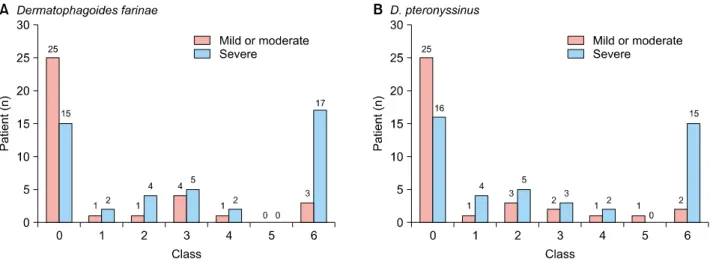
Fig. 2. Comparison of both house dust mite (HDM)-specific IgE levels according to atopic dermatitis (AD) severity. More patients with class 6 both HDM sensitization were found in the severe AD group.
Comparison of HDM-specific IgE levels according to AD severity
In our study, there was no significant relevance between both HDM sensitization and AD severity. However, more patients with class 6 both HDM sensitizations were found in the severe AD group (Fig. 2).
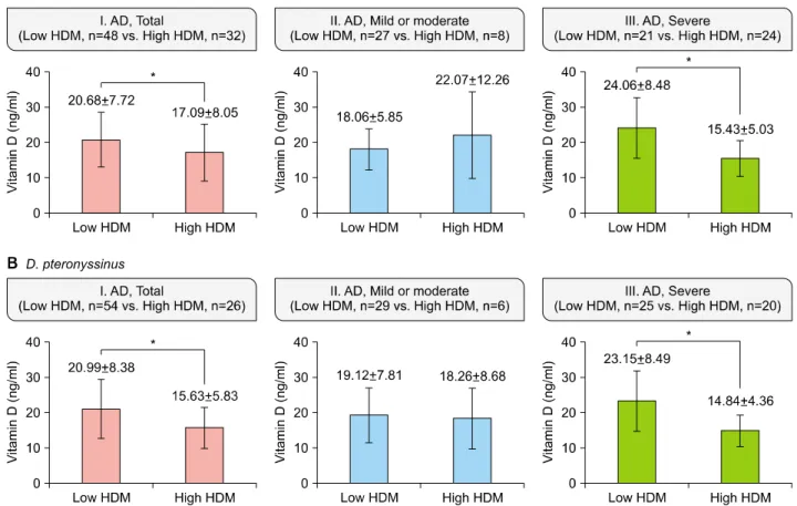
Differences in vitamin D levels according to HDM sensitization
In the severe AD group, significantly lower serum vitamin D levels were found in AD patients with high D. farinae sensitization (p<0.05). However, in AD patients with mild to moderate severity, serum vitamin D levels showed no significant difference between the low and high D. far- inae sensitization groups (p=0.77, Fig. 3A).
Results of D. pteronyssinus showed a similar tendency with those of D. farinae. In the severe AD group, high D.
pteronyssinus sensitization group had lower serum vita- min D levels with statistical significance (p<0.05). How- ever, there is no significant difference of vitamin D levels between the low and high D. pteronyssinus sensitization groups in mild or moderate AD patients (p=0.51, Fig. 3B).
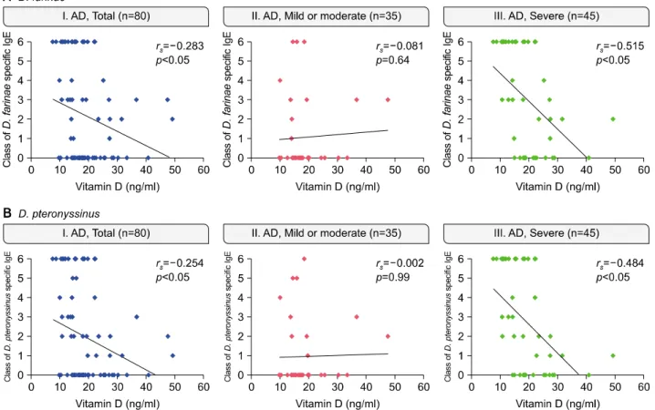
Relationship between vitamin D levels and HDM sensitization
In total AD patients, vitamin D levels showed a negative cor- relation with D. farinae sensitization (rs=−0.283, p<0.05).
In addition, there was a negative correlation between vita- min D levels and D. farinae sensitization in severe AD pa- tients with statistical significance (rs=−0.515, p<0.05).
However, no correlation was found between D. farinae
Fig. 3. Differences in vitamin D levels according to house dust mite (HDM) sensitization (*p<0.05). (A) In the severe atopic dermatitis (AD) group, significantly lower vitamin D levels were found in AD patients with high Dermatophagoides farinae sensitization (p<0.05).
However, vitamin D levels showed no significant difference between the low and high D. farinae sensitization groups in patients with the mild or moderate AD severity (p=0.77). (B) In the severe AD group, high D. pteronyssinus sensitization group had significantly lower serum vitamin D levels with statistical significance (p<0.05). However, in mild or moderate AD group, there is no significant difference of vitamin D levels between the low and high D. pteronyssinus sensitization groups (p=0.51).
sensitization and serum vitamin D levels in AD patients with mild or moderate severity (rs=−0.081, p=0.64; Fig.
4A).
Results of D. pteronyssinus showed the similar findings.
Vitamin D levels showed a negative correlation with D.
pteronyssinus sensitization with statistical significance in total AD patients (rs=−0.254, p<0.05). In severe AD pa- tients, serum vitamin D levels showed significantly neg- ative correlation with D. pteronyssinus sensitization (rs=
−0.484, p<0.05). However, there was no correlation be- tween serum vitamin D levels and D. pteronyssinus sensi- tization in mild or moderate AD patients group (rs=−0.002, p=0.99; Fig. 4B).
Relationship between vitamin D and total IgE levels There was a negative relationship between log trans- formed total IgE levels and serum vitamin D levels in total AD patients (R2=0.119, p<0.05). In addition, a negative correlation between log transformed total IgE levels and
serum vitamin D levels was found in the severe AD group (R2=0.234, p<0.05). However, an association between log transformed total IgE and serum vitamin D levels was not found in AD patients with mild or moderate severity (R2=0.107, p=0.06; Fig. 5).
DISCUSSION
There are many controversies about the association be- tween serum vitamin D levels and AD severity14. Overall, it seems that a predominance of reports points to a neg- ative association between serum vitamin D levels and AD severity4,10,15. However, in our study, serum vitamin D levels were not correlated with AD severity. Most AD pa- tients have increased serum IgE levels, which correlate with disease severity16. Our results also showed a positive correlation between serum IgE levels and AD severity.
Several previous studies reported a positive association between AD severity and HDM sensitization17-19. Howev-
Fig. 4. Relationship between vitamin D levels and house dust mite (HDM) sensitization. (A) Dermatophagoides farinae sensitization showed a negative correlation with vitamin D levels with statistical significance in severe atopic dermatitis (AD) patients (rs=−0.515, p<0.05). (B) There was a negative correlation between serum vitamin D levels and D. pteronyssinus sensitization in severe AD group (rs=−0.484, p<0.05).
Fig. 5. Relationship between vitamin D and total immunoglobulin E (IgE) levels. There was a negative correlation between log transformed total IgE levels and vitamin D levels with statistical significance in total (R2=0.119, p<0.05) and severe atopic dermatitis (AD) group (R2=0.234, p<0.05).
er, we were unable to replicate these findings in our study. We only found more patients with class 6 HDM sensitization in the severe AD group than in the mild or moderate groups.
A few studies examined the relationship between serum vi- tamin D levels and AD severity according to allergen
sensitization. Akan et al.20 showed a negative correlation between AD severity and serum vitamin D levels in the group with allergic sensitization but no correlation in the group without sensitization. This study suggested that vita- min D levels in children are correlated with AD severity but only in patients with allergic sensitizations16. However,
three domains of AD pathogenesis, including the immune system, antimicrobial defense mechanisms, and epidermal barrier integrity10. Specifically, regarding its immunomo- dulatory effects, vitamin D influences both the innate and adaptive immune system. Vitamin D has antimicrobial ef- fects related to macrophages and monocytes, enhancing chemotaxis and the phagocytic capabilities of innate im- mune cells22. In adaptive immunity, vitamin D functions in the differentiation and proliferation of T- and B-cells, leading to a shift from a proinflammatory to a more tolero- genic immune status23. Defected immune systems can in- fluence antimicrobial defense systems and epidermal bar- rier integrity. Therefore, in consideration of the function of vitamin D, there is a possibility that serum vitamin D lev- els are associated with HDM sensitization and ex- aggerated immune response to HDM. Our hypothesis is that low serum vitamin D levels lead to disturbed epi- dermal barrier function, immunologic dysregulation, and impaired cutaneous defense mechanism in patients with an atopic background. Patients with low serum vitamin D levels have an increased risk of HDM sensitization by in- creased penetration of HDM through broken skin barrier.
Then, high HDM sensitization may induce the aggravation of immunologic dysregulation and the development of se- vere AD. Findings of this study suggest that vitamin D lev- el may affect HDM sensitization.
Regarding the relationship between serum total IgE and vi- tamin D levels, a previous study suggested that lower se- rum vitamin D levels were associated with elevated serum IgE levels6,24. Our study also showed that serum vitamin D levels were negatively correlated with total IgE levels in the severe AD group.
The main limitation of this study is that we did not ac- count for clinical factors associated with serum vitamin D levels, such as individual outdoor activity and dietary hab- its that can affect vitamin D homeostasis. Seasonal varia- tions in vitamin D levels were not considered. In addition, we used the Rajka and Langeland score to evaluate AD se-
ACKNOWLEDGMENT
This research was supported by the Basic Science Research Program through the National Research Founda- tion of Korea funded by the Ministry of Education (NRF-2015R1D1A3A01016229 and 2014R1A5A2009242).
CONFLICTS OF INTEREST
The authors have nothing to disclose.
REFERENCES
1. Benson AA, Toh JA, Vernon N, Jariwala SP. The role of vitamin D in the immunopathogenesis of allergic skin diseases. Allergy 2012;67:296-301.
2. Searing DA, Zhang Y, Murphy JR, Hauk PJ, Goleva E, Leung DY. Decreased serum vitamin D levels in children with asthma are associated with increased corticosteroid use. J Allergy Clin Immunol 2010;125:995-1000.
3. Muehleisen B, Gallo RL. Vitamin D in allergic disease:
shedding light on a complex problem. J Allergy Clin Immunol 2013;131:324-329.
4. Peroni DG, Piacentini GL, Cametti E, Chinellato I, Boner AL. Correlation between serum 25-hydroxyvitamin D levels and severity of atopic dermatitis in children. Br J Dermatol 2011;164:1078-1082.
5. El Taieb MA, Fayed HM, Aly SS, Ibrahim AK. Assessment of serum 25-hydroxyvitamin d levels in children with atopic dermatitis: correlation with SCORAD index. Dermatitis 2013;24:296-301.
6. Wang SS, Hon KL, Kong AP, Pong HN, Wong GW, Leung TF. Vitamin D deficiency is associated with diagnosis and severity of childhood atopic dermatitis. Pediatr Allergy Immunol 2014;25:30-35.
7. Oren E, Banerji A, Camargo CA Jr. Vitamin D and atopic disorders in an obese population screened for vitamin D deficiency. J Allergy Clin Immunol 2008;121:533-534.
8. Lee SA, Hong S, Kim HJ, Lee SH, Yum HY. Correlation between serum vitamin d level and the severity of atopic dermatitis associated with food sensitization. Allergy
Asthma Immunol Res 2013;5:207-210.
9. Heimbeck I, Wjst M, Apfelbacher CJ. Low vitamin D serum level is inversely associated with eczema in children and adolescents in Germany. Allergy 2013;68:906-910.
10. Chiu YE, Havens PL, Siegel DH, Ali O, Wang T, Holland KE, et al. Serum 25-hydroxyvitamin D concentration does not correlate with atopic dermatitis severity. J Am Acad Dermatol 2013;69:40-46.
11. Searing DA, Leung DY. Vitamin D in atopic dermatitis, asthma and allergic diseases. Immunol Allergy Clin North Am 2010;30:397-409.
12. Rajka G, Langeland T. Grading of the severity of atopic dermatitis. Acta Derm Venereol Suppl (Stockh) 1989;144:
13-14.
13. Pajno GB, Caminiti L, Vita D, Barberio G, Salzano G, Lombardo F, et al. Sublingual immunotherapy in mite- sensitized children with atopic dermatitis: a randomized, double-blind, placebo-controlled study. J Allergy Clin Immunol 2007;120:164-170.
14. Han TY, Kong TS, Kim MH, Chae JD, Lee JH, Son SJ.
Vitamin D status and its association with the SCORAD score and serum LL-37 level in Korean adults and children with atopic dermatitis. Ann Dermatol 2015;27:10-14.
15. Mesquita Kde C, Igreja AC, Costa IM. Atopic dermatitis and vitamin D: facts and controversies. An Bras Dermatol 2013;88:945-953.
16. Liu FT, Goodarzi H, Chen HY. IgE, mast cells, and eosinophils in atopic dermatitis. Clin Rev Allergy Immunol 2011;41:298-310.
17. Park M, Lee HY, Lee SI, Kim J, Ahn K. Positive conversion of specific IgE against house dust mite in children with atopic dermatitis under 24 months of age. Allergy Asthma Respir Dis 2013;1:350-356.
18. Kim J, Lee S, Woo SY, Han Y, Lee JH, Lee IY, et al. The indoor level of house dust mite allergen is associated with severity of atopic dermatitis in children. J Korean Med Sci 2013;28:74-79.
19. Kimura M, Tsuruta S, Yoshida T. Correlation of house dust mite-specific lymphocyte proliferation with IL-5 production, eosinophilia, and the severity of symptoms in infants with atopic dermatitis. J Allergy Clin Immunol 1998;101:84-89.
20. Akan A, Azkur D, Ginis T, Toyran M, Kaya A, Vezir E, et al.
Vitamin D level in children is correlated with severity of atopic dermatitis but only in patients with allergic sensitizations. Pediatr Dermatol 2013;30:359-363.
21. Baek JH, Shin YH, Chung IH, Kim HJ, Yoo EG, Yoon JW, et al. The link between serum vitamin D level, sensitization to food allergens, and the severity of atopic dermatitis in infancy. J Pediatr 2014;165:849-854.e1.
22. Baeke F, Takiishi T, Korf H, Gysemans C, Mathieu C.
Vitamin D: modulator of the immune system. Curr Opin Pharmacol 2010;10:482-496.
23. Provvedini DM, Tsoukas CD, Deftos LJ, Manolagas SC.
1,25-dihydroxyvitamin D3 receptors in human leukocytes.
Science 1983;221:1181-1183.
24. Ehlayel MS, Bener A, Sabbah A. Is high prevalence of vitamin D deficiency evidence for asthma and allergy risks?
Eur Ann Allergy Clin Immunol 2011;43:81-88.
25. Rullo VE, Segato A, Kirsh A, Sole D. Severity scoring of atopic dermatitis: a comparison of two scoring systems.
Allergol Immunopathol (Madr) 2008;36:205-211.
26. Gånemo A, Svensson Å, Svedman C, Grönberg BM, Johansson AC, Wahlgren CF. Usefulness of Rajka &
Langeland eczema severity score in clinical practice. Acta Derm Venereol 2016;96:521-524.