Copyright © 2014 The Korean Brain Tumor Society and The Korean Society for Neuro-Oncology 39
INTRODUCTION
Intracranial dermoid cysts are rare congenital lesions that have been reported to account for 0.04 to 0.7 percent of all in- tracranial tumors [1]. Intracranial dermoid cysts occur at the midline strand, and two thirds of these tumors are found at the posterior fossa [2,3]. Dermoid cysts are commonly located at intradural portions of the parasellar and midline frontobasal regions [1]; however, they are rarely found in the extradural location. In the present study, we report an intracranial extra- dural dermoid cyst at the lateral sphenoid ridge accompanied by bone erosion.
CASE REPORT
History and examinationA 53-year-old woman was admitted because of progressive headache, nausea, and dizziness lasting one month. On neuro- logical examinations, no deficit was detected. On computed to-
Extradural Dermoid Cyst Located in the Lateral Sphenoid Ridge
Seok-Jin Ko, Kyung-Jae Park, Dong-Hyuk Park, Shin-Hyuk Kang
Department of Neurosurgery, Korea University College of Medicine, Seoul, Korea
Received February 13, 2014 Revised March 31, 2014 Accepted April 1, 2014 Correspondence Shin-Hyuk Kang
Department of Neurosurgery, Korea University College of Medicine, 73 Inchon-ro, Seongbuk-gu, Seoul 136-075, Korea Tel: +82-2-920-5391 Fax: +82-2-929-0629 E-mail: hermes23@kumc.or.kr
Dermoid cysts are rare congenital tumors that occur primarily at the midline at a characteristic intradu- ral location. However, dermoid cysts located at extradural and lateral regions have been rarely reported until now. In the present study, the authors demonstrate the unusual instance of an intracranial extra- dural dermoid cyst at the lateral sphenoid ridge. A 53-year-old woman admitted because of progres- sive headache and dizziness. The patient had no neurologic deficits, and magnetic resonance imaging with no contrast enhancement revealed a mass at the right sphenoid ridge. The mass was accompa- nied with sphenoid bone erosion visible on computed tomography. The patient underwent right pteri- onal craniotomy, and the tumor including the capsule was totally resected. Presence of a dermoid cyst was confirmed with histopathological examination. The patient had no complications during the post- operative period. This study suggests that dermoid cyst should be considered for differential diagnosis of extradural and lateral intracranial masses.
Key Words Dermoid cyst; Extradura; Bone erosion; Brain mass.
mography (CT) scan, a low attenuating lobulated mass was observed at the right sphenoid ridge, and the lesion was ac- companied by bone erosion. There was no extracranial com- munication with the scalp (Fig. 1). Magnetic resonance imag- ing (MRI) of the brain demonstrated a mass of 2.2×1.2×2.9 cm in size, low to iso signal intensity on T2-weighted images, and high signal intensity on T1-weighted images with no gad- olinium enhancement. And cerebrospinal fluid (CSF) space widening around the mass was not found (Fig. 2).
Operative findings
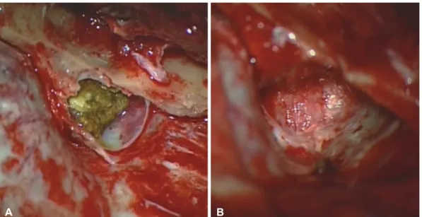
Right pterional craniotomy was performed under general anesthesia. A yellowish and sticky extradural mass containing the capsule was observed at the sphenoidal ridge. The content of the mass was soft and contained desquamated cell debris and hairs. Total resection including the capsule was performed, and the dura was intact after the removal of the mass (Fig. 3).
Postoperative course
During the postoperative period, the patient had no specific neurologic deficit. Total removal of the mass was confirmed on postoperative MRI. Pathological examination revealed squamous epithelial lining of the cyst and some keratinous CASE REPORT Brain Tumor Res Treat 2014;2(1):39-42 / pISSN 2288-2405 / eISSN 2288-2413
http://dx.doi.org/10.14791/btrt.2014.2.1.39
This is an Open Access article distributed under the terms of the Creative Commons Attribution Non-Commercial License (http://creativecommons.org/licenses/by-nc/3.0) which permits unrestricted non-commercial use, distribution, and reproduction in any medium, provided the original work is properly cited.
40 Brain Tumor Res Treat 2014;2(1):39-42 Lateral Temporal Extradural Dermoid Cyst
contents (Fig. 4), and these findings were consistent with a dermoid tumor. The patient was discharged nine days after operation.
DISCUSSION
Dermoid cysts originate from an embryonic ectodermal cell that is involved in the process of primary neurulation during neural tube closure [4-6]. Therefore, the contents of dermoid cysts include elements of the dermis such as hair, hair follicles, apocrine, sebaceous, or sweat gland. In the current study, the authors found hairs during tumor resection and keratinous materials in histopathologic findings.
Dermoid cysts primarily occur in intradural regions; how- ever, rare interdural dermoid cysts have also been reported [7].
In addition, although the posterior fossa is the main location
for dermoid cysts, a few intradural dermoid cysts originating at the temporal skull base has been reported [1]. Extradural dermoid cysts have been observed in the posterior fossa, dip- loe of anterior fontanelle, and orbital region in children [1,5,8].
Only one case of a extradural dermoid cyst has been reported in an adult that was located at the paramedian petrous apex accompanied by osseous erosion [9]. And extradural dermoid cysts at the lateral temporal base have been rarely reported so far. Herein, this study has important implications because it is the infrequent case of an intracranial dermoid cyst that is ex- tradural and located at the lateral sphenoid ridge.
Martínez-Lage et al. [5] demonstrated extradural dermoid cysts of the posterior fossa and hypothesized that dermoid cysts of the midline may originate from the cutaneous ecto- derm that entered during invagination of dura during devel- opment of the falx and tentorium. This hypothesis is difficult
A B
Fig. 1. Computed tomography: soft tissue window (A) and bone window (B) demonstrating a hypodense lesion with bone erosion at right sphenoid ridge, but not communicating with scalp.
Fig. 2. Magnetic resonance imaging showing an expansile and multilobulated extraaxial mass on right lateral sphenoid ridge with high sig- nal intensity on T1-weighted (A), low to iso signal intensity on T2-weighted (B) and no enhancement on contrast enhancing T1-weighted se- quence (C). There was not a cerebrospinal fluid space widening around the mass.
A B C
SJ Ko et al.
41 to apply to the origin of a lateral extradural dermoid cyst be-
cause there is no dural invagination at the lateral temporal re- gion. Two theories for the origin of cells of rare lateral and in- tradural dermoid cysts are presented here. It has been suggested that dermoid cysts may originate from multipotent embryonic cells or from translocation of epithelial cells that have migrated from the otic vesicles or developing neurovasculature [4]. Al- though multipotent stem cells were not found in the dura, the possibility of proliferation of these cells cannot be excluded in the formation of extradural dermoid cyst. In addition, during the formation of extradural neurovasculature, epithelial cell migration may also occur. The origin of dermoid cysts is still unknown because multipotent stem cell proliferation or epi- thelial cell migration occurs in cases of intradural dermoid
cysts, and there are only a few reports on these cases [4,6]. In this study, the dermoid cyst could be completely detached from the dura, and no communication with the scalp was found de- spite its association with the temporal bone erosion on CT scan. The cranium is formed from peripheral mesenchyme during brain development. Although both mesodermal and ectodermal cells are involved in this process, the cartilaginous part of the cranium is formed by the fusion of several cartilag- es originated from ectodermal cells [10]. Therefore, the possi- bility that an extradural and lateral dermoid cyst may originate from remnants of ectodermal cells in the skull should be care- fully evaluated.
Dermoid cysts are mainly considered hypodense because of fat content with a value of around -100 Hounsfield unit on CT
A B
Fig. 3. Intraoperative findings: dermoid contents including capsule at epidural space of right sphenoid ridge (A). Totally resected mass in- cluding capsule and intact dura (B).
A B
Fig. 4. Photomicrographs of a portion of cyst wall (A) and contents (B) demonstrating stratified squamous epithelium (arrow) and keratinous materials (arrowhead) (H&E, original magnification ×400).
42 Brain Tumor Res Treat 2014;2(1):39-42 Lateral Temporal Extradural Dermoid Cyst
scan [1,9]. In addition, capsular calcification can be observed, and most dermoid cysts do not show contrast enhancement.
Dermoid cysts can be detected clearly by high signal intensity on T1-weighted MRI and by low to high signal intensity on T2- weighted images because of the amount of fat present. In some cases of a dermoid cyst, it is difficult to determine whether a lesion is intradural or extradural. Skeletal anomalies such as skull erosion may be a characteristic finding in extradural der- moid cysts [9]. In this study, sphenoid bone erosion was iden- tified on CT scan. In addition, CSF space widening around the mass was not found on MRI. Together with these findings, ex- tradural dermoid cyst should be ruled out for differential diag- nosis when bone erosion and CSF space widening are consid- ered.
As a treatment modality, total removal of a dermoid tumor is desirable. However, in the case of intramedullary dermoid cyst [6] which exists in vital structures, attention toward gross total resection is needed because of the risk of a neurological disorder after surgery. Neuromonitoring is sometimes required during surgery. In this case, the tumor could be easily detached from the surrounding structure, and therefore a total removal of the tumor was possible.
Conclusion
This study provides the unusual description of an extradural dermoid cyst accompanied by bone erosion located in the lat- eral sphenoid ridge of the skull. Although various mechanisms can be involved, the origin of lateral and extradural dermoid cyst remains to be determined. In addition, bone erosion and
CSF space widening should be considered for the possibility of extradural dermoid cysts when doing differential diagnosis of intracranial mass.
Conflicts of Interest
The authors have no financial conflicts of interest.
REFERENCES
1. Rubin G, Scienza R, Pasqualin A, Rosta L, Da Pian R. Craniocerebral epidermoids and dermoids. A review of 44 cases. Acta Neurochir (Wien) 1989;97:1-16.
2. Chu W, Feng H, Zhu G, Ye X, Lin J. Intradural dermoid cyst located on the ventral surface of the brainstem in a child. Surg Neurol 2008;
70:531-5.
3. Guidetti B, Gagliardi FM. Epidermoid and dermoid cysts. Clinical evaluation and late surgical results. J Neurosurg 1977;47:12-8.
4. Eekhof JL, Thomeer RT, Bots GT. Epidermoid tumor in the lateral ventricle. Surg Neurol 1985;23:189-92.
5. Martínez-Lage JF, Ramos J, Puche A, Poza M. Extradural dermoid tu- mours of the posterior fossa. Arch Dis Child 1997;77:427-30.
6. Park JG, Babu R, Kranz PG, McLendon RE, Adamson C. Intraaxial dermoid cyst of the medulla. J Neurosurg 2013;119:442-5.
7. Nakagawa K, Ohno K, Nojiri T, Hirakawa K. [Interdural dermoid cyst of the cavernous sinus presenting with oculomotor palsy: case report].
No Shinkei Geka 1997;25:847-51.
8. Tateshima S, Numoto RT, Abe S, Yasue M, Abe T. Rapidly enlarging dermoid cyst over the anterior fontanel: a case report and review of the literature. Childs Nerv Syst 2000;16:875-8.
9. Ammirati M, Delgado M, Slone HW, Ray-Chaudhury A. Extradural dermoid tumor of the petrous apex. Case report. J Neurosurg 2007;
107:426-9.
10. Moore KL, Persaud TVN, Torchia MG. The developing human: clini- cally oriented embryology. 7th ed. Philadelphia: Elsevier Saunders Co;
2008. p.389-90.