소아 상완골 원위부 골단의 골절 및 분리
전체 글
수치
관련 문서
1 John Owen, Justification by Faith Alone, in The Works of John Owen, ed. John Bolt, trans. Scott Clark, "Do This and Live: Christ's Active Obedience as the
The greater tubercle is palpable on the line from the lateral epicondyle of the distal humerus in the direction of the humeral longitudianl axis and just below the acromion
limit the crack growth by increasing # of site of crazing or
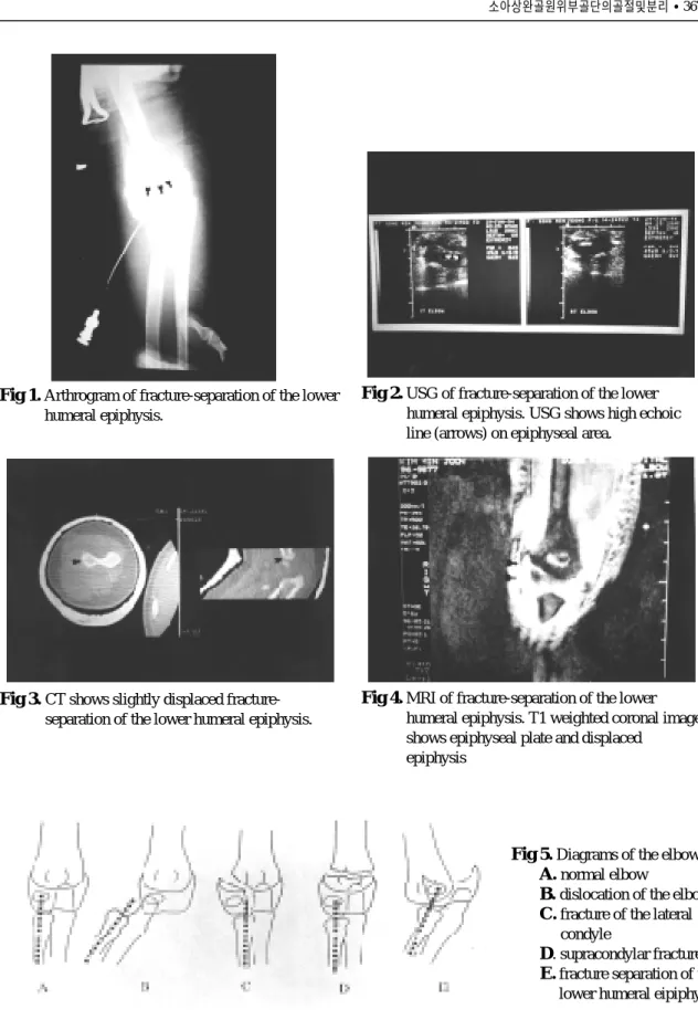
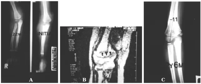
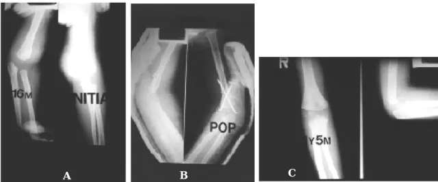
contralateral extremity except 3 patients who had comminuted fracture at the last follow-up. These three cases showed an angular deformity of less than 5 degrees and
Through this result, it was confirmed that brittle fracture did not occur in high Mn steel and ductile fracture occurred in spite of cryogenic
Purpose: Calcaneal fracture is a rare fracture, which accounts for about 2% of all fractures, but is one of the most common fractures in the ankle bone.. There is
This study investigated the loading rate effect on the fracture resistance under cyclic loading conditions to clearly understand the fracture behavior of
Also, the quasi-infraversion is larger for the inferior safe insertion limit direction (Fig. In practical design of a screw guiding device, targeted safe angles can
