www.jkfas.org 나이다.1) 거골은 독특한 해부학적 및 혈류 공급 구조로 외상으로 인한 골절 발생 시 정확한 정복 및 고정이 어려우며, 무혈성 괴사, 지연 및 불유합, 부정 유합, 감염, 외상 후 관절염, 골 소실 등 다양 한 합병증이 비교적 높게 보고되고 있다.2) 거골은 주위 거골하 관 절, 횡족근관절, 족관절 복합체를 연결하는 가장 중요한 뼈로서 손 상 시 심각한 문제를 초래할 수 있다.3) 따라서 거골의 해부학적 구 조를 이해하고 계측하여 손상 발생 시 이를 바탕으로 한 정확한 해 부학적 정복 및 적절한 고정물의 삽입이 요구되나 사체 등을 바탕 으로 한 한국인의 거골 실측에 대한 해부학적 연구는 미미한 실정 이다. 주로 유럽인 및 북미인들을 대상으로 개발된 기존의 고정물 모델이 한국인의 거골 골절의 치료에 사용되기에는 크기가 크거나
서 론
거골은 다양한 주위 뼈들과의 관절성으로 인해 “족부의 보편적 인 관절(the universal joint of the foot)”로 불려질 정도로 해부학 적 및 생역학적으로 중요한 역할을 담당하는 족부의 뼈 가운데 하
This is an Open Access article distributed under the terms of the Creative Commons Attribution Non-Commercial License (http://creativecommons.org/licenses/CC
by-nc/4.0) which permits unrestricted non-commercial use, distribution, and reproduction in any medium, provided the original work is properly cited.
Copyright 2016 Korean Foot and Ankle Society. All rights reserved.ⓒ
Purpose: To investigate the measured values of the talus in Koreans.
Materials and Methods: We measured 88 tali from 44 cadavers that have been donated between December 2012 and December 2015.
Of the cadavers, 27 were male and 17 were female. Their mean age was 73 years. The length and width of the talus were measured using a digital goniometer and vernier caliper.
Results: The values of cadaveric measurement, mean maximal width and length, width and length of the dome anterior, width and
length of the posterior facet, height and length of the trochlear medial facet, and height and length of the trochlear lateral facet were 43.6±2.6 mm, 56.5±3.3 mm, 32.5±2.0 mm, 42.2±2.7 mm, 22.2±2.2 mm, 34.7±2.0 mm, 15.3±1.3 mm, 33.3±2.9 mm, 25.3±3.3 mm, and 30.8±2.4 mm for men and 38.9±1.6 mm, 53.6±2.4 mm, 27.9±2.1 mm, 37.4±3.2 mm, 20.6±0.8 mm, 31.9±1.2 mm, 13.6±2.6 mm, 28.4±2.5 mm, 24.9±2.1 mm, and 28.9±1.4 mm for women, respectively. The size of the talus showed an accuracy of 86% when anteroposterior diameter was greater than 59 mm. A difference in the size of the right and left talus was not observed. The mean inclination and declina- tion angles were 24.4o±4.2o and 28.2o±5.4o for men, and 24.6o±3.6o and 24.7o±6.7o for women (p=0.980, p=0.018), respectively, at least 15o, which showed a big difference for every object up to 37o.Conclusion: This paper, to the best of our knowledge, is the first study to measure the talus in Koreans. There were differences by gen-
der and ethnicity in the in measured talus values. The measurements were smaller than European-Americans and greater than Japanese.Key Words: Talus, Cadaver, Size, Angle, Korean
한국인 사체에서의 정상 거골의 실측
하동준*, 곽희철, 김전교, 김정한, 이창락, 김영준, 이정한
†, 하병호, 김의철
인제대학교 부산백병원 정형외과, *메리놀병원 정형외과, †인제대학교 부산백병원 마취통증의학과
The Measurement of Normal Talus in Korean Cadaver
Dong-Jun Ha*, Heui-Chul Gwak, Jeon-Gyo Kim, Jung-Han Kim, Chang-Rak Lee, Young-Jun Kim, Jeong-Han Lee
†, Byung-Ho Ha, Ui-Cheol Kim
Department of Orthopedic Surgery, Inje University Busan Paik Hospital,
*Department of Orthopedic Surgery, Maryknoll Medical Center,
†Department of Anesthesiology and Pain Medicine, Inje University Busan Paik Hospital, Busan, Korea
Received August 15, 2016 Revised October 9, 2016 Accepted October 18, 2016 Corresponding Author: Jeon-Gyo Kim
Department of Orthopedic Surgery, Inje University Busan Paik Hospital, 75 Bokji- ro, Busanjin-gu, Busan 47392, Korea
Tel: 82-51-890-6996, Fax: 82-51-892-6619, E-mail: bluewhisle@gmail.com Financial support: None.
Conflict of interest: None.
였으며, 평균 73세였다. 남성이 27구로 평균 73세(53∼91세)였으 며, 여성은 17구로 평균 72세(50∼85세)였다. 44구의 사체에서 박 리한 총 88개(우측 43개, 좌측 40개, 우측 부패 1개, 좌측 부패 4개) 모양이 적합하지 않은 경우도 발생한다. 성별에 의한 한국인 거골
의 해부학적 계측치에 있어서도 차이가 있을 수 있으나 이에 대한 기존 해부학적 연구의 보고도 비교적 적다.
이에 본 연구는 한국인 사체의 거골 계측을 시행하면서 족관절 및 거골하 관절면의 다양성을 관찰하고, 성별 비교 및 기존의 외국 보고와의 비교를 통하여 한국인의 거골 실측치를 보고하고자 한 다.
대상 및 방법
본 연구는 본 병원의 임상연구윤리위원회의 면제 승인 이후 진 행되었다. 2012년 12월부터 2015년 12월까지의 기간 중 본 대학교 의과대학 해부학교실에 기증된 총 44구의 한국인 사체를 대상으로 하였다. 병력을 조회하여 족부 및 족관절 부위의 외상력이 없음을 확인하였으며, 3차원 C-arm (Siemens, Berlin, Germany)을 통하여 방사선학적으로 골성 병변이 발견되거나 박리 시 부패가 심한 경 우는 분석에서 제외하였다. 대상군의 연령은 50세에서 91세 사이
A B
Figure 4. (A, B) Actual measurement of head for navicular width (longitudinal length from medial edge of talar head to lateral edge of talar head) and length (longitudinal length from superior edge of talar head to inferior edge of talar head) of talus in cadaver.
Figure 2. Actual measurement of anterior width (longitudinal length from medial edge of talar dome to lateral edge of talar dome surface at the highest) and length (longitudinal length from anterior edge of talar dome to posterior edge of talar dome surface) of talus in cadaver.
Figure 1. Actual measurement of total width (longitudinal length from lateral process to talus head) and total length (longitudinal length from talus head to lateral tubercle) of talus in cadaver.
Figure 3. Actual measurement of posterior facet width (longitudinal length from medial edge to lateral edge of posterior facet at posterior calcaneal articular facet surface) and length (longitudinal length from anterior edge to posterior edge of posterior facet at posterior calcaneal articular facet surface) of talus in cadaver.
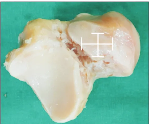
www.jkfas.org 서 가장 긴 거리를 0.1 mm 단위로 측정하였다. 거골의 경사각 및 편각의 측정 시 전자측각기(digital goniometer; Bluebird, Seoul, Korea) (Fig. 11B)를 사용하여 0.1도 단위로 측정하였다. 경사각은 내측 원개면의 장축과 전방 관절면의 수선이 이루는 각도를 측정 하였고, 편각의 경우에는 거골 원개의 장축과 거골 목의 외측면이 이루는 각도를 측정하였다.
모든 측정은 2인의 정형외과의(B.H.H, J.G.K.)에 의하여 이루어 졌으며, 측정자 간 측정오차를 최소화하기 위해 한 관찰자(J.G.K.) 가 모든 길이와 각도에 대하여 2회 측정하였고, 전체 변수들의 평 균값을 산출하여 측정값으로 정하였다. 측정된 각 변수들에 대해 정규성 검정을 실시하였고, 정규 분포에 따른 값들에 대해서는 독 립 t검정을 시행하였으며, 정규 분포를 따르지 않은 비모수인 변수 의 거골을 측정 대상으로 하였다.
측정한 지표로는 거골 전체 전후 직경과 좌우 직경(Fig. 1), 거골 원개 전후 직경과 좌우 직경(Fig. 2), 거골후방 관절면 전후 직경과 좌우 직경(Fig. 3), 거골두의 전후 직경과 좌우 직경(Fig. 4), 거골 전방 관절면 전후 직경과 좌우 직경(Fig. 5), 거골 중간 관절면 전후 직경과 좌우 직경(Fig. 6), 원개 내측 관절면 길이와 높이(Fig. 7), 거골원개 외측 관절면 길이와 높이(Fig. 8)를 측정하였고, 거골 경 사각(inclination angle) (Fig. 9), 거골 편각(declination angle) (Fig.
10)을 측정하였다. 길이는 관절의 경우 줄 자를 이용해 가장 긴 거리를 측정하였으며, 그 외 길이와 폭의 경우 길이 지표의 측정 시 측경양각기(vernier calipers; Mitutoyo Philosophy, Kawasaki, Japan) (Fig. 11A)를 사용하여 거골 외측돌기의 첨부를 문 상태에
Figure 7. Actual measurement of trochlear medial facet height (longitu- dinal length from inferior edge of trochlear for medial malleolus to su- perior edge of trochlear for medial malleolus) and length (longitudinal length from anterior edge of trochlear for medial malleolus to posterior edge of trochlear for medial malleolus) of talus in cadaver.
Figure 8. Actual measurement of trochlear lateral facet height (longi- tudinal length from inferior edge of trochlear for lateral malleolus to superior edge of trochlear for lateral malleolus) and length (longitudinal length from anterior edge of trochlear for lateral malleolus to posterior edge of trochlear for lateral malleolus) of talus in cadaver.
Figure 5. Actual measurement of anterior facet width (longitudinal length from anterior edge of anterior calcaneal articular facet to poste- rior edge of anterior calcaneal articular facet) and length (longitudinal length from anterior edge of anterior calcaneal articular facet to poste- rior edge of anterior calcaneal articular facet) of talus in cadaver.
Figure 6. Actual measurement of middle facet width (longitudinal length from medial edge of middle calcaneal articular facet to lateral edge of middle calcaneal articular facet) and length (longitudinal length from anterior edge of middle calcaneal articular facet to poste- rior edge of middle calcaneal articular facet) of talus in cadaver.
방 관절면 좌우 직경 남성 22.2±2.2 mm, 여성 20.6±0.8 mm, 전 후 직경 남성 34.7±2.0 mm, 여성 31.9±1.2 mm, 거골 중간 관절 면 좌우 직경 남성 13.8±0.9 mm, 여성 13.9±1.0 mm, 전후 직경 은 남성 22.0±2.1 mm, 여성 21.8±1.1 mm, 거골 원개 내측 관 절면 높이 남성 15.3±1.3 mm, 여성 13.6±2.6 mm, 길이는 남성 33.3±2.9 mm, 여성 28.4±2.5 mm, 거골 원개 외측 관절면 높이 남성 25.3±3.3 mm, 여성 24.9±2.1 mm, 길이는 남성 30.8±2.4 mm, 여성 28.9±1.4 mm로 측정되었고(Tables 1∼3), 전방 관절면 의 형태에 있어서는 종골 관절면과 만나는 거골 목의 중앙 하부 전 방 내외측 관절면이 후방으로 두드러지게 연결된 경우가 전체 44 구의 사체 가운데 23구에서 관찰되었다. 경사각은 남성 24.4o± 4.2o, 여성 24.6o±3.6o, 편각의 경우 남성 28.2o±5.4o, 여성 24.7o± 6.7o, 최소 15o, 최대 37o로 개체마다 큰 차이를 보였다(Tables 1∼
3).
본 연구는 거골의 계측에 있어 남녀 차이를 보여주고 있으며, 좌 우 방향에 따른 거골의 계측에 있어 각 항목별 크기 차이가 거의 없었다. 남녀 계측의 차이가 있는 경우 대부분 통계적으로 유의하 들에 대해서는 Mann Whitney U test를 이용하여 분석하였다. 유의
수준은 p<0.05일 때 통계적으로 유의한 것으로 판단하였다. 모든 통계적인 검정은 IBM SPSS version 22.0 (IBM Co., Armonk, NY, USA) 프로그램을 통해 분석을 시행하였다. 2인의 측정자 간/측정 자 내 신뢰도 분석을 위하여 interclass/intraclass correlation 분석을 시행하였다.
결 과
측정한 지표로는 평균±표준편차로 표시하여 거골 전체 좌우 직 경 남성 43.6±2.6 mm, 여성 38.9±1.6 mm, 전후 직경 남성 56.5
±3.3 mm, 여성 53.6±2.4 mm, 거골 원개 좌우 직경 남성 32.5±
2.0 mm, 여성 27.9±2.1 mm, 전후 직경 남성 42.2±2.7 mm, 여성 37.4±3.2 mm, 거골 전방 관절면 좌우 직경 남성 13.6±2.0 mm, 여성 13.8±1.4 mm, 전후 직경 남성 33.7±1.9 mm, 여성 29.3±
3.9 mm, 거골두의 좌우 직경 남성 13.5±1.7 mm, 여성 13.7±0.7 mm, 전후 직경 남성 20.4±2.3 mm, 여성 21.0±1.4 mm, 거골 후
A B
Figure 11. (A) Actual measurements of talus were done in cadaver by vernier calipers (Mitutoyo Philosophy, Kawasaki, Japan). (B) Actual measure- ments of talus were done in cadaver by digital goniometer (Bluebird, Seoul, Korea).
Figure 9. Actual measurement of inclination angle (γ; angle of between longitudinal line [α] of medial trochlear and perpendicular line of talar head [β]) of talus in cadaver.
Figure 10. Actual measurement of declination angle (γ; angle of be- tween longitudinal line of superior trochlear [α] and longitudinal line of talar neck [β]) of talus in cadaver.
www.jkfas.org 목 관절을 통해서 족부로 전달하는 중요한 골격 구조로, 크게 몸 통, 목, 머리의 세 부위로 구성되어 있다. 몸통은 위쪽면으로 원개, 후방 아랫면으로 종골 관절면으로 구성되어 있고, 목은 거골의 앞 쪽에 위치하면서 몸통과 머리 사이에 위치한다.4)
서양인의 거골 형태에 있어 Williams 등5)은 160구의 백인 미국 인 사체를 대상으로 연구하여 전후 직경 남성 평균 61 mm, 여성 평균 54 mm, 좌우 폭 남성 44 mm, 여성 39 mm로 보고하고 있으 게 나타났다. Interclass와 Intraclass correlation에 있어서는 대부
분 지표에 있어 0.95 이상의 비교적 높은 수준으로 나타났다(Table 4).
고 찰
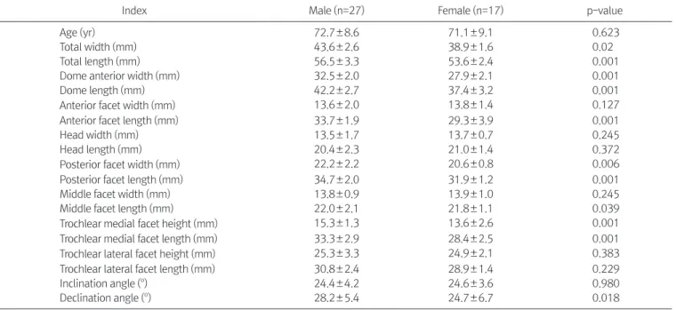
거골은 7개의 tarsal bone 중 하나로 신체의 체중을 지지하고 발 Table 1. Comparison Measurements between Male and Female
Index Male (n=27) Female (n=17) p-value
Age (yr) 72.7±8.6 71.1±9.1 0.623
Total width (mm) 43.6±2.6 38.9±1.6 0.02
Total length (mm) 56.5±3.3 53.6±2.4 0.001
Dome anterior width (mm) 32.5±2.0 27.9±2.1 0.001
Dome length (mm) 42.2±2.7 37.4±3.2 0.001
Anteriorfacet width (mm) 13.6±2.0 13.8±1.4 0.127
Anterior facet length (mm) 33.7±1.9 29.3±3.9 0.001
Head width (mm) 13.5±1.7 13.7±0.7 0.245
Head length (mm) 20.4±2.3 21.0±1.4 0.372
Posteriorfacet width (mm) 22.2±2.2 20.6±0.8 0.006
Posterior facet length (mm) 34.7±2.0 31.9±1.2 0.001
Middle facet width (mm) 13.8±0.9 13.9±1.0 0.245
Middle facet length (mm) 22.0±2.1 21.8±1.1 0.039
Trochlear medialfacet height (mm) 15.3±1.3 13.6±2.6 0.001
Trochlear medial facet length (mm) 33.3±2.9 28.4±2.5 0.001
Trochlear lateralfacet height (mm) 25.3±3.3 24.9±2.1 0.383
Trochlear lateral facet length (mm) 30.8±2.4 28.9±1.4 0.229
Inclination angle (o) 24.4±4.2 24.6±3.6 0.980
Declination angle (o) 28.2±5.4 24.7±6.7 0.018
Values are presented as mean±standard deviation.
Table 2. Comparison Measurements in Males between Left Talus and Right Talus
Index Right Left p-value
Age (yr) 72.5±8.3 72.9±9.1 0.648
Total width (mm) 43.5±2.6 43.8±2.5 0.399
Total length (mm) 56.6±3.5 56.5±3.1 0.684
Dome anteriorwidth (mm) 32.5±2.1 32.6±1.9 0.599
Dome length (mm) 42.5±2.6 41.9±2.8 0.003
Anteriorfacet width (mm) 13.4±2.0 13.9±2.0 0.676
Anterior facet length (mm) 13.4±2.0 13.9±2.0 0.499
Head width (mm) 13.5±1.8 13.7±1.6 0.883
Head length (mm) 20.4±2.4 21.0±2.4 0.773
Posterior facet width (mm) 22.2±2.4 22.1±2.0 0.865
Posterior facet length (mm) 34.7±2.1 34.7±2.0 0.995
Middle facet width (mm) 13.9±1.0 13.8±0.9 0.839
Middle facet length (mm) 22.0±2.7 22.3±1.1 0.901
Trochlear medialfacet height (mm) 15.4±1.3 15.2±1.2 0.803
Trochlear medialfacet length (mm) 33.3±3.1 33.2±2.9 0.782
Trochlear lateralfacet height (mm) 25.2±3.4 25.5±3.3 0.801
Trochlear lateralfacet length (mm) 30.4±2.5 31.2±2.2 0.796
Inclination angle (o) 23.5±4.4 25.3±3.9 0.005
Declination angle (o) 27.3±5.2 28.9±5.6 0.037
Values are presented as mean±standard deviation.
골 크기는 전후 길이 남성 57 mm, 여성 54 mm, 폭 남성 44 mm, 여 성 39 mm로 유럽인에 비해 전후 길이는 다소 작은 값으로 나타났 으며 폭은 비슷한 값을 나타냈고, 일본인에 비해서는 전후 길이 및 폭 모두 큰 값을 나타냈다. 이러한 차이는 2009년 경제협력 개발 기구(Organization for Economic Co-operation and Development, OECD) 발표 국가별 신장 크기에서 한국인 남성 173.8 cm, 이탈리 아 남성 176 cm, 일본 남성 170.7 cm로 나타난 것과 관련해 신장과 연관성이 있는 것으로 생각된다. 남녀 차이에 있어서는 2015년 발 표한 국민건강보험공단 가입자 통계자료에서 남성 170.5 cm, 여성 156.9 cm로 나타난 것과 관련해 신장과의 연관성이 있을 것으로 판단되고, 기존의 외국의 논문에 있어서 신체지수와 계측치를 같 이 발표한 자료가 없어 직접적인 비교의 어려움이 있다.
거골 골절은 전체 골절의 0.1%로 발생 빈도는 떨어지나 근육이 나 건의 부착이 없고 관절면이 전체의 60%를 차지하여 적절한 혈 류공급을 제공할 수 있는 면적이 적고 역행성으로 골두에서 체부 로 공급하기 때문에 전위 골절 발생 시 무혈성 괴사가 발생할 확률 이 높고, 불유합으로 인해 외상성 관절염 등이 잘 발생한다. 거골 골절 중 목의 골절은 전체의 50%를 차지하며 치료가 어렵고 결과 가 좋지 않다. 조기에 정확한 정복과 내고정, 적극적인 재활이 강 조되고 있으며, 현재 금속나사가 널리 사용되고 있으나 골절선에 직각으로 삽입하기가 어려우며 이로 인해 나사못 고정술 후 거골 목의 단축, 거골 체부의 붕괴를 일으키는 경우가 있다.4) 따라서 거 골 목의 골절 시 내고정물을 이용해 내고정술을 시행하며 경사각 과 편각의 값을 통해 복원각도를 정하는 데 도움이 될 수 있다. 거 골의 경사각 및 편각에 대해 국내에는 이에 대한 연구 자료는 없는 며, 서양인 이외에 Sakaue6)는 143구의 일본인 사체 연구에 있어 전
후 직경 남성 평균 51 mm, 여성 평균 46 mm, 좌우 폭은 남성 평균 41 mm, 여성 평균 37 mm로 보고하고 있다. Gualdi-Russo7)는 현 대 북부 이탈리아인 남성 62명, 여성 56명을 대상으로 한 연구에 서 전후 길이 남성 평균 56 mm, 여성 평균 49 mm, 좌우 폭 남성 평 균 43 mm, 여성 평균 38 mm를 보고하고 있다. 한국인의 사체 거 Table 4. Interclass and Intraclass Correlation Coefficients (ICC) of the Measurements
Index Inter-observer Inter-observer Total width 0.98 (0.97∼0.99) 0.99 (0.97∼1.00) Total length 0.97 (0.95∼0.98) 0.99 (0.96∼1.00) Dome anteriorwidth 0.98 (0.96∼1.00) 0.98 (0.96∼0.99) Dome length 0.96 (0.94∼0.97) 0.97 (0.95∼0.98) Anteriorfacet width 0.99 (0.98∼1.00) 0.99 (0.97∼0.99) Anteriorfacet length 0.98 (0.96∼0.99) 0.96 (0.94∼0.99) Head width 0.96 (0.95∼0.99) 0.98 (0.95∼0.98) Head length 0.98 (0.97∼0.99) 0.96 (0.94∼0.98) Posteriorfacet width 0.97 (0.96∼0.99) 0.99 (0.97∼1.00) Posteriorfacet length 0.98 (0.98∼0.99) 0.99 (0.96∼0.99) Middle facet width 0.96 (0.95∼0.98) 0.96 (0.95∼0.98) Middle facet length 0.99 (0.98∼1.00) 0.97 (0.95∼0.99) Trochlear medialfacet height 0.99 (0.98∼1.00) 0.98 (0.94∼1.00) Trochlear medialfacet length 0.97 (0.95∼0.98) 0.97 (0.96∼0.98) Trochlear lateralfacet height 0.98 (0.94∼0.98) 0.98 (0.97∼1.00) Trochlear lateralfacet length 0.98 (0.96∼0.99) 0.99 (0.98∼1.00) Inclination angle 0.99 (0.97∼1.00) 0.98 (0.95∼0.98) Declination angle 0.95 (0.94∼0.98) 0.95 (0.93∼0.97) Values are presented as agreement ICC (95% confidence interval).
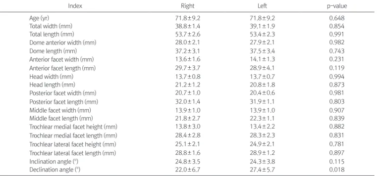
Table 3. Comparison Measurements in Females between Left Talus and Right Talus
Index Right Left p-value
Age (yr) 71.8±9.2 71.8±9.2 0.648
Total width (mm) 38.8±1.4 39.1±1.9 0.854
Total length (mm) 53.7±2.6 53.4±2.3 0.991
Dome anteriorwidth (mm) 28.0±2.1 27.9±2.1 0.982
Dome length (mm) 37.2±3.1 37.5±3.4 0.743
Anteriorfacet width (mm) 13.6±1.6 14.1±1.3 0.231
Anteriorfacet length (mm) 29.7±3.7 28.9±4.1 0.119
Head width (mm) 13.7±0.8 13.7±0.7 0.994
Head length (mm) 21.2±1.2 20.8±1.8 0.873
Posteriorfacet width (mm) 20.7±1.0 20.4±0.6 0.981
Posteriorfacet length (mm) 32.0±1.4 31.9±1.1 0.803
Middle facet width (mm) 13.9±1.0 13.9±1.0 0.907
Middle facet length (mm) 21.8±2.7 22.3±1.1 0.839
Trochlear medialfacet height (mm) 13.8±3.0 13.4±2.2 0.882
Trochlear medialfacet length (mm) 28.4±2.8 28.3±2.3 0.831
Trochlear lateralfacet height (mm) 25.1±2.1 24.9±2.1 0.781
Trochlear lateralfacet length (mm) 28.8±1.6 28.9±1.2 0.897
Inclination angle (o) 24.8±3.5 24.3±3.8 0.115
Declination angle (o) 22.0±6.7 27.4±5.7 0.018
Values are presented as mean±standard deviation.
www.jkfas.org 실정이며, 본 연구에서는 Sarrafian’s anatomy에서 제시하는 방법
으로 측정하였으며 경사각 평균 24도, 편각 평균 27도로 측정되었 고, 국외에서 Testut8)은 경사각 25도, 편각 22도, Paturet9)은 경사각 25도, 편각 20∼30도로 발표하였고, Sarrafian’s anatomy에서는 경 사각 24도, 편각 24도,10) Sewell11)은 1,006명의 이집트인에서 편각 7
∼43도, 평균 18도로 발표하였다. 한편으로 일정 크기 이상에서 남 자 또는 여자로 분류하는 방법으로 Robinson 등12)은 15명의 미국 인을 대상으로 한다면 컴퓨터 단층촬영(multi-detector computed tomography)을 이용한 거골 크기 측정에서 전후 직경 58 mm 이상 일 때 남성, 이하일 때 여성으로 분류하였고 이때 87%의 정확성이 있다고 보고하였다. 본 연구에서는 전후 직경이 59 mm 이상을 남 자로 분류할 때 86%의 정확성으로 나타나 Robinson 등12)과 비교하 였을 때 비슷한 수치를 가지는 것으로 확인되었다.
본 연구의 한계점으로 각 인종별, 국가별 거골에 대한 다양한 부 위의 크기 측정에 대한 국내외 자료가 많지 않아 기존 자료와 비 교하는 데 한계가 있으며, 각 자료마다 발표하고 있는 값 또한 상 당한 차이를 보이고 있다. 또한 해부학교실에 기증된 사체 연구 로 대상군 수가 부족하고 고령이라는 점으로 한국인의 거골 크기 를 대표하기는 어렵고, 남녀별로 신장과 체중의 전수 조사가 이루 어지지 않아 키와 몸무게를 거골 크기와 연관 짓기에 어려웠다. 측 정 과정에 있어서 2인의 관찰자가 각자 측정 후 측정 오차에 대해 서 계측한 모든 값이 0.95 이상으로 신뢰할 수 있는 값을 얻었다 (Table 4). 추후 대상군 숫자를 늘려 더 많은 데이터를 통해 대표 크기를 설정할 수 있을 것으로 기대된다. 또한 3차원 컴퓨터 단층 촬영 등을 이용한 계측보고가 증가하고 있어 사체연구를 통한 실 측과 방사선학적 계측을 비교를 통해 사체연구의 한계를 극복하는 데 도움이 될 것으로 생각된다.
결 론
거골은 족부와 족관절 복합체의 중심을 이루는 해부학적 및 생 역학적으로 중요한 뼈이며, 본 연구는 사체를 바탕으로 거골을 실
측하여 기존에 보고되지 않은 한국인의 해부학적 특성에 대해 보 고한 선행 연구이다. 기존 외국의 거골 측정치 및 성별에 의한 차 이를 발견할 수 있었으며, 백인의 유럽인 또는 미국인에 비해 작은 계측치를 확인하였고, 일본인보다는 큰 계측치를 확인하였다.
REFERENCES
11 Crossan ET. Fractures of the tarsal scaphoid and of the os calcis1 Surg Clin North Am1 1930;10:14771
21 Art W, Roy WS, Alireza B, John GA, Donald RB. Ankle fractures1 In: Coughlin MJ, Saltzman CL, Anderson RB, editors1 Mann’s surgery of the foot and ankle1 9th ed1 Philadelphia: Saunders;
20141 p121021
31 Sarrafian SK. Anatomy of the foot and ankle1 Philadelphia: Lip- pincott; 19831
41 Na WC, Lee SH, Lee JY, Lee SJ, Kim B. The result of open reduc- tion and mini-plate fixation for displaced talar neck fracture1 J Korean Fract Soc1 2015;28:215-221
51 Williams PL, Bannister LH, Berry MM, Collins P, Dyson M, Dussek JE, et al. Gray’s anatomy1 38th ed1 London: Churchill Livingstone; 19991
61 Sakaue K. Sex assessment from the talus and calcaneus of Japa- nese1 Bull Natl Mus Nat Sci Ser D1 2011;37:35-481
71 Gualdi-Russo E. Sex determination from the talus and calcaneus measurements1 Forensic Sci Int1 2007;171:151-61
81 Testut L. Traité d’anatomie humaine1 7th ed1 Paris: Doin; 19211 p13681
91 Paturet G. Traité d’anatomie humaine1 Paris: Masson; 19511 p15731
101 Gautham K, Clarista MQ, Sheela N, Vidyashambhava P. Mor- phometric analysis of the human tali1 CIBTech J Surg1 2013;2:64- 81
111 Sewell RBS. A study of the astragalus1 J Anat Physiol1 1904;39:
74-881
121 Robinson C, Eisma R, Morgan B, Jeffery A, Graham EA, Black S, Rutty GN. Anthropological measurement of lower limb and foot bones using multi-detector computed tomography1 J Forensic Sci1 2008;53:1289-951

![Figure 10. Actual measurement of declination angle (γ; angle of be- be-tween longitudinal line of superior trochlear [α] and longitudinal line of talar neck [β]) of talus in cadaver.](https://thumb-ap.123doks.com/thumbv2/123dokinfo/5295756.152263/4.918.87.837.883.1084/figure-actual-measurement-declination-longitudinal-superior-trochlear-longitudinal.webp)