서 론
전방접근 경추간판 제거 및 추체간 유합술(anterior cer- vical discectomy and fusion: ACDF)은 경추에 발생할
CLINICAL ARTICLE
J Korean Neurotraumatol Soc 2011;7:63-67 ISSN 1738-8708
수 있는 다양한 퇴행성 질환 및 외상 후 상태에 보편적으 로 적용 할 수 있는 신뢰할 만한 술식이다.3,7,11,14,16) 이 술식 은 전방 장골 마루에서 자가골 이식을 이용한 경추간판 제 거 및 골유합술의 형태로 수십 년 동안 발전해 왔다. 이 술 식을 통해 높은 골유합률 달성이 가능해졌으나 골 제공부 위 합병증은 이 술식을 고려하는데 있어 중요한 문제다. 이 러한 합병증으로는 혈종형성, 지속적인 골 제공부위 통증, 감염, 대퇴부 감각이상증이 있다. 이를 예방하기 위해 동종 이식편을 대안으로 고려하였으나 전염성 질환의 전파 외에 골유합률은 낮은 반면, 척추간 거리침하율은 높은 것이 문 Received: March 29, 2011 / Revised: August 22, 2011
Accepted: August 22, 2011
Address for correspondence: Sang Ryong Jeon, MD, PhD Department of Neurological Surgery, Asan Medical Center, Uni- versity of Ulsan College of Medicine, 88 Olympic-ro 43-gil, Song- pa-gu, Seoul 138-736, Korea
Tel: +82-2-3010-3550, Fax: +82-2-476-6738 E-mail: [email protected]
Hydroxyapatite Block을 이용한 전방접근 경추간판 제거 및 추체간 유합술의 임상, 방사선학적 결과분석
울산대학교 의과대학 서울아산병원 신경외과학교실
하진경 .박진훈 .전상용
Clinical and Radiological Outcome Analysis on Anterior Cervical Discectomy and Interbody Fusion Using Hydroxyapatite Block
Jin Gyeong Ha, MD, Jin Hoon Park, MD and Sang Ryong Jeon, MD
Department Neurological Surgery, Asan Medical Center, University of Ulsan College of Medicine, Seoul, Korea
Objective: Although use of autologous iliac bone graft in anterior cervical discectomy and fusion (ACDF) for cervical de- generative diseases remain standard surgical procedure, donor site morbidity are still concerns. Several synthetic graft materials have been developed to prevent this complication. This is retrospective study of clinical and radiological out- comes of ACDF using synthetic hydroxyapatite (HA) block to evaluate the efficacy. Methods: From May 2009 to June 2010, twenty-one patients (M 11 ; F 10) were enrolled in this study and 35 segments were involved. All patients were per- formed ACDF using HA block and plating system. Indications of surgery were radiculopathy caused by degenerative cervical spondylosis with or without myelopathy. The mean period of clinical follow-up was 6.6 months (range from 6 to 12 months). The change of Visual Analogue Scale (VAS) and Neck Disability Index (NDI) at 6 month were used for clin- ical outcome analysis. Cervical spine radiographs including dynamic views were obtained at postoperative 3 day, 1, 3, 6 months in all patients to measure the change of disc height, overall and segmental lordosis, segmental motion. Computed tomography was done at postoperative 6 month in all patients to confirm the radiological fusion. Results: Mean VAS and NDI score changed from 8.2 to 2.7 and from 23.4 to 10.5, respectively. The mean disc height change was from 4.5 to 7.1.
Cervical lordosis and segmental lordosis changes were from 24.4 to 24.5 and from 3.93 to 4.57, respectively. Complete in- terbody fusion was achieved in 95.2% of patients. There was one case of non-fusion patient. Conclusion: HA block is very efficient graft material in achieving cervical fusion, maintaining intervertebral disc height for ACDF. Further follow up study should be needed to evaluate the efficacy of this material. (J Korean Neurotraumatol Soc 2011;7:63-67)
KEY WORDS: Bone fusion ㆍCervical lordosis ㆍDisc height ㆍHydroxyapatite block ㆍCervical intervertebral disk.
제점으로 지적되었다. 지금까지 hydroxyapatite (HA) block 을 이용한 ACDF를 시행한 후 임상결과를 보고한 바가 있 고 100% 골유합률을 보고한 문헌도 있다.10,13) 본 저자들도 HA block를 이용한 ACDF 술식의 임상적, 방사선학적 결 과를 기존 연구결과와 비교 분석하고자 한다.
대상 및 방법
환자군
본 후향적 연구는 2009년 5월부터 2010년 6월까지 HA block을 이용하여 ACDF를 시행한 환자를 대상으로 이루 어졌다. 이 기간 동안 21명의 환자들은 경추 신경근증 혹은 척수증으로 인해 HA block을 이용한 1~2 level의 ACDF 를 시행하였다. 환자군은 11명의 남성과 10명의 여성으로 구성되어 있으며 평균연령 53.5세 (범위, 27~75세)였다.
수술 적응증은 척수증 또는 점진적인 신경학적 악화소 견을 동반한 신경근증이나 3개월 이상의 보존적 치료에 호전을 보이지 않은 신경근증을 포함하였다. 경부종양, 경 부감염, 3 level 이상의 경추 추간판 병변, 심각한 내과적 유병상태는 수술 적응증에서 배제하였다. 임상증상과의 상 관관계를 확인하기 위해 술 전 magnetic resonance image (MRI)를 시행하여 수술 범위를 선정하였다. 모든 수술은 동일 신경외과의에 의해 시행되었고 HA block 사용에 대 해서는 환자의 동의를 전제로 하였다. 술 후 1, 3, 6개월에 외래 내원하여 신경학적 검사 및 영상검사를 시행하였다.
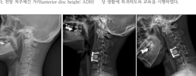
추적관찰 기간은 6개월에서 12개월이었으며 평균 6.6개월 이었다. 골유합 여부를 평가하기 위해 CT 스캔을 사용하여 방사선투과성 간극이 없으며 전, 후방의 연결골 형성을 확 인하였다. 전방 척추체간 거리(anterior disc height: ADH)
는 수술대상이 되는 운동관절의 척추체 상,하방 끝면의 앞 쪽지점 사이의 거리로, 후방 척추체간 거리(posterior disc height: PDH)는 상, 하방 끝면의 뒤쪽지점 사이의 거리로 정의하였고 평균 척추체간 거리(mean disc height: MDH) 는 전후방 척추체 간 거리의 평균으로 정의하였다 (Figure 1A). 또한 경부 굴신전 단순촬영을 시행하여 척추불안정성 여부를 확인하였다 (Figure 1B).
수술기법
모든 환자는 앙와위 자세를 취한 후 경부신전을 유지하 였고 횡단절개를 가한 후 전방 경추간판을 접근하였다. 견 인유발 손상을 예방하기 위해 Platysma 근육을 광범위하 게 노출한 후 Fluoroscopy를 이용하여 수술 level을 확인 한 후 척수와 신경근에 적절한 감압을 하기 위해 수술용 현 미경과 고속 드릴을 이용하여 경추간판을 제거하고 후방 의 비후성 골극을 제거하였다.
추체삽입 나사견인기를 이용하여 척추체간 공간을 견인 하여 공간을 확보하였다. 연골을 제거하여 상, 하방 종판을 노출시키고 적절히 연마한 후 척추체간 공간에 HA block 을 삽입하였다. 표피층을 봉합하기 전에 fluoroscopy를 이 용한 측위 사진을 시행 후 이식체가 적절한 위치에 삽입되 었는지를 확인하였다.
수술 후 관리
환자들은 수술 후 회복실에서 1~2시간 가량 경과관찰 후 일반병동으로 옮겨졌다. 24시간 경과하여 필라델피아 경부 보조기를 착용한 채로 보행을 시작하였고 3개월간 착용하도 록 교육하였다. 평균 7.7일 경과 후 퇴원하였으며 퇴원 후 정 상 생활에 복귀하도록 교육을 시행하였다.
FIGURE 1. A: Mean height=(A+B)/2, A=anterior disc height, B=posterior disc height. B: C spine Stress view.
A B
A B
임상지표의 평가
수술 전후 Visual Analogue Scale (VAS)과 Neck Dis- ability Index (NDI)의 변화를 평가하였으며 경추 전만굴 곡의 변화와 수술대상부위의 전만굴곡의 변화를 아울러 평가하였다. 1분절 수술과 2분절 수술에 따른 수술 전, 후 6개월 경과 VAS, NDI 변화에 대해 유의한 차이가 있는지 비모수적 방법으로 검정하였다 (SPSS Ver. 18).
결 과
전체 35분절에 수술이 시행되었다. 1분절 수술은 7예, 2 분절 수술은 14예였다. 수술대상 분절 별로 나누어 보면 C3-4, C4-5, C5-6, C6-7에 해당하는 경우는 각각 4예, 9예, 14예, 8예였다. 이러한 임상적, 방사선학적 특징은 Table 1
에 요약하였다.
수술 전 VAS 8.2에서 수술 시행 후 4.5로 감소하였고 3개 월, 6개월 외래 내원시 3.4, 2.7로 추가 감소하여 통증은 호전 되었다 (Figure 2A). NDI는 수술 전 23.4에서 15.8로 감소하 였고 3개월, 6개월 외래 내원시 12.4, 10.5로 추가 감소하여 통증에 의한 일상생활제한 정도도 호전되었다 (Figure 2B).
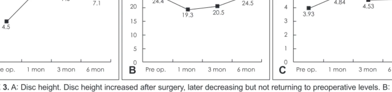
평균 척추체간 거리는 술전에 비해 증가하였다. 단순방사 선 검사로 분석한 수술 전 평균 척추체간 거리는 4.5에서 수술 시행 후 7.8로 증가하였고 3개월, 6개월 외래 내원시 7.6, 7.1로 변화되었다 (Figure 3A). 경추 전만굴곡은 수술 전 24.4도에서 수술 시행 후 19.3도로 감소하였고 3개월, 6 개월 외래 내원시 20.5도, 24.5도로 변화되었다 (Figure 3B).
수술대상 분절 전만굴곡은 수술 전 평균 3.93도에서 수술 시행 후 4.84도로 증가하였고 3개월, 6개월 외래 내원시 4.53도, 4.57도로 변화되었다 (Figure 3C). 수술 후 6개월 경과 CT로 분석할 때 골유합률은 95.2%였다 (Figure 4).
수술 전 VAS, 수술 후 6개월 경과 VAS에 수술 분절 수 가 미치는 영향을 비모수적 방법으로 검정한 결과 유의한 차이를 보이지 않았다 (Table 2). NDI 변화에 대해서도 동 일한 분석을 시행하였을 때 2 level 수술을 시행 한 것이 1 level 수술에 비해 6개월 후 NDI의 호전에 더 큰 영향을 미 치지 못했다 (Table 3).
수술 시 혹은 수술 후 합병증은 발생하지 않았다.
고 찰
ACDF는 경추간판 유발 신경근증과 척수병증을 앓고 있는 환자들에게 유용한 외과술식이다. 그동안 장골이식 TABLE 1. Baseline demographic and clinical characteristics
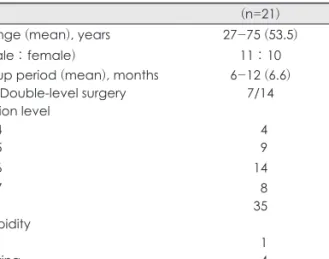
(n=21) Age range (mean), years 27-75 (53.5)
Sex (male : female) 11 : 10
Follow up period (mean), months Single-/Double-level surgery Operation level
6-12 (6.6) 7/14 C3-4
C4-5
04 09
C5-6 14
C6-7 Total Comorbidity
08 35 DM
Smoking Osteoporosis
01 04 00
Osteoporosis: Bone mineral density < T-2.5. DM: diabetes mel- litus
Radiculopathy (VAS)
8.2 Neck disability index (NDI)
A
9 8 7 6 5 4 3 2 1
0 Pre op. 1.5 mon 3 mon 6 mon 4.5
3.4
2.7
B
25
20
15
10
5
0 Pre op. 1 mon 3 mon 6 mon 23.4
15.8
12.4
10.5
FIGURE 2. VAS scores (A) and NDI (B) scores were decreased through the follow-up period since surgery. VAS: Visual Analogue Scale, NDI: Neck Disability Index.
편을 이용한 술식은 방사선학적으로, 임상적으로 만족스 러운 결과를 보여왔다. 그러나 골이식편의 파손, 공여부위 의 합병증, 수술시간 연장 등이 단점으로 지적되고 있다. 이 에 여러 골 대체제를 사용한 많은 연구가 진행되었고, 좋 은 결과들이 보고되었다.2,10,13) 본 연구결과도 이와 비슷한 임상적, 방사선학적 결과를 보여주고 있다. 환자의 주 증상 이었던 신경근증은 수술 직후 빠르게 호전을 보였으며 이
후 완만하나 지속적으로 호전양상을 보였다. 환자의 기능 적인 회복을 알 수 있는 NDI도 수술 후 6개월 동안 지속 적으로 감소하는 소견을 보이고 있다.
기존의 많은 논문들이 골 대체제를 사용하여 다양한 골 유합률 (86.5~100%)을 보고하였다.8,9,12,15) 본 연구에서 6개 월 동안의 95.2%는 위의 결과와 비교하였을 때 상당히 좋 은 경과로 생각되어진다. 그러나 기존의 보고에서 임상적 결과는 우수한 반면 방사선학적으로는 수술 후 척추체간 거리 침하와 후만변형을 보고하고 있다.12) 하지만, 본 연구 결과는 수술 후 형성된 척추체간 거리와 각도를 수술 후 6개 월간 잘 유지하고 있는 것을 확인할 수 있었다. 다만 6개월 은 비교적 짧은 기간의 추적관찰이므로 향후에도 지속적 인 추적관찰이 필요할 것이다.
HA는 인산칼슘의 수산기 화합물[Ca10(PO4)6(OH)2]이며 골매트릭스의 주 구성성분이다.1,4-6) 골전도 (Osteoconduc- tion)가 진행하게 됨에 따라 보형물은 연결골로 둘러싸이게 되고 이를 통해 인접한 척추체와 골유합이 일어나게 된다.
HA는 균일한 화학조성을 가지기 때문에 최적의 생체역학 형상이 가능하며 이를 통해 기계적 붕괴에 저항성이 있고 면 역반응으로부터 안전하다. HA의 이러한 생물학적 장점에 도 불구하고 기존의 연구에서는 내압장력에 취약점이 있었 고17) plate로 고정한 후에도 24%에서 이식체 붕괴를 보고 하였다. 하지만 본 연구결과에서는 이식체 붕괴를 보이는 소견은 없었다.
본 연구에서는 평균 추적기간이 6.6개월로서 척추체 사
9 8 7 6 5 4 3 2 1
0 Pre op. 1 mon 3 mon 6 mon 4.5
7.8 7.6
7.1
Disc height 30
25 20 15 10 5
0 Pre op. 1 mon 3 mon 6 mon 24.4
19.3 20.5
24.5
Cervical iordosis 6
5 4 3 2 1
0 Pre op. 1 mon 3 mon 6 mon 3.93
4.84 4.53 4.57
Segmental iordosis
C B
A
FIGURE 3. A: Disc height. Disc height increased after surgery, later decreasing but not returning to preoperative levels. B: Cervical lordosis. Cervical lordosis decreased after surgery, later returning to preoperative levels. C: Segmental lordosis. Segmental lordosis increased immediately after surgery, later decreasing but not returning to preoperative levels.
FIGURE 4. A 52-year-old man who present with cervical radicu- lopathy and underwent ACDF with HA block. C spine CT scan at A immediately postoperative, B postoperative 6 months visits.
Bone briding was formed between vertebral endplates (arrows).
ACDF: anterior cervical discectomy and fusion, HA: hydroxyap- atite.
A B
TABLE 2. Non-parametric analysis on surgical outcome (VAS) Variables 1 level (n=7) 2 level (n=14) p value Pre OP VAS
(mean) 05.9 005.2
Post OP 6M VAS
(mean) 03.0 003.6
VAS change
(rank sum) 81.0 150.0 0.799*
*this p-value was calculated from the Man-Whitney U test. OP:
operative, M: months, VAS: Visual Analogue Scale
TABLE 3. Non-parametric analysis on surgical outcome (NDI) Variables 1 level (n=7) 2 level (n=14) p value Pre OP NDI
(mean) 22.7 025.4
Post OP 6M NDI
(mean) 09.3 010.7
NDI change
(rank sum) 78.0 153.0 0.917*
*this p-value was calculated from the Man-Whitney U test. OP:
operative, NDI: Neck Disability Index
이공간 침하, 인접 분절 퇴행 등의 이상소견 발생 등을 확 인하기에는 추적기간이 비교적 짧고 평가대상 환자의 수가 21명으로 증례의 수가 비교적 적은 점이 단점으로 더 많은 증례와 장기 추적관찰이 향후 필요할 것으로 보인다.
결 론
본 연구에서는 HA block을 이용하여 1내지 2분절 전방 접근 경추간판 제거술 및 골유합술을 시행한 환자들을 대 상으로 임상적, 방사선학적 결과를 후향적 분석하였다. 수 술 후 6개월간 임상적 결과는 호전되었고, 방사선학적 골 유합률은 95.2%였으며, 수술 후 6개월간 척추체간 거리와 각도는 비교적 잘 유지됨을 알 수 있었다. 다만, 향후 더 많 은 증례와 장기간의 추적관찰을 통해 추가분석이 필요할 것이다.
중심 단어: 골유합・경추전만・척추체간 거리・수산화 인회 석블록 경추 추간판 .
■ The authors have no financial conflicts of interest.
REFERENCES
1) Alvarez JA, Hardy RW Jr. Anterior cervical discectomy for one- and two-level cervical disc disease: the controversy surrounding the question of whether to fuse, plate, or both. Crit Rev Neurosurg 9:234-251, 1999
2) Bärlocher CB, Barth A, Krauss JK, Binggeli R, Seiler RW. Com- parative evaluation of microdiscectomy only, autograft fusion, polymethylmethacrylate interposition, and threaded titanium cage fusion for treatment of single-level cervical disc disease: a prospective randomized study in 125 patients. Neurosurg Focus 12:E4, 2002
3) Bohlman HH, Emery SE, Goodfellow DB, Jones PK. Robinson anterior cervical discectomy and arthrodesis for cervical radicu- lopathy. Long-term follow-up of one hundred and twenty-two patients. J Bone Joint Surg Am 75:1298-1307, 1993
4) Connolly PJ, Esses SI, Kostuik JP. Anterior cervical fusion: out-
come analysis of patients fused with and without anterior cervical plates. J Spinal Disord 9:202-206, 1996
5) Denissen HW, de Groot K. Immediate dental root implants from synthetic dense calcium hydroxylapatite. J Prosthet Dent 42:551- 556, 1979
6) el Deeb M, Waite DE, Mainous EG. Correction of the deficient alveolar ridge. Clin Plast Surg 16:733-748, 1989
7) Geer CP, Papadopoulos SM. The argument for single-level ante- rior cervical discectomy and fusion with anterior plate fixation.
Clin Neurosurg 45:25-29; discussion 21, 1999
8) Gercek E, Arlet V, Delisle J, Marchesi D. Subsidence of stand- alone cervical cages in anterior interbody fusion: warning. Eur Spine J 12:513-516, 2003
9) Hacker RJ. Threaded cages for degenerative cervical disease. Clin Orthop Relat Res :39-46, 2002
10) Hwang SL, Lin CL, Lieu AS, Lee KS, Kuo TH, Hwang YF, et al.
Three-level and four-level anterior cervical discectomies and ti- tanium cage-augmented fusion with and without plate fixation. J Neurosurg Spine 1:160-167, 2004
11) Madawi AA, Powell M, Crockard HA. Biocompatible osteocon- ductive polymer versus iliac graft. A prospective comparative study for the evaluation of fusion pattern after anterior cervical discectomy. Spine (Phila Pa 1976) 21:2123-2129; discussion 2129- 2130, 1996
12) Matge G. Anterior interbody fusion with the BAK-cage in cervical spondylosis. Acta Neurochir (Wien) 140:1-8, 1998
13) McConnell JR, Freeman BJ, Debnath UK, Grevitt MP, Prince HG, Webb JK. A prospective randomized comparison of coralline hydroxyapatite with autograft in cervical interbody fusion. Spine (Phila Pa 1976) 28:317-323, 2003
14) Pintar FA, Maiman DJ, Hollowell JP, Yoganandan N, Droese KW, Reinartz JM, et al. Fusion rate and biomechanical stiffness of hy- droxylapatite versus autogenous bone grafts for anterior discec- tomy. An in vivo animal study. Spine (Phila Pa 1976) 19:2524- 2528, 1994
15) Schmieder K, Wolzik-Grossmann M, Pechlivanis I, Engelhardt M, Scholz M, Harders A. Subsidence of the wing titanium cage after anterior cervical interbody fusion: 2-year follow-up study. J Neu- rosurg Spine 4:447-453, 2006
16) Schneider JR, Bright RW. Anterior cervical fusion using preserved bone allografts. Transplant Proc 8:73-76, 1976
17) Zdeblick TA, Cooke ME, Kunz DN, Wilson D, McCabe RP. An- terior cervical discectomy and fusion using a porous hydroxyap- atite bone graft substitute. Spine (Phila Pa 1976) 19:2348-2357, 1994