| Abstract |
1)PURPOSE: The aim of this study was to investigate the influence of hip abduction velocity and position change on the relative onset times of the gluteus medius, the tensor fascia latae, and the quadratus lumborum in healthy subjects.
METHODS: For this study, 15 healthy young adults were recruited. The subjects were asked to move their hip joints up to 35 degrees of abduction at a speed of 70⁰/sec and 17.5⁰/sec in the supine and side-lying positions. Electromyography data was collected for the gluteus medius, tensor fascia latae, and quadratus lumborum to determine the onset times.
RESULTS: There were significant differences between the fast speed (70⁰/sec) and the slow speed (17.5⁰/sec) in hip abduction in a supine position and in a side-lying position, relatively. The onset time of the gluteus medius was faster than that of the tensor fascia latae and the quadratus lumborum in the side-lying position at the speed of 70⁰/sec and 17.5⁰/sec.
CONCLUSION: The findings of this study indicated that hip abduction in a side-lying position is an effective method
†Corresponding Author : [email protected]
This is an Open Access article distributed under the terms of the Creative Commons Attribution Non-Commercial License (http://creativecommons.org/licenses/by-nc/3.0) which permits unrestricted non-commercial use, distribution, and reproduction in any medium, provided the original work is properly cited.
to recruit the gluteus medius earlier than the tensor fascia latae and the quadratus lumborum.
Thus, the exercise position is considered necessory in the purpose of rehabilitation for gluteus medius muscle strengthening program.
Key Words: Hip abduction velocity, Position, Gluteus medius, Onset time
Ⅰ. Introduction
The abductor muscle in the hip joints serves as a major muscle providing hip stability while walking and a solution to pathologic problems (Beasley, 1956). In addition, the muscle presents stability of the pelvis and external stability of the hip joints in one leg standing during which bearing three times more weight is applied to the opposite hip joint and stance phase in walking (Lyons et al., 1993). The gluteus medius (GMed) is important in maintaining a standing position and controls movement of the hip. It also stabilizes the pelvis while mobilizing the lower extremities (Hwang-Bo et al., 2016; Lee et al., 2015). Because the GMed and the quadratus lumborum (QL) are closely linked with joint support, an imbalance in muscle recruitment
Research Article Open Access
Influence of Hip Abduction Velocity and Position on the Onset Times of Gluteus Medius and Tensor Fascia Latae Relative to Quadratus Lumborum
in Healthy Subject: A Pilot Study
Jung-Bin Kim, PT, MSc⋅Chang-Kyo Yun, PT, MSc⋅Gak Hwang-Bo, PT, PhD
†Department of Rehabilitation Science, Graduate School, Daegu University Received: July 14, 2016 / Revised: July 25, 2016 / Accepted: August 11, 2016
ⓒ 2016 J Korean Soc Phys Med
patterns causes movement damage, and when the GMed is weakened, the compensation movement of the QL induces external pelvic inclination and external flexion of the lumbar to cause external instability and motor injury (Sahrmann, 2002). The weakened and contracted GMed causes habitual use including wrong movement patterns of the tensor muscle of fascia lata during hip joint abduction (Travell and Simons, 1983). Attenuation of changes in the length of the posterior GMed, and reduction and strengthening of the Tensor fascia latae (TFL) lead to muscular imbalance, as a result of which the muscle recruitment patterns of the TFL are activated faster than those of the posterior GMed in the case of hip joint abduction (Dennis and Kathryn, 2003).
The activities of the muscles are related to measurement postures and movement speed (Hodges and Richardson, 1999). Kwon and Koh (2002) reported that patients with backache showed a delay in the onset time of contraction of the gluteus maximus muscle when compared to that of the semitendinous muscle. These patients also showed a delay in the onset time of relative contraction of the backbone erectors against the semitendinous muscle (Kwon and Koh, 2002). Movement speed affects forms of motility control forms and the magnitude of the force (Vogt and Banzer, 1997). According to Hodges and Richardson (1997), in the case of muscular contraction in the flexor muscles of the upper limbs, and the trunk muscles in flexion of the shoulder joints, the onset time of muscular contraction is significantly more delayed in slow movements than in fast and normal movements (Hodges and Richardson, 1997). Such results can be explained with differences in structure and speed of the muscles consisting of fast twitch fibers and slow twitch fibers and differences in recruitment based on sizes of motor nerves. In general concentric contraction, muscular strength is more reduced as muscular contraction is faster and muscular strength is more increased as muscular contraction is slower is slower, indicating that muscular viscosity and resistance due to the viscosity
increase as speed increases (Cha et al., 1999).
There have been many researches on onset time of muscular contraction and recruitment patterns of the the glutaeus maximus, the backbone erectors, and the hamstring in hip joint extension in a prone posture (Chang et al., 2006). However, Few researchers have focused on the onset time of muscular contraction relating to hip joint abduction, which causes mechanical interaction between the spine and the lower limbs and provides pelvic stability when walking.
Based on published reports and clinical experience, we hypothesized that movement velocity and position is give effect to the onset time of muscular contraction. Therefore, the purpose of this study was to measure the onset time of muscular contraction of the GMed, TFL and QL in hip abduction at fast and slow speeds in the supine, and side-lying positions of normal subjects in order to compare the onset time of muscular contraction
Ⅱ. Methods
1. Subjects
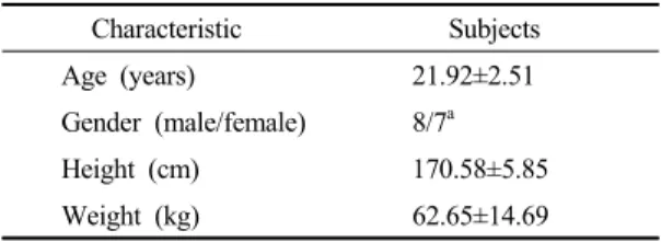
15 healthy young subjects were recruited from the university students who volunteered to participated in this study. The exclusion criteria were past or present neurologic, musculoskeletal, or cardiopulmonary diseases that could interfere with hip abduction (Table 1). This study was conducted in the subjects provided informed consent prior to their participation.
Characteristic Subjects
Age (years) 21.92±2.51
Gender (male/female) 8/7a
Height (cm) 170.58±5.85
Weight (kg) 62.65±14.69
Values are expressed as mean±standard deviation.
aValues are numbers. *p<.05.
Table 1. General characteristics of the subjects
2. Outcome measures: Electromyography analysis for muscle onset times
We collected electromyographic (EMG) data using a data acquisition system (BIOPAC MP150WSW, Santa Barbara, USA). The subjects’ skin was first cleansed with rubbing alcohol, and disposable Ag-AgCI surface electrodes were then positioned at and interelectrode distance of 2 cm. EMG data were collected as follows for the muscles on the dominants side as the lower extremity. GMed electrodes were placed parallel to the muscle fibers, over the proximal and one third of the distance between the iliac crest and the greater trochanter.
TFL electrodes were placed parallel to the muscle fibers approximately 2 cm below the anterior superior iliac spinae.
QL electrodes were placed 4cm lateral to the vertebral ridge or belly of the erector spinae muscle, at the erector spinae muscle, and at a slightly oblique angle at half the distance between the twelfth rib and the iliac crest (Lee et al., 2014;
Kasman et al., 1998). We amplified and digitized EMG signals using AcqKnowledge 3.9.1 software, with bandpass filter of (30-450 Hz) and a bandstop filters of (60 Hz), as well as sampling at 1000 Hz. The foot switch side of the popliteal region was used to determine the time (reference time: zero) of leg lifting. A plus value for onset time means later onset of muscle action than the reference time. A minus value of onset time means earlier onset of muscle action than the reference time. A period of two seconds after the start of the EMG data collection was established as the baseline period. After the EMG signals collected during the two seconds were full-wave rectified, the means and standard deviation were evaluated from the signals during the .5 of a second that was the most stable during the baseline period. Mean +2 standard deviation was established as the threshold value of the onset time of muscular contraction. The point exceeding the threshold was automatically calculated and cases of muscular contraction not longer than 25ms were not considered actual muscular contraction (Oh et al., 2006).
3. Experimental procedures
We used an electronic metronome (SEIKO QUARTZ METRONOME, SEIKO, JAPAN) and a goniometer (TSD 130B, BIOPAC System Inc. U.S.A) to control the abduction speed and angle in hip abduction. The subjects were asked to move their hip joints up to 35 degrees of abduction at a speed of 70⁰/sec and 17.5⁰/sec over .5 and 2 seconds using the electronic metronome (Chang et al., 2006). The subjects performed abduction of the right hip joint at 35 degrees in a supine and a side-lying position. To control range, a target bar was placed at the level of 35 degrees hip abduction using a goniometer, so that the side of the popliteal region on the lower extremity’s dominant side touched the target during hip abduction. Before the measurement was taken, the subjects performed three practices in order to adapt to the abduction and the speed of the metronome. The order in which the exercises were preformed were selected randomly. The subjects performed the exercises in all the positions using both speeds repeatedly five times, and had a three-minute rest between each measurement in order to avoid fatigue.
4. Data processing and statistical analysis We used repeated two-way ANOVA to identifying differences in the relative onset time of the muscular contraction in the GMed, the TFL, and the QL at the fast and slow speed in a supine and side-lying position.
Turkey-HSD was used for post analysis. Result of statistics, there were no interaction. The SPSS version 17.0 for Windows was used for statistical processing of the data.
For all tests, the statistical significance level was ɑ=.05.
Ⅲ. Results
The onset time of muscular contraction of the tensor
fascia lata muscle of against the time of the gluteus medius
muscle in hip joint abduction in a supine position was
.45±.29 seconds earlier at fast speed (70⁰/sec) and 1.05±.62 seconds earlier at low speed (17.5⁰/sec). In hip joint abduction in a side lying position, the onset time of muscular contraction of the quadratus lumborum muscle against the time of the gluteus medius muscle was .18±.13 seconds delayed at fast speed and .24±.21 seconds delayed at slow speed; the onset time of muscular contraction of the tensor muscle of fascia lata was .14±.11 seconds delayed at fast speed and .29±.25 seconds delayed at slow speed.
For hip abduction in a supine position, the onset time of muscular contraction in the TFL and QL compared to the GMed onset time was earlier at the speed of 70⁰/sec and 17.5⁰/sec. In hip abduction in a side-lying position, the onset time of muscular contraction of the GMed compared to the TFL and QL times was earlier at speeds of 70⁰/sec and 17.5⁰/sec (Table 2).
Ⅳ. Discussion
This aim of this study was identify differences in patterns of muscular contraction onset and of relative muscular contraction onset in the GMed, TFL and QL in hip joint abduction at fast and slow speeds in the supine and side-lying position of normal subjects. The results showed that in the side-lying position, the muscle contraction occurred in the order GMed, TFL and QL during hip joint abduction at the fast speed, while contraction occurred in the order GMed, QL, and TFL at the slow speed. In the supine position, the TFL was the first to contract, followed by the GMed. Maximum force can be exercise in concentric contraction at slow speed, while faster contraction can
produce stronger force in eccentric movement (Wilmore and Costill, 2001). In this study, we controlled the speed of hip joint abduction as a common condition based on the research reporting that movement speed affects forms of motility control (Hodges and Richardson, 1999) and that reactions of the agonistic muscles vary in exercises with different eccentric speeds (Kulig et al., 2001). The relative onset time of muscular contraction in the QL, the TFL, and the GMed based on hip joint abduction speeds showed that there were significant differences in both positions due to the effects of the speed; changes in position also displayed significant differences at the slow speed.
Hodges and Richardson (1997) reported that no differences were observed in the onset time of muscular contraction between movements at fast speed and those at slow speed in the upper limbs while reactions were delayed at slow speed. In this study, there were differences in the onset time of muscular contraction in hip joint abduction both at the fast and slow speeds, regardless of positions. Such differences may be caused by varying conditions of the upper and lower limbs. Movements of the upper limbs are wide in terms or working range. Also, angular speed is easy to maintain in these limbs, and detailed movements can be controlled because of the small muscular fibers.
However, the muscles in the lower limbs are not wide in terms of working range, and it is difficult to perform uniform motion. Also, the bigger fibers in the muscles induce powerful, large movements (Horak et al., 1984).
In this study, no differences were found in the onset time of muscular contraction based on positions―supine or side-lying, but there are few previous researches to support the results. There is little previous research in
Muscle Supine(70⁰/sec) Supine(17.5⁰/sec) Side lying(70⁰/sec) Side lying(17.5⁰/sec) pTensor fascia latae -.45±.29 -1.05±.62 .14±.11 .29±.25 .02
Quadratus lumborum -.22±.06* -.37±.16* .18±.13 .24±.21 .00
Values are expressed as mean±standard deviation.
*p<.05.