108
서 론
혈관성 평활근종(vascular leiomyoma)은 평활근종의 아형으로 드문 양성 종양이다.1) 혈관의 평활근(smooth muscle)에서 기원하며 조직학적으로는 평활근 세포와 혈관 내피(vascular endothelium)로 구성된다.1,2) 전신 의 피부 및 피하조직에서 발생할 수 있으며,2) 주로 하지 에서 통증을 호소하는 고형성의 피부 종물로 발생한 다.3,4) 8.5%에서 10%의 혈관성 평활근종이 두경부 영역 에서 발생하며, 대부분 서서히 자라는 무통성의 연성
종물 형태로 나타난다.2-4) 두경부 영역 중 비강(nasal cav- ity), 이개(auricle), 구순(lip) 및 경부 등에서 발생하며,5) 구강(oral cavity) 내 경구개(hard palate)에 발생하는 경 우는 매우 드물다.6,7) 저자들은 구강 내 부위 중 경구개에 발생한 혈관성 평활근종 1예를 경험하였기에 이를 문헌 고찰과 함께 보고하는 바이다.
증 례
64세 남자 환자가 1개월 전 우연히 발견한 경구개 부 위의 연성 종물을 주소로 내원하였다. 이학적 검사 상 상절치 1 cm 좌후방의 경구개 중앙 부위에 약 1×1 cm 크기의 구형 종물이 관찰되었으며(Fig. 1), 통증이나 압 통을 동반하지 않았다. 그 외 구강, 비강 및 후두에 특별 한 이상 소견은 관찰되지 않았으며, 다른 경부 림프절 비대는 관찰되지 않았다. 종물 이외의 전신적 신체 증 상의 호소는 없었으며, 과거력 및 가족력 상 당뇨와 고
경구개에 발생한 혈관성 평활근종 1예
대구가톨릭대학교 의과대학 이비인후-두경부외과학교실
고재진
·강승현
·진효승
·김정규
A Case of Vascular Leiomyoma of the Hard Palate
Jae Jin Ko, MD, Seung Hyun Kang, MD, Hyo Seung Jin, MD and Jeong Kyu Kim, MD, PhD Department of Otorhinolaryngology-Head and Neck Surgery, School of Medicine,
Catholic University of Daegu, Daegu, Korea - ABSTRACT -
Vascular leiomyoma is an uncommon benign tumor that originates from the smooth muscle in the blood vessel walls. Vascular leiomyoma can occur in the head and neck region, but very rarely involves the hard palate. Vascu- lar leiomyoma usually presents as small painless mass, and the diagnosis is usually confirmed after histopatholog- ic study with immunohistochemical stain. The treatment of choice is complete surgical excision. Recently, we ex- perienced a case of vascular leiomyoma on the hard palate and we report this case with review of literature. (J Clinical Otolaryngol 2015;26:108-111)
KEY WORDS:Vascular leiomyomaㆍOral cavityㆍHard palate.
臨床耳鼻:第 26 卷 第 1 號 2015
• • • • • • • • • • • • • • • • • • • • • • • • • • • • • • • • • • • • • • • • • • • • • • • • • • • • • • • • • • • • • • • • • • • • • • • • • • • • • • • • • • • • • • • • • • • • • • • • • • • • • • • • • • • • • • • • • • • • • • • • • • • • • • • • • • • • • • • • • • • • • • • • • • • • • • • • • • • • • • • • • • • • • • • • • • • • • • • • • • • • • • • • • • • • • • • • • • • • • • • • • • • • • • • • • • • • • • •
J Clinical Otolaryngol 2015;26:108-111 증 례
논문접수일 :2014년 12월 9일 논문수정일 :2015년 2월 25일 심사완료일 :2015년 4월 27일
교신저자 :김정규, 705-718 대구광역시 남구 두류공원로 17길 33 대구가톨릭대학교 의과대학 이비인후-두경부외과학교실 전화 :(053) 650-4071・전송:(053) 650-4533
E-mail:[email protected]
고재진 외 : 경구개에 발생한 혈관성 평활근종
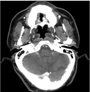
109 혈압 이외의 특이 사항은 없었다. 경부 전산화 단층촬 영 상 경구개 좌중앙 부위에 0.8×0.6 cm 크기의 균일 한 조영 증가 양상을 보이는 원형 병변이 확인되었으 며, 주변 조직과의 경계가 명확하였다(Fig. 2). 이학적 검사 및 경부 전산화 단층촬영소견을 종합하면 양성 병 변이 의심되며 조직검사를 시행할 경우 남은 조직이 많 지 않을 것으로 판단되어, 전신 마취 후에 입안으로 접 근하여 종물의 완전 적출술을 시행하였다. 종물 적출시 점막은 약 1 mm의 경계를 두고 절개를 시행하였으며, 심부는 경구개의 골막을 경계로 종물을 적출하였다. 종 물은 딱딱하지 않고 부드러운 편이었으며 출혈은 많지 않았다. 적출 후 결손 부위는 봉합이나 피판을 시행하지 않고 그대로 두었으며, 드레싱 없이 식이를 허용하면서 관
찰하였다. 수술 후 시행한 병리조직학적 검사에서 방추 상 평활근 섬유다발이 혈관 세포들로 둘러싸인 양상이 관찰되었고, 평활근 액틴 면역조직염색(smooth muscle actin immunostaining)에서 종양세포가 갈색으로 염색되 어(Fig. 3) 혈관성 평활근종으로 진단되었다. 수술 1주 일 후 결손부위에 특별한 합병증은 발생하지 않았으며 (Fig. 4A), 1개월 후 추적관찰 시 구강점막 상피의 재생으 로 결손 부위가 완전히 치유된 소견을 보였다(Fig. 4B).
1년 후 외래에서 추적관찰 시 재발 소견은 관찰되지 않 았다.
Fig. 1. Preoperative view. A well-defined mucosal mass with central ulceration is located on the left-side hard palate.
Fig. 2. Axial CT scan of hard palate. A well-defined, strong enhanced mass is shown on the left sided hard palate.
A B
Fig. 3. Histopathologic findings. Proliferation of thick walled vessels with patent lumens is identified. Inner layer of mus- cle is arranged in an orderly circumferential fashion, and outer layer shows less well-ordered arrangement merging with peripheral muscle fiber (H&E, ×100)(A). Immunohistochemical stains for smooth muscle actin shows diffuse posi- tive reaction to actin filament of smooth muscle fibers (anti-actin immunohistochemical stain, ×200)(B).
J Clinical Otolaryngol 2015;26:108-111
110
고 찰
평활근종(leiomyoma)은 평활근에서 기원하는 양성 종양이며, 평활근이 존재하는 신체의 어느 부위에서나 발생할 수 있다. 평활근종이 주로 발생하는 부위는 자 궁체부와 같은 비뇨 생식기계이며 그 외 위장관이나 피 하조직에도 생기는 경우가 있으나, 평활근의 분포가 상 대적으로 적은 두경부 영역에서는 발생 빈도가 2% 이 하로 드물다.5) 평활근종은 조직학적으로 고형 평활근종 (solid leiomyoma), 혈관평활근종, 상피양 평활근종(ep- ithelioid leiomyoma)으로 분류된다.1,8,9) 그 중 혈관평활근 종은 혈관 중간막(tunica media)에서 기원하는 것으로 알 려져 있다.8) 대부분의 혈관평활근종은 피부에서 발생하 며, 두경부 영역에서는 구강과 비강 내 점막에서도 발생 한다. 또한 이하선 공간(parotid space), 악하선 공간(subm- andibular space), 경동맥초(carotid sheath), 안면골(facial bone), 인두뒤 공간(retropharyngeal space)에서도 발생 한다.12) 이개(auricle)와 구강 및 비강이 두경부 영역에서 가장 호발하는 부위로 알려져 있다. 구강 내에서는 입술 과 구개, 혀에서 가장 호발한다.3) 전체 혈관평활근종 중 구강 내에서 발생하는 경우는 2.7% 정도이며, 호발 부위 는 상하구순이 48.6%로 가장 많고, 그 다음이 구개부위, 협부점막과 설근, 하악 순으로 발생한다고 하였다. 두경 부에 발생한 혈관평활근종의 경우 남녀비가 1.3 : 1로 남 자에서 더 호발하며, 연령대는 40대에서 60대까지로 평 균 발생연령은 48세였다.13)
혈관평활근종의 임상증상 중 동통이 가장 흔하게 보 고되지만, 두경부 영역에서 발생한 경우 무통성의 종물 을 증상으로 호소하는 경우가 가장 많다. 두경부에 발생 하는 혈관평활근종은 크기가 대부분 2 cm 이하로 서서 히 크기가 증가하는 양상을 보인다.3,5,13) 본 증례에서도 환자는 무통성 종물로 내원하였으며, 진찰 시 크기는 약 1×1 cm로 관찰되었다.
혈관평활근종은 병력 및 이학적 검사, 세침흡인검사, CT나 MRI 등의 방사선학적 검사 등에서 특징적인 소견 을 보이지 않아 확진이 어렵고, 술후 병리조직학적 검사 에 의해 확진된다.7,12) H&E 염색의 광학현미경 소견으로 대부분 진단이 가능하며, 여러 방향으로 주행하는 호산 성의 방추상 세포가 평활근 섬유다발을 구성하고 이를 발달된 혈관세포가 둘러싸는 양상을 확인할 수 있다.7,16) 평활근세포에 특이 염색되는 desmin, vimentin, Masson’s trichrome, actin과 myosin 등에 의해 평활근종을 확진 할 수 있으며, 혈관내피세포에 특이염색되는 VIII 인자, CD31을 통해 혈관종, 혈관섬유종, 섬유종 등과 같은 방 추형세포 종양과 감별할 수 있다.14-16) 평활근 기원의 악 성 종양인 평활근육종과의 감별은 세포의 유사분열 정도 나 세포 형태로 추정할 수 있다.13-15) 본 증례에서는 적출 된 종물의 광학현미경 소견 상 혈관성 평활근종의 양상을 확인하였으며, 유사분열이나 세포 이상의 증거가 관찰되 지 않아 악성 종양을 배재할 수 있었다.
혈관평활근종의 치료는 수술적 완전 절제가 필요하 며, 적절하게 제거된 경우 재발은 극히 드물고 예후는 좋 은 것으로 알려져 있다.3,12) 본 증례는 1×1 cm 정도 크기 Fig. 4. Postoperative view. Surgical wound was left without suture, graft or flap to heal spontaneously. Surgical wound was examined at 1 week after operation (A), and at 1 month after operation (B).
A B
고재진 외 : 경구개에 발생한 혈관성 평활근종
111 의 종물을 적출 후 결손 부위에 봉합이나 피판 없이 1개 월 후 자연치유가 이루진 것을 확인하였으며, 현재까지 합병증 및 재발 없이 추적 관찰 중이다.
중심 단어:혈관성 평활근종・구강・경구개.
REFERENCES
1) Enzinger FM, Lattes R, Torloni H. Histological typing of soft tissues and tumors. Geneva: World Health Organiza- tion;1969.
2) Duhig JT, Ayer JP. Vascular leiomyoma: a study of sixty one cases. Arch Pathol 1959;68:424-30.
3) Hachisuga T, Hashimoto H, Enjoji M. Angioleiomyoma: a clinico-pathologic reappraisal of 562 cases. Cancer 1984;54 (1):126-30.
4) Morimoto N. Angiomyoma (vascular leiomyoma): a clini- copathologic study. Med J Kagoshima Univ 1973;24:663- 5) Nam OH, Kim MS, Fung WC, Ahn SH, Ro HS, Chang EY, 83.
et al. A case of oral leiomyoma. J Korean Assoc Oral Max- illofac Surg 2002;28(6):484-7.
6) Yoon TM, Yang HC, Choi YD, Lee DH, Lee JK, Lim SC, et al. Vascular leiomyoma in the head and neck region: 11 years experience in one Institution. Clin Exp Otorhinolar- yngol 2013;6(3):171-5.
7) Kim SC, Kim SB, Han WJ, Park SY. A case report of leio- myoma of the hard palate. Korean J Otolaryngol-Head
Neck Surg 2005;48(12):1522-5.
8) Natiella JR, Neiders ME, Greene GW. Oral leiomyoma. Report of six cases and a review of the literature. J Oral Pathol 1982;
11(5):353-65.
9) Stout AP. Leiomyoma of the oral cavity. Am J Cancer 1938;
34:31-36.
10) Park WI, Shim JS, Joo JB, Cho JE. A case of angioleiomyo- ma of the nasal septum. J Clinical Otolaryngol 2013;24(2):
247-50.
11) Choi JW, Park HS, Park BS, Koo SK. A case of angiomy- oma of the inferior turbinate. J Clinical Otolaryngol 2011;
22(1):98-101.
12) Wang CP, Chang YL, Sheen TS. Vascular leiomyoma of the head and neck. Laryngoscope 2004;114(4):661-5.
13) Brooks JK, Nikitakis NG, Goodman NJ, Levy BA. Clico- pathologic characterization of oral angioleiomyoma. Oral Surg Oral Med Oral Pathol Oral Radiol Endod 2002;94(2):
221-7.
14) Jang TY, Park JS, Yun YS, Jung DH, Han JY. 2 cases of leiomyoma of the nasal cavity. Korean J Otolaryngol-Head Neck Surg 1999;42(1):110-3.
15) Yeo CK, Park JY, Kwon SW, Kim IS. Leiomyoma of the nasal septum-report of a case. Korean J Otolaryngol-Head Neck Surg 2001;44(8):890-2.
16) Maeda Y, Hirota J, Osaki T, Hayashi K, Sonobe H, Otsuki Y, et al. Angiomyoma of th upper lip: report of a case with electron microscopic and immunohistochemical observa- tion. Br J Oral Maxillofac Surg 1989;27(3):236-42.