서 론
고관절 부위의 이상으로 비교적 흔하게 증상이 발생되 며, 고관절 동통의 형태 역시 다양하다. 이때 고관절 및 골 반의 복잡한 해부학적 구조로 인해 정확한 동통의 원인을 찾는 것이 어려울 수 있으며 감별해야 할 질환도 매우 다 양하다. 고관절 동통은 고관절 자체의 질환 또는 근골격계 를 침범하는 전신질환의 일부로 인하여 나타날 수 있으며 그 외 고관절 주위로의 방사통으로 인하여, 또는 고관절 주위의 연부 조직 이상으로 인하여 나타나는 등 매우 다양 한 원인이 있다.
이 중 고관절 주위의 연부 조직 이상으로 인해 발생되는 동통의 경우, 그 빈도가 상대적으로 드물고 임상 증상이 특이하지 않은 경우가 많다. 정확한 진단과 치료를 위해서 는 다양한 고관절의 특성을 이해하고 이에 알맞은 진찰이 나 검사 등이 필요하다. 감별 진단을 위한 영상의학적 방 법으로는 단순 방사선, 초음파 (ultrasonography), 전산 화 단층 촬영 (CT), 자기 공명 영상 (MRI) 등이 있다.
여기서는 고관절 동통을 유발할 수 있는 대표적인 연부 조직 질환의 진찰 방법 및 동통의 특징, 진단 그리고 치료 에 대하여 여러 문헌들을 고찰하고 기술하고자 한다.
발음성 고관절 (Snapping hip)
발음성 고관절은“snapping hip”또는“coxa saltans”
라고 하며 주로 고관절을 굴곡, 내전, 내회전 할 때 구축된 장경대가 대퇴전자부의 상부경계 위를 미끄러지면서 청 진, 시진, 촉진할 수 있는 탄발 (snapping)을 유발한다.
발음성 고관절을 일으키는 원인은 크게 관절 외부의 원 인 (extra-articular type)과 관절 내부의 원인 (intra- articular type)의 두 가지로 나눌 수 있으며, 관절 외부의 원인은 다시 external type (또는 lateral type)과 internal type (또는 medial type)으로 나눌 수 있다15,56). 특징적으 로 수의적으로 동통 없이 간헐적으로 생길 수도 있고, 불 수의적으로 동통을 동반하고 만성적으로 발생하기도 한 다. Zoltan64)등은 동통이 전자부 점액낭염의 진행과 관계 있다고 믿었으며 특정 동작에서 대퇴전자부에 걸친 무디 고, 쑤시는 동통이 예리하고(sharp) 강한(intense) 동통으 로 진행한다고 기술하였다. Larsen34)등은 장경대 이완술 을 시행하는 과정에서 전자부 점액낭을 따라 장경대의 섬 유화와 퇴행성 변화가 일부에서 발견되었다고 보고하였 으나 항상 그런 것은 아니며, Binnie6)등은 고관절의 탄발 음과 전자부 점액낭에 생긴 염증은 서로 관계가 없다고 보 고하였다. 일부 저자들은 장경대, 대전자 후방, 대둔근의
Submitted: April 22, 2009 1st revision: May 21, 2009 2nd revision: May 29, 2009 Final acceptance: May 29, 2009
�Address reprint request to Kee-Haeng Lee, MD
Department of Orthopaedic Surgery, Holy Family Hospital, The Catholic University of Korea, 2, Sosa-dong, Wonmi-gu, Bucheon, Kyounggi-do 420-717, Korea
TEL: +82-32-340-2114 FAX: +82-32-340-2255 E-mail: [email protected]
Soft Tissue Disease around the Hip
Kee-Haeng Lee, MD
Department of Orthopedic Surgery, Holy Family Hospital, The Catholic University of Korea, Bucheon, Korea
Hip pain is a common but nonspecific symptom that has many etiologies. Due to the complex anatomy of the hip and pelvis, locating the exact origin of pain may be difficult. Soft tissue diseases around the hip are relatively rare and their clinical symptoms are often nonspecific. For such patients, it is necessary to understand various hip diseases, and do proper history taking and examination to correctly diagnose and treat soft tissue diseases of the hip. Advances in imaging studies such as MRI and ultrasonography can provide accurate information on soft tissues. This article reviews the soft tissue diseases of the hip that are experienced by clinics. It may be helpful in the differential diagnosis of hip pain arising from soft tissue structures and in the treatment of such diseases.
Key Words: Hip, Soft tissue disease
전방 경계에 압통이 있다고 보고하였다6,64).
역동적 초음파 (dynamic sonography)를 이용하여 대 부분의 환자에서 관절 외부의 원인 (extra-articular type) 을 감별 할 수 있다11,46). Pessler46)등은 24명 26예의 발음
성 고관절증에 대하여 역동적 초음파를 시행한 결과 23명 24예에서 특징적으로 구조적 전위가 발생하는 구조를 발 견하였다고 보고하였으며, 탄발을 유발시키면서 초음파 검사를 시행하게 되면 건의 회전운동 또는 직선운동을 관
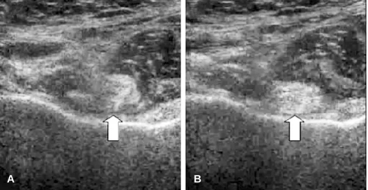
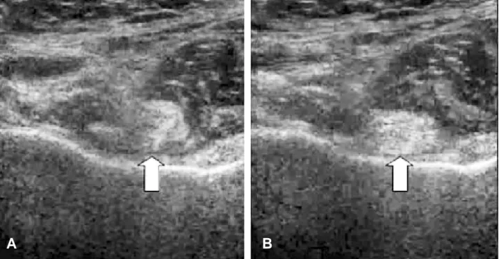
Fig. 1. 55-year-old female with greater than 10-year history of hip pain with catching and a snap, especially with rotation, with a dynamically confirmed snapping hip. (A) Frame captures from a sonographic cine clip just prior to snapping shows the iliopsoas tendon (arrow) in a vertical orientation but beginning to rotate. (B) In the frame immediately after the snap, the iliopsoas tendon (arrow) is now rotated into a horizontal position and opposed to the acetabular rim18).
A B
Fig. 2. (A) Forty-three year old man without snapping hip. T2-weighted axial MR image reveals normal iliotibial band (short arrow) and gluteus maximus muscle. (B) Forty-four year old man with snapping hip. Thickened iliotibial band and anterior border of the gluteus maximus insertion (white black arrow) is noted in the space between the greater trochanter iliotibial band36).
A B
찰할 수 있다(Fig. 1). 자기 공명 영상에서는 T2 강조 영상 에서 일반적으로 비대한 장경대와 대둔근건은 염증과 체 액 저류로 인해 고신호 강도로 나타나며(Fig. 2), 초음파 역시 인대의 비대와 점액낭의 크기를 재는데 유용하며 자 세 변화에 따른 장경대의 위치 파악도 가능하다.
관절의 탄발이나 발음(popping)을 듣거나 느끼는 경우 는 흔하나 수술이 필요할 정도의 동통이나 장애가 있는 경 우는 드물다. 대부분의 환자에게 병의 원인을 인지시키고 탄발을 유발하는 동작을 피하는 것으로 증상이 개선된다.
그러나 일부 소수의 환자에서 지속적인 동통, 기능 제한, 장거리 보행시 증상의 악화와 보존적 치료에 실패를 보이 는 경우 수술적 치료의 적응증이 될 수 있다. external type의 경우 장경대의 두꺼워진 후방 경계나 대둔근의 전 방 경계가 대퇴전자부 위를 미끄러지면서 발생하게 되는
데2,14)일반적으로 통증이 없기 때문에 수술이 필요한 경우
는 드물고, 통증이 있는 경우에도 물리 치료, 국소 주사 요 법, 활동 수정(activity modification) 등으로 조절하며 경 과도 양호하다.
보존적 치료 방법으로는 활동제한, 온열, 비스테로이드 성 약물과 물리치료가 있다. 스테로이드의 주입이 보존적 치료의 한 요소가 될 수 있으며 보통 1회 이상의 주입이 필요하다. Gordon24)등은 51명의 환자를 추적한 결과 49 명에서 스테로이드와 마취제를 같이 주입한 경우에는 평 균 1.5회, 마취제만 주입한 경우에는 1.8회의 주입으로 양 호 이상의 결과를 얻었다. 대부분의 외부형 고관절 탄발음
은 6개월에서 12개월의 보존적인 치료로 호전된다.
장경대를 수술한 첫 보고는 장경대가 대전자에 부착된 곳을 이완 해준 것이다. 수술적 치료가 필요한 경우 국소 마취 하에 긴장된 장경대를 찾아 길이를 연장시키거나 두 꺼워진 장경대 후방부를 전방으로 이동시키는 방법 등이 이용된다. Orlandi44)등은 20명의 환자군에서 대전자에 장 경대의 재접합을 시도하여 16명에서 완전한 통증 해소를 얻었으나 2명에서는 고관절 내회전시 통증이 있었으며 2 명에서는 재발하였다고 보고하였다. 여러 저자들은 또 단 순절개와 재접합 외에도 연장술 및 복원술을 시도하였다.
Sarkis50)등은 4명의 환자 모두에서 대전자 위를 주행하는 장경대를 앞쪽으로 옮겨 주어 탄발음 및 동통의 소실과 함 께 효과적인 길이 연장 효과를 얻었다고 하였으며, Larsen34)등은 평균 2년 동안 관절강 주위 탄발음을 가진 31명의 환자를 대상으로 27명에서는 대둔근의 부착부위 에서 장경인대의 후방 1/2의 절제술을 시행하였으며, 4명 에서는 장경대의 후방피판을 근막의 전외측면에 봉합해 주었으며, 이들 31명 중 8명에서는 전자부 점액낭의 절제 술을 병행하였다.
수술 소견에서 31명중 30명에서는 장경대의 비정상적 인 비후가 발견되었으며, 4년 추시상에서 22명(71%)에서 완전한 증상 소실, 6명(19%)에서 통증 없는 탄발음, 3명 (10%)에서 통증을 동반한 탄발음의 결과를 얻었다. 통증 이 남아있는 3명 중 2명은 재수술하여 좋은 결과를 얻었 다. Brignall9)등은 외부형 탄발음성 고관절이 있는 8명에
Fig. 3. (A) With flexion of the hip. the iliopsoas tendon shifts laterally in relation to the center of the femoral head. (B) With extension of the hip. the iliopsoas tendon shifts medially in relation to the center of the femoral head2).
A B
서 장경대의 Z-성형술을 시행하여 보고하였는데, 모든 환 자들은 탄발음의 감소를 보였고 수술 후 평균 3년 안에 동 통이 없어졌다고 하였다. 이 중 2명의 환자 3례에서 운동 시 때때로 대전자 위의 통증이 있었다 하였으며 수술 후 4 개월 뒤 한명의 환자가 재수술을 받았고 그 후 6년 동안 동통을 느끼지 않았다고 하였다. 국내 보고로 Kyung33)등 은 5명 7예에서 Z-성형술을 시행하여 수술하여 1예에서 운동시 약간의 동통을 호소하였으나, 모든 환자에서 탄발 음이 소실되어 만족할 만한 결과를 얻었다고 보고하였고 Lee37) 등은 4명 6예에서 Z-성형술을 시행하여 모든 환자 에서 탄발음이 소실되어 만족할 만한 결과를 얻었다고 보 고하였다.
internal type의 경우 장요근건이나 장요근 밑에 있는 점액낭에 의해 발생되며, 고관절 굴곡시 장요근건이 대퇴 골두의 외측으로 이동했다가 고관절 신전하면 건이 내측 으로 미끄러지면서 탄발이 유발된다12) (Fig. 3). 진단에는 단순 방사선과 초음파를 이용하거나46), 관절 내부의 이상 을 감별하기 위해 자기 공명 영상이 유용하며39), 비후된 장요근 점액낭의 확인에는 iliopsoas bursography도 도 움이 된다. Bernard5) 등은 증상이 있는 internal type의 54명의 환자에서 단순 방사선과 초음파를 이용한 경우 83%에서 진단할 수 있었으며 추가로 MRI를 시행한 경우 100%에서 진단할 수 있다고 보고하였다. 따라서 초기 진 단 방법으로는 단순 방사선과 초음파를 이용하고 추가로 자기 공명 영상을 시행할 것을 추천하였다. 치료는 장요근 건의 계단식 연장술(step cut lengthening)이나 수술적 이완술을 시행할 수 있다. Jacobson과 Allen27)은 증상이 있는 18명 20예에서 장요근건의 연장술을 시행하여 14예 에서 재발 없이 치료되었다고 보고하였으며, Taylor와 Clarke60)는 통증이 있는 16예에서 수술적 이완술을 시행 하여 10예에서 통증 및 탄발음 소실을 얻었으며, 5예에서 는 통증이 소실되었다고 보고하였으며, 합병증으로는 대 퇴부로 가는 신경에 손상을 주어 대퇴 전외측부의 감각 소 실을 유발 할 수 있다고 하였다.
관절 내부의 원인 (Intra-articular type)의 경우 골연골 종증, 관절내 유리체, 비구순 파열, 고관절 아탈구, 골절 골편 등이 유발 원인이다. 진단은 주로 자기 공명 관절 조 영술(MR arthrography)을 이용한다. Toomayan61) 등은 비구순 파열 진단에서 통상적인 자기 공명 영상을 시행하 였을 경우 민감도가 25%인 것에 비해 자기 공명 관절 조 영술(MR arthrography)을 시행하였을 경우에는 민감도 가 92%라고 보고하였으며, Schmid52) 등은 고관절 연골 결손에서 자기 공명 관절 조영술을 시행하여 진단하였을 경우 민감도는 79%, 특이도는 77%라고 보고하였다. 치료 는 관혈적 또는 관절경을 이용하여 유발 인자를 제거하거 나 복원해 주는 방법이 있다62).
고관절 주위 건염 및 점액낭염 (Tendinitis & Bursitis)
1. 대전자 동통 증후근
(Greater trochanteric pain syndrome)
대전자 동통 증후근(GTPS)은 가장 흔한 regional pain syndrome 중 하나로 측와위에서 대퇴전자부 위로 촉진 시 압통을 호소하는 질환을 의미한다40). 외전근건의 이상 이 소위 GTPS의 흔한 원인이며 중년 또는 노년의 여성에 서 호발 한다32).
GTPS의 유병률에 대한 자료는 적으나, 만성 하부요통 을 호소하는 환자의 20~35%가 신체검사를 통해 GTPS를 진단받는다. Kingzett-Taylor32) 등은 하부요통이 있는 250명의 환자를 대상으로 시행한 자기 공명 영상 소견 중 35명에서 중둔근건과 소둔근건의 건염 및 건파열을 발견 하였고 이중 8명은 완전 파열, 14명은 부분 파열을 보였다 고 보고하였다. GTPS는 주로 중둔근건과 소둔근건을 침 범하며 이들 근육들이 주요 기능 중 하나인 고관절의 내회 전을 담당하기 때문에‘고관절 회전근개 (hip rotator cuff)’라고 하며, 건염, 부분 또는 완전 파열, 견열 등을 나 타낸다32). 견관절의 회전근개와 비교하여 고관절의 회전 근개의 구성은 내회전을 하는 견갑하근은 장요근으로 소 전자에 부착하며, 두개의 외전근인 극상근과 극하근은 대 전자부에 부착하는 중둔근과 소둔근, 상완이두건장두는 대퇴골두를 가로지르는 대퇴직근으로 고관절의 회전근개 가 구성된다. 고관절 회전근개 파열의 가장 많은 유형은 중둔근 앞쪽건의 부분파열이 있는 경우와 중둔근의 파열 과 소둔근의 부분파열이 동반된 경우 이다38). Bunker10)등 은 대퇴경부 골절이 있는 50명의 환자중 11명에게 중둔근 과 소둔근의 만성파열이 있는 것에 대하여 보고하였다.
GTPS는‘전자 점액낭염 (trochanteric bursitis)’으로 불 려지기도 하는데 일반 점액낭염과의 차이점은 염증에 의 한 통증이 아니며 원인 역시 근막 동통과 연관되어 발생하 게 된다38). 고관절의 생역학적 불균형으로 인하여 중둔근 및 소둔근의 반복적인 미세 외상이 건, 근육, 연부조직의 퇴행성 변화를 가져오는 것으로 생각되고 있다. 따라서 고 관절의 퇴행성 관절염, 척추 병변, 하지 길이 부동 등이 병 을 유발할 수 있다. Segal53)등은 여성, 장경대 압통, 슬관 절 퇴행성 관절염, 하부 요통이 있는 경우 GTPS의 발병과 연관이 있다고 보고하였다.
임상증상으로는 고관절의 측부로 만성적, 간헐적인 통증 을 보이며, 경우에 따라서는 급성으로 예리하고 강한 통증 을 호소하기도 한다. 신체검사가 진단에 매우 중요하며, 수 동적 고관절 운동은 모두 정상이나 능동적으로 고관절을 90도 굴곡하고 외회전시 대퇴전자부 측면에 통증을 호소하 며 저항을 준 상태에 외전시 50~70% 환자에서 통증을 나
타낸다. 또한 신체검사에서 중둔근 보행(Trendelenburg gait) 양성 소견을 보이기도 한다. Bird7)등은 24명의 환자 를 대상으로 11명에 대하여 자기 공명 영상을 시행한 결과 3/4에서 중둔근 보행 양성 소견을 보였다고 하였다.
초음파를 이용하여 GTPS를 진단할 수 있는데 중둔근건 과 소 둔 근 건 의 대 퇴 전 자 부 부 착 부 위 가 표 재 성 (superficial position)으로 위치하기 때문에 초음파가 유 용하며 부분 또는 완전 파열이 있는 경우 90%의 민감도와 95%의 특이도를 보인다16). 자기 공명 영상에서는 파열된 건 주위로 고신호 강도(high signal intensity) 및 액체 신 호(fluid signal), 얇아지거나 두꺼워진 건을 확인 할 수 있 다(Fig. 4)6). Bunker10)등은 전형적인 원형 또는 타원형의 중둔근 및 소둔근건 파열이 주로 대전자부 상부 1 cm 이 내에서 발견된다고 보고하였으며, Cvitanic17) 등은 자기 공명 영상이 GTPS를 진단하는데 91% 높은 정확성을 가 진다고 보고하였다. 핵의학 검사 상 대퇴전자부의 흡착 (uptake)이 증가된 소견을 볼 수 있다. 치료는 대개 보존 적 치료에 잘 반응 하나 증상이 지속되거나 재발한 경우 수술적 복원술 또는 재접합술이 필요한 경우가 있다10). Gordon24) 등은 51명의 환자에서 국소 마취제 주사(local anesthetic injection)와 글 루 코 코 티 코 이 드 주 사 (glucocorticoid injection)를 시행한 결과 49명의 환자에 서 양호(good) 또는 우수(excellent)한 결과를 보였으며, Swezey58)등은 트리암시놀론(triamcinolone) 40 mg을 투
여한 결과 60%의 환자에서 증상의 호전을 보였다고 보고 하였다. 최근 연구에서는 70~100%의 환자에서 부신 피질 호르몬(corticosteroid)를 주사한 결과 증상의 호전을 보이 며 이 중 25%는 10개월 이내에 증상의 재발을 보인다고 하 였다51). Mohammad40) 등은 12~40 mg 베타메타손 (betamethasone) 또는 40~80 mg 메칠프레드니솔론 (methylprednisolone)을 4~6 mL 1% 리도카인(lidocaine) 과 혼합 주사하여 60%의 환자에서 증상의 호전을 보였으 며, 스테로이드의 용량이 많을수록 증상 호전의 정도가 더 높았다고 보고하였다.
수술적 치료가 필요한 경우로는, 첫째 만성 또는 반복적 인 증상 재발하거나, 둘째 방사선학적 검사에서 건 파열이 확인된 경우, 셋째 주입검사에서 양성인 경우, 넷째 중둔 근의 퇴축이나 지방 변성이 없는 경우 시행하게 된다. 주 입검사는 초음파 유도하에 procain을 주사하여 시행하게 되며 통증이 소실되거나 증상이 현저히 개선되는 경우 양 성으로 판단하게 된다. 통증이 재발 할 경우 수술적 치료 의 적응이 된며, 보통 재접합술이 선호된다. 괴사 또는 변 성된 부분을 제거한 후 남아있는 tendon stump를 비흡수 성 봉합사를 이용하여 뼈에 재접합하게 된다. 접근을 위해 절개를 가한 장경대는 봉합하지 않고 열어 놓는다. 대부분 작은 병변이라도 건과 인접해 있는 점액낭의 경우 파열되 어있기 때문에 관절경을 이용하거나 관혈적으로 점액낭 제거술을 동시에 시행한다. Lequesne38) 등은 7명의 환자 에 대해서 재접합술을 시행한 결과 6예에서 증상의 완전 소실을 보였고 나머지 1예에서는 증상의 호전을 보였다고 보고하였다. 그 외에도 여러 저자들이 보고한 결과 예후는 양호하였다.
2. 점액낭염(Bursitis)
고관절 주위 점액낭염의 경우 해부학적 구조 및 위치에 따라 둔부하 점액낭염(subgluteal bursitis), 전자 점액낭 염(trochanteric bursitis), 좌둔 점액낭염(ischiogluteal bursitis), 장치점액낭염(iliopectineal bursitis)으로 나눌 수 있다47)(Fig. 5).
둔부하 점액낭은 해부학적으로 대둔근과 대퇴전자부 및 단외회전근(short external rotator muscle)사이에 위치한 다. 발생원인에 따라 크게 감염성 및 비감염성 점액낭염으로 나눠지며, 감염성 타입의 경우 화농성 고관절염과 감별을 요 하며 주로 고관절의 후방과 심부조직에 동통이 있고 전신적 인 감염 징후(infection sign)가 있으면 의심해 볼 수 있다.
관절천자를 시행하거나 초음파를 이용하여 위치를 확인하 고 직 후외측 절개(straight posterolateral incision)를 이용 하여 배액을 시행하여 치료한다. O’Conner43) 등은 일반적 인 점액낭의 치료에 있어서“PRICEMM”(protection, rest, ice, compression, elevation, medication, modalities) 란 Fig. 4. On the fat-suppressed T2-weighted turbo spin echo
image the gluteus medius tendon demonstrates extensive thickening with increased signal (white arrowheads). In addition, bursitis of the medial subgluteal bursa is visible (black arrowhead)5).
치료 지침을 보고하여 좋은 결과를 보고 하였는데, 이와 같이 비감염성 타입의 경우 항염증약물, 물리치료, 국소 주사 등을 이용하여 치료한다.
전자 점액낭은 대둔건 부착 부위와 외측 광근 사이에 위
치한다. 원인으로는 주로 급성 화농성 감염에 의한 경우가 흔하며 드물게 결핵28), 류마티스 관절염59)에 의한 경우도 있다. 점액낭을 압박하는 외전 및 외회전 자세에서 통증이 악화되며 자기 공명 영상에서 액체가 찬 점액낭(fluid filled bursa), 중격 및 벽 조영증강(septations and wall enhancement) 소견을 관찰 할 수 있다. 감염성 타입 일 경우 항생제 치료와 함께 점액낭 절제 및 외과적 배액을 시행한다. Gerber23)등에 의하면 점액낭과 관절낭 사이의 밸브 기전에 의하여 관절액 교환을 역동적으로 조절한다 고 하였으며, 잦은 재발을 보이는 경우 이러한 일방향 밸 브 (check valve) 기전의 제거를 위한 낭종 절제술과 관 절낭 봉합술을 고려해야 한다고 하였다.
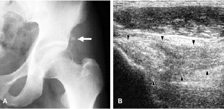
좌둔 점액낭은 좌골 조면과 대둔근 사이에 위치한다. 소 위“Weaver’s bottom”이라고 하여 지속적인 자극 (irritation) 에 의해 유발되며 오랫동안 앉아서 일하는 직 업 (미싱공, 재단사)에서 주로 발생한다. 방사선학적 검사 에서는 단순방사선 촬영은 대부분 음성이며 전산화 단층 촬영(CT), 자기 공명 영상(MRI)을 이용할 수 있으나 Zeiss63)등은 자세한 이학적 검사로 추간판 탈출증이나 직 장의 질환을 감별하여 이러한 고가의 검사는 필요가 없다 고도 하였다. 초음파 검사도 점액낭염 검사로 유용하게 사 용되기도 하지만 골 및 주위의 연부 조직과의 관계는 명확 히 알 수 없는 단점이 있으며 전산화 단층 촬영은 골조직 과의 관계는 명확히 알 수 있으나 종물의 성격을 알 수 없 는 단점이 있다. 자기 공명 영상 소견 상 T1 강조영상에서 는 저신호 강도와 T2 강조영상에서는 고신호 강도를 보이 는 내부의 균일한 포함물이 관찰되며 신체 검사 상 좌골 조면의 바로 아래 종물이 만져지면서 통증 및 압통을 호소 할 경우 대둔근 좌골 점액낭염을 의심해야 한다. 고관절 Fig. 5. Schematic diagram of bursae around greater trochanter40).
Fig. 6. 25-year-old female with lung tranplant with hip pain. (A) Coronal T2 fat-suppressed images show a large left iliopsoas bursal collection (arrow) (B) Static sonographic images prior to injection of the bursa show the hypoechoic bursal collection with area of debris medial to the iliopsoas tendon and lateral to the femoral vessels (arrow)18).
A B
굴곡 시 동통이 증가되고 보행 시 동통 감소를 위해 보폭 이 짧아 진다. 감별 진단해야 할 질환으로는 수핵 탈출증 이나 혈관 정맥염 등이 있다. 대부분 보존적 치료로 증상 이 호전되며 급성 화농성 감염 시에는 외과적 배액술을 시 행한다. 피부 절개 시 Boutin8)등은 횡절개로 접근하여야 좌골 신경, 후대퇴피신경(posterior femoral cutaneous nerve), 회음신경(perineal nerve) 등을 안전하게 보호할 수 있다고 하였다.
장치 점액낭은 고관절 주변에서 가장 큰 점액낭으로 고 관절낭 전방에 위치하고, 장요근의 후방을 따라 골반까지 퍼져 있으며, 9~15%는 고관절과 통해있다56). 증상으로는 고관절 전방부 통증 및 장골서혜부 종양, 고관절 굴곡 자 세 및 신전 제한, 스카르파 삼각(scarpa triangle) 주위에 압통이 있다. 주로 고관절 염증으로 인하여 이차적으로 발 생한다. 전산화 단층 촬영, 자기 공명 영상, 초음파를 이용 해 진단하게 되며 관절천자를 시행하여 확진하게 된다 (Fig. 6). 치료는 경피적 도관 배액술이나 외과적 배액술 을 시행하게 된다.
3. 고관절 주위 석회화 건염
(Calcific tendinitis around hip joint)
고관절 주위 석회화 건염의 경우 서혜부 통증이나 하지 방사통 같은 비전형적인 증상이나 드문 발생빈도로 인해 정확한 진단이 어려울 수 있다29). 조직학적으로 관절 주위 조직 및 건의 석회 인산염(calcific phosphate)이 특징적 이며, 임상 증상은 갑작스럽게 발생하여 서서히 소실되는
통증을 보인다. 급성기에는 국소 염증 반응 및 미열을 나 타내며 만성 통증과 압통 또는 증상이 없는 경우도 있다.
외전건에 발생할 경우 요추간판 탈출증과 유사한 대퇴 우 회측 통증을 유발한다. 40~70세 사이에 주로 발생하며 대 퇴골의 대둔결절의 대둔건 부착 부위에 가장 호발한다. 그 외에도 및 대퇴 직근(rectus femoris), 외측 광근(vastus lateralis), 이상근(piriformis), 장요근(iliopsoas), 대내전 근(adductor magnus), 대퇴이두근(biceps femoris), 중 둔근(gluteus medius), 소둔근 건(gluteus minimus tendon)에도 발생될 수 있다.
원인은 정확하지는 않으나 유전적 요인 및 당뇨, 갑상선 질환, 결핵 등과 연관이 있다. 발생기전은 근 부착 부위의 반복적 운동, 만성적 과부하, 국소 허혈에 의한 연골변성, 석회 침착 등이 제시되었으며 석회 침착물의 분해가 심한 통증을 유발한다고 알려져 있다22). 감별해야 할 질환으로 는 비구 부골, 견열 골절, 대퇴 직근의 종자골, 화골성 근 염, 악성 종양 (피질골에 인접한 골막 연골종, 연골 육종, 활막 육종) 등이 있다.
단순방사선 검사상 전후면 또는 frog-leg lateral view 를 시행하여 비정형성 형태의 석회 결절을 확인 할 수 있 으며 건 주위 피질골의 미란 또는 천공성 병소도 나타날 수 있다. 전산화 단층 촬영을 시행하여 악성 종양과 감별 할 수 있으며25), 자기 공명 영상을 시행하여 염증의 정도 를 확인하며 이 것은 통증의 정도와 비례하게 된다30). 단 순 방사선 검사상 석회화 건염은 비정형성 형태의 석회화 병변으로 나타난다. 전산화 단층 촬영이 석회화 병변의 발 생 위치를 찾는데 민감도가 높으며, 자기공명영상검사를
Fig. 7. 34-year-old man with calcific tendinitis of the left rectus femoralis. (A) Radiography of the pelvis shows small calcifications adjacent to the acetabulum, suggesting calcifications in the orgin rectus femoris tendon. (B) Sonographic image of the left rectus femoris tendon shows thickening of the tendon and tiny calcific spots without shadowing35).
A B
통해서 관절 및 연부 조직의 변화를 확인할 수 있다. 최근 에는 초음파를 이용한 석회화 건염 진단 및 치료에 대하여 보고되고 있는데, 특히 초음파는 건의 정확한 상태에 대해 검사 할 수 있는 장점이 있으며, 초음파 소견 상 건의 비 후, 후방음향음영을 동반하거나 또는 동반하지 않은 난원 형의 고에코 음영, 색 도플러 검사 상 혈류 증가를 보인다 (Fig. 7). Chiou15)등은 color 또는 doppler 초음파를 견 관절의 석회화 건염 환자에서 시행한 결과 임상증상이 심 할 수록 혈류가 더 증가하는 소견을 보여 서로 연관성이 있다고 보고하였다.
치료는 비스테로이드 소염제, 진통제를 투약하거나 전 산화 단층 촬영 유도57)또는 초음파 유도55)하에 부신 피질 호르몬과 국소 마취제의 혼합 주사를 시행하여 통증의 빠 른 소실을 얻을 수 있다. Sarkar30)등은 전산화 단층 촬영 유도하에 메칠프레드니솔론 (methylprednisolone) 80 mg과 0.5% bupivacaine 2 ml를 조합하여 국소 주사하여 대퇴 직근의 reflected head에서 발생했던 석회화 건염 6 예중 5예에서 추가적인 시술 없이 즉각적인 증상의 소실 을 얻었고, 모든 예에서 방사선 추시상 4개월 내에 석회화 병변이 완전히 소실되었다는 보고하였으며, 국내에서는 Ahn1) 등은 고관절 주위 대퇴 직근 석회화 건염 환자들에 서는 골절 침대에 환자를 앙와위로 눕히고 방사선 투시기 유도하에 석회화 병변의 실질부에 도달하여 흡입 후 methylprednisolone 80 mg과 0.5% bupivacaine 2 ml 혼합물을 주입하여 빠른 증상의 소실을 보고하였다. 반면 Faure19) 등은 미세 결정에 의한 염증 치료를 목적으로 부
신피질 호르몬을 투여할 수 있는데 감염의 위험성에 주의 해야 한다고 하였으며, 건 내의 주사는 국소 괴사를 유발 할 수 있다고 하였다. 그 외에도 급성 석회화 건염에서 다 발성 탐침술(multiple needling)의 효과가 보고되고 있다.
치료로 관혈적 또는 관절경을 이용하여 수술적 제거술 을 시행하기도 한다. Kandermir31)등은 내시경을 이용하 여 고관절 주위 만성 석회화 건염을 효과적으로 치료하였 다고 보고하였다. 주로 보존적 치료에 오랫동안 반응이 없 거나 주사 요법이 실패한 경우, “needle phobia”가 있는 경우, 악성 종양과 감별이 필요한 경우 수술적 제거술을 시행하게 된다.
이상근 증후근(Piriformis syndrome)
이상근 증후군은 드문 질환이며 둔부의 압통, 동통, 하 지의 감각이상 및 방사통으로 인해 요추의 추간판 탈출증 으로 종종 오인되어 잘못된 치료를 할 수가 있으며 그 치 료방법도 추간판 탈출증과 다르기 때문에 좌골신경통을 유발하는 타 질환과의 감별이 필수적이다.
이상근 증후군은 대좌골 절흔(greater sciatic notch) 부 위의 좌골신경 근위부에서의 포착증후군으로 설명할 수 있다. 1947년 Robinson48)은 처음으로 이상근 증후군이라 고 명명하였으며, 특징적인 임상양상을 보고하였다. 첫째, 둔부나 천장관절부위 외상력, 둘째, 보행시 악화되는 천장 관절, 대좌골 절흔, 이상근 주위의 동통 및 하지로의 방사 통, 셋째, 물건을 들거나 허리를 굽힐때 악화되는 증상, 넷
Fig. 8. Magnetic resonance image demonstrates the slightly hypertrophied right piriformis muscle (arrow) of the right hip. (A) axial image, (B) coronal image37).
A B
째, 증세가 악화될 때 촉지되는 심한 압통이 발생하는 이 상근 주위의 종물, 다섯째, 하지 직거상 검사상 양성소견, 여섯째 이환기간에 따른 둔부의 위축소견이다. 이상근은 천 추 의 골 반 면 , 대 좌 골 절 흔 의 상 연 , 천 결 절 인 대 (sacrotuberous ligament)의 골반면에서 기시하여 대좌 골 절흔을 통과하여 원형의 근건형태로 대전자의 상연에 부착하고 제 5요추신경, 제 1천추신경, 제 2천추신경의 지 배를 받는다. 일반적으로 좌골 신경은 이상근 전방으로 상 하로 주행하지만 10~21%에서 해부학적 기형이 있는 것 으로 보고되고 있다26). 이상근은 골반내에서 기시하여 대 좌골 절흔을 통과하기 때문에 근육과 좌골신경이 근접하 고 접촉이 되면 좌골신경통(sciatic pain)이 발생할 수 있 으며, 이를 이상근 증후군이라 부른다.
이상근 증후군의 원인으로는 둔부 외상에 의해 속발된 이상근과 근막의 염증 및 부종으로 인한 좌골신경의 압박, 이상근의 유발점 증후군, 이상근의 기형으로 인한 좌골신 경의 압박 등으로 생각할 수 있다. 이상근 증후군을 진단 하는 데에는 많은 어려움이 있으며, 좌골신경통의 발생으 로 인해 추간판 탈출증으로 종종 오인되기 때문에 추간판 탈출증에 대한 치료를 받거나 다른 치료를 받으며 여러 병 원을 방문하는 경우가 대부분이다. 이상근 증후군을 확진 하기 위해서는 임상적 소견 및 자기공명영상검사, 근전도 등 여러 가지 검사를 종합적으로 고려하여 판단하여야 한 다. 이상근 증후군의 진찰 소견으로는 둔부의 대 좌골 절 흔 부분의 압통, 고관절 외전 및 내회전시에 증가하는 동 통, 하지직거상 검사상 제한된 소견 등이 관찰된다.
Freiberg씨 징후 및 고관절을 굴곡한 상태에서 외전하려 고 하는 것을 방해할 때 근력약화와 동통, 기능의 감소가 나타나는 Pace씨 징후 등의 소견을 보이며 환자를 앙와위 로 눕혔을 때 이환된 부위의 외회전으로 인해 비대칭적인 모습을 보이는 이상근(Piriformis) 징후21)를 관찰 할 수 있 다. 컴퓨터 단층촬영 및 자기공명영상 검사상 이상근의 비
대45,49)를 보이거나(Fig. 8) 비골신경을 자극하여 그에 따르
는 마미 활동 전위(Cauda equina action potential)의 변 화를 관찰41)하여 이상근 증후근을 의심할 수 있으나 확실 하게 진단할 수 있는 방법은 없으며, 상기에 서술한 질환 들과 감별하면서 임상적 및 방사선학적 소견을 종합하여 진단을 하는 것이 필수적이다42).
이상근 증후군은 대부분의 경우에 있어서 물리치료, 비 스테로이드성 항염증제의 투여, 경직장 마사지(transrectal massage) 등의 보존적 치료로 치료가 되나3), 보존적 치료 에 효과가 없는 경우 마취약제나 스테로이드 제제의 국소 주사를 시행할 수도 있다. 최근에는 초음파 유도하에 국소 주사를 시행하거나, Botulinum toxin의 이상근 내 근육 주사가 동통감소의 효과가 있다고 보고하는 저자들도 있
다12,20). 수술적 요법으로는 이상근의 유리술 또는 절제술,
섬유대 및 혈관의 제거, 신경해리술(neurolysis) 등이 있
으며 예후는 양호한 것으로 보고되고 있다. 근절제술의 경 우 60~80%, 유착박리술의 경우 70~100% 까지 성공율이 보고되고 있다4).
결 론
고관절 동통은 그 형태와 원인들이 매우 다양하며, 그 중 연부 조직 질환으로 인한 고관절 동통은 그 빈도가 드 물고 임상 증상이 특이하지 않아 정확한 진단과 치료를 위 해서는 고관절 주위 연부조직들의 특성을 이해하고 이에 알맞은 진찰이나 검사 등이 필요하다. 앞에서 고찰해 본 연부조직 질환들에 대한 적절한 진찰 및 검사를 바탕으로 정확한 진단방법을 이해하고, 이를 바탕으로 올바른 치료 를 시행함으로써 연부 조직 질환으로 인한 고관절 동통에 대하여 바르게 대처해야 할 것이다.
REFERENCES
01. Ahn GY, Jang JH, Yun HH.Calcific Tendinitis of the Rectus Femoris Around the Hip Joint. J Korean Hip Soc 18: 73-78, 2006.
02. Allen WC, Cope R. Coxa saltans. The snapping hip revisited. J Am Acad Orthop Surg, 3: 303-308, 1995.
03. Barton PM. Piriformis syndrome: A rational approach to management. Pain, 47: 345-352, 1991.
04. Benson ER, Schutzer SF. Posttraumatic piriformis syndrome: Diagnosis and results of operative treatment. J Bone Joint Surg, 81-A: 941-949, 1999.
05. Bernard M, Christian WAP, Juerg H. Hip pain in adults:
MR imaging appearance of common causes. Eur Radiol 17: 1746-1762, 2007.
06. Binnie JF. Snapping hip (Hanche a resort; Schnellend Hefte). Ann Surg, 75-A: 909-910, 1993.
07. Bird PA, Oakley SP, Shnier R, Kirkham BW. Prospective evaluation of magnetic resonance imaging and physical examination findings in patients with greater trochanteric pain syndrome. Arthritis Rheum, 44: 2138-45, 2001.
08. Boutin FJ Sr, Boutin RD, Boutin FJ Jr. Operative orthopaedics, Philadelphia, J.B.Lippincott Co: 3419- 3432, 1993.
09. Brignall CG, Stainsby GD. The snapping hip. J Bone Joint Surg, 73-B: 253-254, 1991.
10. Bunker TD, Esler CN, Leach WJ. Rotator-cuff tear of the hip. J Bone Joint Surg, 79-B: 618-620, 1997.
11. Cardinal E, Kenneth B, Capello W, Duval N. US of the snapping iliopsoas tendon. Radiology, 198: 521-522, 1996.
12. Childers MK, Wilson DJ, Gnatz SM, Conway RR, Sherman AK. Botulinum toxin type A use in piriformis muscle syndrome : a pilot study. Am J Phys Med Rehabil, 81: 751-759, 2002.
13. Choi JC, Kim YH, Na HY, et al. Treatment of Calcific
Tendinitis around Hip Joint. J Korean Hip Soc 17: 83-87, 2005.
14. Choi YS, Lee SM, Song BY, et al. Dynamic sonography of external snapping hip syndrome. J Ultrasound Med, 21:
753-758, 2002.
15. Chiou HJ, Chou YH, Wu JJ, Hsu CC, Huang DY, Chang CY. Evaluation of calcific tendonitis of the rotator cuff:
role of color Doppler ultrasonography. J Ultrasound Med, 21: 289-295, 2002.
16. Connell DA, Bass C, Sykes CA, Young D, Edwards E.
Sonographic evaluation of gluteus medius and minius tendinopathy. Eur Radiol, 13: 1339-1347, 2003.
17. Cvitanic O, Henzie G, Skezas N, Lyons J, Minter J. MRI diagnosis of tear of the hip abductor tendons.(gluteus medius and gluteus minimus) AJR Am J Roentgenol, 182:
137-143, 2004.
18. Donna G. Blankenbaker, Michael J. Tuite. The painful hip.: new concepts Skeletal Radiol, 35: 352-370, 2006.
19. Faure G, Daculsi G. Calcified tendinitis: a review. Ann Rheum Dis, 42(suppl): 49-53, 1983.
20. Fishman LM, Anderson C, Rosner B. BOTOX and Physical therapy in the treatment of piriformis syndrome.
Am J Phys Med Rehabil, 81: 936-942, 2002.
21. Foster MR. Piriformis syndrome. Orthopedics, 25: 821- 825, 2002.
22. Gartner J, Simons B. Analysis of calcific deposits in calcifying tendonitis. Clin Orthop Relat Res, 254: 111- 120, 1990.
23. Gerber NJ, Dixon AS. Synovial cyst and juxtaarticular bone cysts. Semin Arthritis Rheum, 3: 323-327, 1974.
24. Gordon EJ. Trochanteric bursitis and tendinitis. Clin orthop Relat Res, 20: 193-202, 1961.
25. Harris JH Jr, Coupe KJ, Lee JS, Trotscher T. Acetabular fractures revisited: Part 2, a new CT-based classification.
AJR Am J Roentgenol, 182: 1367-1375, 2004.
26. Indrekvam K, Sudmann E. Piriformis muscle syndrome in 19 patients treated by tenotomy-α1 to 16-years follow-up study. Int Orthop, 26: 101-103, 2002.
27. Jacobson T, Allen WC. Surgical correction of the snapping iliopsoas tendon. Am J Sports Med 18: 470-4, 1990.
28. Jaovisidha S, Chen C, Ryu KN, Siriwongpairat P, Pekanan P, Sartoris DJ, et al. Tuberculous tenosynovitis and bursitis?: imaging findings in 21 cases. Radiology, 201:
507-513, 1996.
29. Johnson GS, Guly HR. Acute calcific periarthritis outside the shoulder: A frequently misdiagnosed condistion. J Accid Emerg Med, 11: 198-200, 1994.
30. Sarkar JS, Haddad FS, Crean SV, Brooks P. Acute calcific tendinitis of the Rectus Femoris. J Bone Joint Surg, 78-B:
814-816, 1996.
31. Kandemir U, Bharam S, Philippon MJ, Fu FH.
Endoscopic treatment of calcific tendinitis of gluteus medius and minimus arthroscopy, 19: E4, 2003.
32. Kingzett-Taylor A, Tirman PF, Feller J, McGann W,
Prieto V, Wischer T, et al. Tendinosis and tear of gluteus medius and minimus muscles as a cause of hip pain.: MR imaging finding. AJR Am J Roentgenol, 173: 1123-1126, 1999.
33. Kyung HS, Kim SY, Jung HS, Kim YG. Treatment of snapping hip caused by a tight iliotibial tract. J Korean Orthop Soc Sports Med, 2: 158-162, 2003.
34. Larsen E, Gebuhr P. Snapping hip after total hip replacement. J Bone Joint Surg, 70-A: 919-920, 1986.
35. Lee HS, Lee YH, Sung NK, et al. Sonographic findings of calcific tendinitis around the hip. J Korean Soc Ultrasound Med , 24: 139-144, 2005.
36. Lee KH, Kim YS, Sung MS, et al. External snapping hip treated by z-plasty of the iliotibial band. J Korean Hip Soc, 17: 52-57, 2005.
37. Lee SU, Kim KT, Cho YJ, Ryu KN, Chun YS. Sciatic pain caused by piriformis syndrome. J Korean Orthop Assoc, 40: 143-148, 2005.
38. Lesquesne M. From “periarthritis”to hip “rotator cuff”
tears. Trochanteric tendinobursitis. Joint Bone Spine, 73:
344-348, 2006.
39. Miller TT. Abnormalities in and around the hip: MR imaging versus sonography. Magn Reson Imaging Clin N Am, 13: 799-80, 2005.
40. Mohammad IS, Eric LM. Trochanteric Bursitis.(Greater trochanter pain syndrome) Mayo clic Proc, 71: 565-569, 1996.
41. Nakamura H, Seki M, Konishi S, Yamano Y, Takaoka K.
Piriformis syndrome diagnosed by cauda equine action potentials: report of two cases. Spine, 28: E37-40, 2003.
42. Nicola AD, Brian DB. Assessment and differential diagnosis of the painful hip. Clinical Orthopedics and Related Research, 406: 11-18, 2003.
43. O’Connor FG, Sobel JR, Nirschl RP. Five-step treatment for overuse injuries. Physician Sports Med, 20: 128-130, 135-136, 139-142, 1992.
44. Orlandi S, Ossola A, Pellegrini F. L’anca a scatto extra- articolare. Arch Sci Med, 138: 599-602, 1981.
45. Pamela MB. Piriformis syndrome: A rational approach to management. American Academy of Physical Medicine and Rehabilitation in Seattle, WA, on 2 November, 1988.
46. Pelsser V, Cardinal E, Hobden R, Aubin B, Lafortune M.
Extra articular snapping hip: sonographic findings. AJR Am J Roentgenol, 176: 67-73, 2001.
47. Pfirrmann CWA, Chung CB, Theumann NH, Trudell DJ, Resnick D. Greater trochanter of the hip: Attachment of the abductor mechanism and a complex of three bursae- MR imaging and MT bursography in cadavers and MR imaging in asymptomatic volunteers. Radiology, 221: 469- 477, 2001.
48. Robinson DR. Piriformis syndrome in relation to sciatic pain. Am J Surg, 73: 355-358, 1947.
49. Rossi P, Cardinali P, Serrao M, Parisi L, Bianco F, DeBac S. Magnetic resonance imaging findings in piriformis syndrome: a case report. Arch Phys Med Rehabil, 82:
Tendinitis around Hip Joint. J Korean Hip Soc, 17: 83-87, 2005.
14. Choi YS, Lee SM, Song BY, et al. Dynamic sonography of external snapping hip syndrome. J Ultrasound Med, 21:
753-758, 2002.
15. Chiou HJ, Chou YH, Wu JJ, Hsu CC, Huang DY, Chang CY. Evaluation of calcific tendonitis of the rotator cuff:
role of color Doppler ultrasonography. J Ultrasound Med, 21: 289-295, 2002.
16. Connell DA, Bass C, Sykes CA, Young D, Edwards E.
Sonographic evaluation of gluteus medius and minius tendinopathy. Eur Radiol, 13: 1339-1347, 2003.
17. Cvitanic O, Henzie G, Skezas N, Lyons J, Minter J. MRI diagnosis of tear of the hip abductor tendons.(gluteus medius and gluteus minimus) AJR Am J Roentgenol, 182:
137-143, 2004.
18. Donna G. Blankenbaker, Michael J. Tuite. The painful hip.: new concepts Skeletal Radiol, 35: 352-370, 2006.
19. Faure G, Daculsi G. Calcified tendinitis: a review. Ann Rheum Dis, 42(suppl): 49-53, 1983.
20. Fishman LM, Anderson C, Rosner B. BOTOX and Physical therapy in the treatment of piriformis syndrome.
Am J Phys Med Rehabil, 81: 936-942, 2002.
21. Foster MR. Piriformis syndrome. Orthopedics, 25: 821- 825, 2002.
22. Gartner J, Simons B. Analysis of calcific deposits in calcifying tendonitis. Clin Orthop Relat Res, 254: 111- 120, 1990.
23. Gerber NJ, Dixon AS. Synovial cyst and juxtaarticular bone cysts. Semin Arthritis Rheum, 3: 323-327, 1974.
24. Gordon EJ. Trochanteric bursitis and tendinitis. Clin Orthop Relat Res, 20: 193-202, 1961.
25. Harris JH Jr, Coupe KJ, Lee JS, Trotscher T. Acetabular fractures revisited: Part 2, a new CT-based classification.
AJR Am J Roentgenol, 182: 1367-1375, 2004.
26. Indrekvam K, Sudmann E. Piriformis muscle syndrome in 19 patients treated by tenotomy-α1 to 16-years follow-up study. Int Orthop, 26: 101-103, 2002.
27. Jacobson T, Allen WC. Surgical correction of the snapping iliopsoas tendon. Am J Sports Med 18: 470-4, 1990.
28. Jaovisidha S, Chen C, Ryu KN, Siriwongpairat P, Pekanan P, Sartoris DJ, et al. Tuberculous tenosynovitis and bursitis?: imaging findings in 21 cases. Radiology, 201:
507-513, 1996.
29. Johnson GS, Guly HR. Acute calcific periarthritis outside the shoulder: A frequently misdiagnosed condistion. J Accid Emerg Med, 11: 198-200, 1994.
30. Sarkar JS, Haddad FS, Crean SV, Brooks P. Acute calcific tendinitis of the Rectus Femoris. J Bone Joint Surg, 78-B:
814-816, 1996.
31. Kandemir U, Bharam S, Philippon MJ, Fu FH.
Endoscopic treatment of calcific tendinitis of gluteus medius and minimus arthroscopy, 19: E4, 2003.
32. Kingzett-Taylor A, Tirman PF, Feller J, McGann W,
Prieto V, Wischer T, et al. Tendinosis and tear of gluteus medius and minimus muscles as a cause of hip pain.: MR imaging finding. AJR Am J Roentgenol, 173: 1123-1126, 1999.
33. Kyung HS, Kim SY, Jung HS, Kim YG. Treatment of snapping hip caused by a tight iliotibial tract. J Korean Orthop Soc Sports Med, 2: 158-162, 2003.
34. Larsen E, Gebuhr P. Snapping hip after total hip replacement. J Bone Joint Surg, 70-A: 919-920, 1986.
35. Lee HS, Lee YH, Sung NK, et al. Sonographic findings of calcific tendinitis around the hip. J Korean Soc Ultrasound Med , 24: 139-144, 2005.
36. Lee KH, Kim YS, Sung MS, et al. External snapping hip treated by z-plasty of the iliotibial band. J Korean Hip Soc, 17: 52-57, 2005.
37. Lee SU, Kim KT, Cho YJ, Ryu KN, Chun YS. Sciatic pain caused by piriformis syndrome. J Korean Orthop Assoc, 40: 143-148, 2005.
38. Lesquesne M. From “periarthritis”to hip “rotator cuff”
tears. Trochanteric tendinobursitis. Joint Bone Spine, 73:
344-348, 2006.
39. Miller TT. Abnormalities in and around the hip: MR imaging versus sonography. Magn Reson Imaging Clin N Am, 13: 799-80, 2005.
40. Mohammad IS, Eric LM. Trochanteric Bursitis.(Greater trochanter pain syndrome) Mayo clic Proc, 71: 565-569, 1996.
41. Nakamura H, Seki M, Konishi S, Yamano Y, Takaoka K.
Piriformis syndrome diagnosed by cauda equine action potentials: report of two cases. Spine, 28: E37-40, 2003.
42. Nicola AD, Brian DB. Assessment and differential diagnosis of the painful hip. Clinical Orthopedics and Related Research, 406: 11-18, 2003.
43. O’Connor FG, Sobel JR, Nirschl RP. Five-step treatment for overuse injuries. Physician Sports Med, 20: 128-130, 135-136, 139-142, 1992.
44. Orlandi S, Ossola A, Pellegrini F. L’anca a scatto extra- articolare. Arch Sci Med, 138: 599-602, 1981.
45. Pamela MB. Piriformis syndrome: A rational approach to management. American Academy of Physical Medicine and Rehabilitation in Seattle, WA, on 2 November, 1988.
46. Pelsser V, Cardinal E, Hobden R, Aubin B, Lafortune M.
Extra articular snapping hip: sonographic findings. AJR Am J Roentgenol, 176: 67-73, 2001.
47. Pfirrmann CWA, Chung CB, Theumann NH, Trudell DJ, Resnick D. Greater trochanter of the hip: Attachment of the abductor mechanism and a complex of three bursae- MR imaging and MT bursography in cadavers and MR imaging in asymptomatic volunteers. Radiology, 221: 469- 477, 2001.
48. Robinson DR. Piriformis syndrome in relation to sciatic pain. Am J Surg, 73: 355-358, 1947.
49. Rossi P, Cardinali P, Serrao M, Parisi L, Bianco F, DeBac S. Magnetic resonance imaging findings in piriformis syndrome: a case report. Arch Phys Med Rehabil, 82:
519-521, 2001.
50. Sarkis F, Chicote-campos F. Die schnappende hufte.
Orthop Praxis, 14: 618-624, 1978.
51. Schapira D, Nahir M, Scharf Y. Trochanteric bursitis: A common clinical problem. Arch Phys Med Rehabil, 67:
815-817, 1986.
52. Schmid MR, Notzli HP, Zanetti M, Wyss TF, Hodler J.
Cartilage lesions in the hip: diagnostic effectiveness of MR arthrography. Radiology, 226: 382-386, 2003.
53. Segal NA, Felson DT, Torner JC, et al., Multicenter Osteoarthritis Study Group: Greater trochanteric pain syndrome epidemiology and associated factors. Arch Phys Med Rehabil, 88: 988-992, 2007.
54. Seo YJ, Chang JD, Chang SK, Lee GK. Calcific tendinitis of the Rectus femoris: Case Report. J Korean Orthop Assoc, 39: 343-346, 2004.
55. Shabshin N, Rosenverg ZS, Cavalcanti CF. MR imaging of iliopsoas musculotendinous injuries. Magn Reson Imaging Clin N Am, 13: 705-716, 2005.
56. Steiner E, Steinbach LS, Schnarkowski P, et al. Ganglia and cysts around joints. Radiol Clin North Am, 34: 395- 425, 1996.
57. Sung YB, Hwang SH, Ahn JK, et al. Calcific tendinitis of the rectus femoris: a case report. J Korean Hip Soc, 13:
70-72, 2001.
58. Swezey RL. Pseudo-radiculopathy in subacute trochanteric bursitis of the subgluteus maximus bursa.
Arch Phys Med Rehabil, 57: 387-390, 1976.
59. Tanaka H, Kido K, Wakisaka A, Mine T, Kawai S.
Trochanteric bursitis in rheumatoid arthritis. J Rheumatol, 29: 1340-1341, 2002.
60. Taylor G.R, Clarke NM. Surgical release of the ‘snapping iliopsoas tendon’. J Bone Joint Surg, 77-B: 881-883, 1995.
61. Toomayan GA, Holman, WR, Major NM, Kozlowicz SM, Vail TP. Sensitivity of MR arthrography in the evaluation of acetabular labral tears. AJR Am J Roentgenol, 186:
449-453, Feb, 2006.
62. Yamamoto Y, Hamada Y, Ide T, Usui I. Arthroscopic surgery to treat intra-articular type snapping hip.
Arthroscopy, 21: 1120-1125, 2005.
63. Zeiss J, Coombs Rj, Booth RL, Saddemi SR. Chronic bursitis presenting as a mass in the pes anserinus bursa:
MR diagnosis. J Comput Assist Tomogra, 17: 137-140, 1993.
64. Zoltan DJ, Clancy WG, Keene JS. A new operative approach to snapping hip and refractory trochanteric bursitis in athletes. Am J sports Med, 14: 201-204, 1986.
519-521, 2001.
50. Sarkis F, Chicote-campos F. Die schnappende hufte.
Orthop Praxis, 14: 618-624, 1978.
51. Schapira D, Nahir M, Scharf Y. Trochanteric bursitis: A common clinical problem. Arch Phys Med Rehabil, 67:
815-817, 1986.
52. Schmid MR, Notzli HP, Zanetti M, Wyss TF, Hodler J.
Cartilage lesions in the hip: diagnostic effectiveness of MR arthrography. Radiology, 226(2): 382-386, 2003.
53. Segal NA, Felson DT, Torner JC, et al., Multicenter Osteoarthritis Study Group: Greater trochanteric pain syndrome epidemiology and associated factors. Arch Phys Med Rehabil, 88: 988-992, 2007.
54. Seo YJ, Chang JD, Chang SK, Lee GK. Calcific tendinitis of the Rectus femoris: Case Report. J Korean Orthop Assoc, 39: 343-346, 2004.
55. Shabshin N, Rosenverg ZS, Cavalcanti CF. MR imaging of iliopsoas musculotendinous injuries. Magn Reson Imaging Clin N Am, 13: 705-716, 2005.
56. Steiner E, Steinbach LS, Schnarkowski P, et al. Ganglia and cysts around joints. Radiol Clin North Am, 34: 395- 425, 1996.
57. Sung YB, Hwang SH, Ahn JK, et al. Calcific tendinitis of the rectus femoris: a case report. J Korean Hip Soc, 13:
70-72, 2001.
58. Swezey RL. Pseudo-radiculopathy in subacute trochanteric bursitis of the subgluteus maximus bursa.
Arch Phys Med Rehabil, 57: 387-390, 1976.
59. Tanaka H, Kido K, Wakisaka A, Mine T, Kawai S.
Trochanteric bursitis in rheumatoid arthritis. J Rheumatol, 29: 1340-1341, 2002.
60. Taylor G.R, Clarke NM. Surgical release of the ‘snapping iliopsoas tendon’. J Bone Joint Surg, 77-B: 881-883, 1995.
61. Toomayan GA, Holman, WR, Major NM, Kozlowicz SM, Vail TP. Sensitivity of MR arthrography in the evaluation of acetabular labral tears. AJR Am J Roentgenol, 186:
449-453, Feb, 2006.
62. Yamamoto Y, Hamada Y, Ide T, Usui I. Arthroscopic surgery to treat intra-articular type snapping hip.
Arthroscopy, 21: 1120-1125, 2005.
63. Zeiss J, Coombs Rj, Booth RL, Saddemi SR. Chronic bursitis presenting as a mass in the pes anserinus bursa:
MR diagnosis. J Comput Assist Tomogra, 17: 137-140, 1993.
64. Zoltan DJ, Clancy WG, Keene JS. A new operative approach to snapping hip and refractory trochanteric bursitis in athletes. Am J sports Med, 14: 201-204, 1986.