https://doi.org/10.5624/isd.2020.50.1.31
Introduction
The treatment of head and neck cancer is based on 3 mo- dalities, which can be combined or applied individually:
radiotherapy, chemotherapy, and surgery. Radiotherapy, in which ionizing radiation is used to eliminate or control the neoplasm, has been adopted as a standard treatment in the early stages of head and neck cancer. Advanced cases, in contrast, commonly require chemotherapy and/or sur- gery.1,2
The deleterious effects of radiation on bone tissue have been closely examined with the goal of elucidating the
mechanisms leading to osteoradionecrosis,3 but conclusive evidence is not yet available. Histomorphometric analysis of biopsies has proven to be a valuable laboratory method to evaluate bone quality, while clinical methods include digital radiography, ultrasonography, and computed tomog- raphy.4
Panoramic radiographs, especially digital panoramic ra- diographs, are quick and easy to obtain, inexpensive, and comfortable for the patient, and they typically involve the administration of low doses of radiation. In addition, dig- ital radiography has been considered a useful tool for the determination of systemic bone quality through the assess- ment of mandibular bone mass and morphology.5 Despite conflicting results reported in the literature and limitations related to technical aspects and the reproducibility of the method, digital panoramic radiography has been described as capable of predicting osteopenia and osteoporosis after
Impact of radiotherapy on mandibular bone: A retrospective study of digital panoramic radiographs
Luiz Felipe Palma 1,2,*, Ricardo Yudi Tateno 2, Cíntia Maria Remondes 3, Marcelo Marcucci 3, Arthur Rodriguez Gonzalez Cortes 2,4
1Discipline of Descriptive and Topographic Anatomy, Department of Morphology and Genetics, Federal University of São Paulo, São Paulo, Brazil
2MSc Dentistry Program, Ibirapuera University, São Paulo, Brazil
3Stomatology and Oral and Maxillofacial Surgery Center, Heliópolis Hospital, São Paulo, Brazil
4Department of Dental Surgery, Faculty of Dental Surgery, University of Malta, Msida, Malta
ABSTRACT
Purpose: The purpose of this study was to investigate the impact of radiotherapy on mandibular bone tissue in head and neck cancer patients through an analysis of pixel intensity and fractal dimension values on digital panoramic radiographs.
Materials and Methods: Thirty patients with radiographic records from before and after 3-dimensional(3D) conformational radiotherapy were selected. A single examiner carried out digital analyses of pixel intensity values and fractal dimensions, with the areas of interest unilaterally located in the right angle medullary region of the mandible below the mandibular canal and posterior to the molar region.
Results: Statistically significant decreases were observed in the mean pixel intensity(P=0.0368) and fractal dim- ension(P=0.0495) values after radiotherapy.
Conclusion: The results suggest that 3D conformational radiotherapy for head and neck cancer negatively affected the trabecular microarchitecture and mandibular bone mass.(Imaging Sci Dent 2020; 50: 31-6)
KEY WORDS: Radiotherapy; Panoramic Radiography; Mandible
Copyright ⓒ 2020 by Korean Academy of Oral and Maxillofacial Radiology
This is an Open Access article distributed under the terms of the Creative Commons Attribution Non-Commercial License(http://creativecommons.org/licenses/by-nc/3.0) which permits unrestricted non-commercial use, distribution, and reproduction in any medium, provided the original work is properly cited.
Imaging Science in Dentistry·pISSN 2233-7822 eISSN 2233-7830 Received October 31, 2019; Revised December 31, 2019; Accepted January 22, 2020
*Correspondence to : Dr. Luiz Felipe Palma
MSc Dentistry Program, Ibirapuera University, Av. Interlagos, 1329 - Chácara Flora, São Paulo, SP, 04661-100, Brazil
Tel) 55-11-99-100-8038, E-mail) luizfelipep@hotmail.com
correlating those conditions with systemic bone mineral density values obtained through dual-energy X-ray absorp- tiometry.6
A digital image is composed of an array of small areas known as pixels, to which specific numeric values are assigned.5 Pixel intensity analysis of a digital panoramic radiograph can be based on a grayscale that ranges from 0(black) to 255(white),7 in which pixel intensity is consid- ered an indirect measurement of local bone mass5. Fractal analysis is a mathematical model that describes and ana- lyzes complex forms and structural patterns, numerically expressed as fractal dimensions.5 Although several methods of fractal analysis have been described,8 the box-counting technique has been the most widely applied and is suited for binary images9 such as radiographs. This method can reveal complex interconnections of the trabecular bone, consequently indicating bone quality.10
Thus, the aim of the present study was to evaluate the impact of radiotherapy on mandibular bone tissue in head and neck cancer patients through an analysis of pixel in- tensity and fractal dimension values on digital panoramic radiographs.
Materials and Methods
Sample characteristics, eligibility criteria, and ethical issues
This retrospective study was conducted using digital panoramic radiographs from the electronic collection of the Stomatology and Oral and Maxillofacial Surgery Cen- ter at Heliópolis Hospital, obtained from patients between 2015 and 2018. To be considered eligible, individuals were required to be at least 18 years of age, to have a history of 3-dimensional(3D) conformational radiotherapy for head and neck cancer(involving the cervicofacial and supracla- vicular fossa fields), and to have panoramic radiographic records available from before and after radiotherapy. As a local protocol, all initial radiographic images were taken during the oral interventions administered in the prepara- tion for oncologic treatment, ranging from 1 to 3 weeks prior to radiotherapy.
The following exclusion criteria were applied: systemic diseases known to alter bone metabolism(e.g., osteopo- rosis, thyrotoxicosis, or hyperparathyroidism), use of any type of medication with known effects on bone metabolism (e.g., corticosteroids, antiangiogenic agents, or antiresorp- tive agents such as bisphosphonates or denosumab), poor positioning during the radiographic examination, poor- quality images, and the presence of extensive mandibu-
lar pathologies(e.g., condensing osteitis, bone sclerosis, fibrous dysplasia, periapical lesions, or pathological frac- tures) in the areas of interest.
This study was approved by the Research Ethics Commit- tee at Ibirapuera University(CAAE 91440318.3.0000.5597) and Heliópolis Hospital(CAEE 91440318.3.3001.5449) and was in compliance with the Declaration of Helsinki.
Participants’ privacy and confidentiality of information were ensured in all phases of the study.
Digital panoramic radiography
Digital panoramic radiographs were obtained using a PaX-400C device(Vatech, Hwaseong, Korea) with an ex- posure time of 14s and an electric current of 8mA. As suggested by the manufacturer, male and female subjects received peak kilovoltages of 68kVp and 67kVp, respec- tively. The images were stored digitally as 8-bit grayscale bitmap(.bmp) files with a resolution of 2900×1532 pixels.
Pixel intensity and fractal dimension analyses The pixel intensity and fractal dimension analyses were adapted from the original studies by White and Rudolph11 and Tosoni et al.5 and were conducted using the image processing software ImageJ 1.52a(National Institutes of Health, Bethesda, MD, USA).
Initially, a fixed selection tool was used(100×100 pix- els) to determine the region of interest(ROI) on each ra- diograph. This region was located unilaterally in the right angle region of the mandible(below the mandibular canal and posterior to the molar region), parallel to the posterior margin of the mandibular ramus, and included only trabec- ular bone(Fig. 1).
The mean pixel intensity value of each ROI was deter- mined using the software histogram, and fractal dimension analysis was then performed. For this purpose, the image of the ROI was duplicated. This copy of the image was blurred using a Gaussian filter with a 35-pixel radius to re- tain only large density variations and was subtracted from the original image, generating a third image. A constant of 128(corresponding to the half-value of the gray levels on the 8-bit scale) was added at each pixel position on the third image to ensure that variations in brightness corre- sponding to specific features(trabeculae and medullary spaces) were visible. The image was then converted into a binary image at a brightness threshold value of 128 to fa- cilitate image segmentation into components that visually resembled trabeculae and medullary spaces. The image was then subjected to erosion and dilation to reduce noise and was inverted to reveal the trabeculae. Next, the image
was skeletonized, or eroded until only the center pixel line remained, and superimposed over the original image to certify that the structures corresponded to the trabeculae.
Finally, the box-counting method was applied with box siz- es of 2, 3, 4, 6, 8, 12, 16, 32, and 64 pixels to determine the fractal dimension(Fig. 2).
Statistical analysis
The analyses were conducted by a trained observer, who was a dentist with expertise in oral radiology. Intra-exam- iner reproducibility was assessed by reanalysis of 10% of the sample, performed 2 weeks after the initial analysis
to reduce memory bias, and evaluated using the intraclass correlation coefficient(ICC). Another trained observer car- ried out analyses of 10% of the sample, and inter-examiner reproducibility was determined using the ICC as well.
The characteristics of the subjects and their oncological treatment were analyzed descriptively. The data distribution was assessed using the Shapiro-Wilk test, and the pixel in- tensity and fractal dimension values before and after radio- therapy were analyzed using the Wilcoxon sign-rank test.
The statistical analysis was carried out using BioEstat 5.3 software(Instituto de Desenvolvimento Sustentável Ma- mirauá, Amazonas, Brazil) with a 5% significance level.
Fig. 1. Region of interest selection.
Fig. 2. Fractal dimension analysis process. A. Region of interest. B. Result of blurring of (A). C. Result of subtracting (B) from (A). D. Re- sult of adding 128 to each pixel position of (C). E. Result of conversion of (D) into a binary image. F. Result of erosion and dilation of (E). G.
Result of inversion of (F). H. Result of skeletonizing of (G). I. Result of superpositioning (H) over (A).
(A) (B) (C) (D) (E)
(F) (G) (H) (I)
Results
Thirty individuals were selected for the study, resulting in a total of 60 digital panoramic radiographs. Most of these individuals were male(80%), had a primary tumor located in the pharynx(43.4%), and had received a total ra- diation dose of 70Gy(66.7%). The most common interval separating the pre- and post-radiotherapy radiographs was 12 to 24 months(43.4%). More detailed information on the sample is provided in Table 1.
The normal distribution was rejected for the pixel in- tensity data before(P =0.088) and after radiotherapy (P=0.3081) and for the fractal dimension data after radio- therapy(P=0.3851). Intra- and extra-examiner reproduc- ibility were confirmed for both pixel intensity(ICC, 0.89;
P =0.001) and fractal dimension(ICC, 0.81; P =0.01) evaluations.
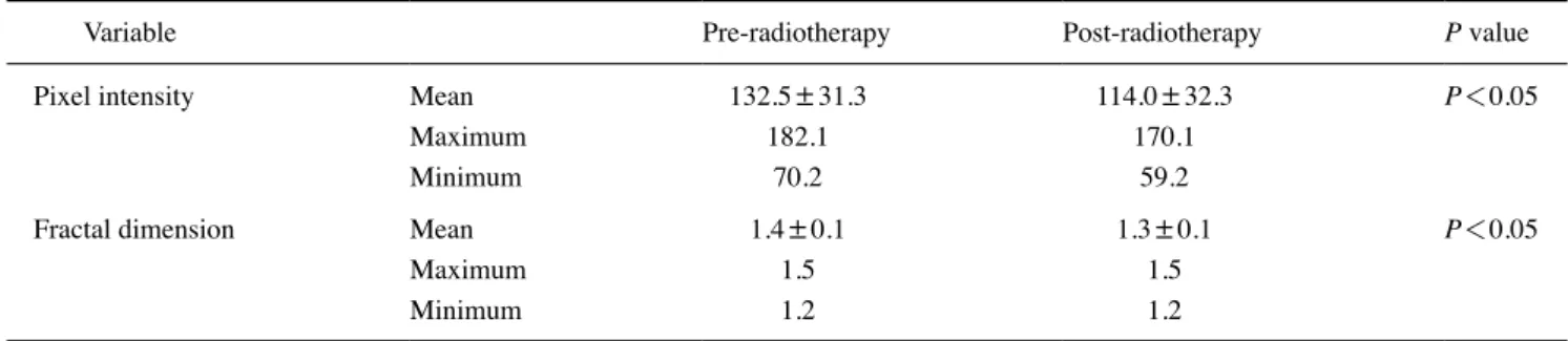
The mean values of pixel intensity before and after ra- diotherapy were 132.5±31.3 and 114.0±32.3, respec-
tively, constituting a statistically significant difference (P=0.0368). The mean fractal dimension values pre- and post-radiotherapy were 1.4±0.1 and 1.3±0.1, respective- ly, similarly presenting a statistically significant difference (P=0.0495). Detailed information on the analyses is pro- vided in Table 2.
Discussion
Bone quality is determined by the tissue microarchitec- ture and the degree of mineralization. Low bone quality has been directly associated with the failure of dental implants, since primary stability and osseointegration are influenced to a large extent by the characteristics of the surrounding bone.4 Evidence has shown that patients with osteoporosis experience more rapid development and greater severity of periodontal disease, as well as a lower accumulated long- term survival rate of dental implants12 and a higher risk of fractures, than individuals without osteoporosis.13
Table 1. Demographic and clinical characteristics of the participants
Characteristic Patients
n %
Sex Male 24 80%
Female 6 20%
Primary tumor location Pharynx 13 43.3%
Oral cavity 9 30%
Larynx 4 13.3%
Parotid gland 3 10%
Occult primary 1 3.3%
Total dose of radiation ≤59Gy 1 3.3%
60 to 69Gy 9 30%
70Gy 20 66.7%
Time period between pre-
and post-radiotherapy radiographs 3 to 12 months 12 40%
12 to 24 months 13 43.3%
24 to 35 months 5 16.7%
Table 2. Mean pixel intensity and fractal dimension values on pre- and post-radiotherapy radiographs in head and neck cancer patients
Variable Pre-radiotherapy Post-radiotherapy P value
Pixel intensity Mean 132.5±31.3 114.0±32.3 P<0.05
Maximum 182.1 170.1
Minimum 70.2 59.2
Fractal dimension Mean 1.4±0.1 1.3±0.1 P<0.05
Maximum 1.5 1.5
Minimum 1.2 1.2
The present study investigated the characteristics of mandibular bone tissue on digital panoramic radiographs, which are commonly used imaging exams in dental prac- tice. Scientific evidence supports the use of digital pan- oramic radiography for the analysis of bone tissue, despite limitations inherent to the technique concerning tissue properties, such as software and image receptor configura- tion, parameters of the radiographic device(such as peak kilovoltage, exposure time, and current), and overlap of tissues and structures.6 Dual-energy X-ray absorptiometry is a costly gold-standard exam for the diagnosis of osteo- porosis. Panoramic radiographs can detect a reduction in mandibular mineral density at a lower cost, which allows early systemic diagnosis and prompt referral to a special- ized professional.14
A pixel is a single digital point with a variable intensity, constituting the smallest unit of an image that can be rep- resented or controlled. In a grayscale radiographic image, the value of each pixel provides information about inten- sity alone.15 For pixel intensity analysis for the indirect measurement of local bone mass, some authorities have suggested methods to normalize variations in noise and image production that could influence the comparison be- tween radiographs obtained with the same devices and pa- rameters, with these methods including density markers,5 binarization, and skeletonization.15 Herein, the direct use of histograms in ImageJ seemed to be satisfactory, making the procedures even quicker and simpler.
Analysis of the fractal dimension is a low-cost, easily ap- plicable, non-invasive, and accurate method for quantifying the interface pattern of trabecular bone and bone marrow, with higher values indicating a more complex structure.10 It is a technique used to identify scale-invariant structures that are not affected by the conditions under which radio- graphic images are produced.8,9 However, a range of dental radiographic processing methods have been developed as adaptations of the original article by White and Rudolph,11 which was used as a reference in our study.
Another conflicting point in the literature relates to the mandibular site used to determine the ROI and its size. The anterior region of the mandible seems to produce relatively inaccurate images due to the overlap of the cervical spine and relatively high likelihood of blurring.16 In our study, we used the angle of the mandible below the mandibular canal and posterior to the molar region to avoid interfer- ence caused by masticatory stress, as proposed by Oliveira et al.,15 and to provide bone structure even in fully edentu- lous aged individuals with extremely atrophic mandibles.
However, this area is subject to the overlapping of other an-
atomical structures, such as the airways, tonsils, soft palate, tongue, and hyoid bone.5 To date, ROI dimensions have not been standardized. The protocol proposed by Tosoni et al.5 (100×100 pixels) was satisfactory for all individuals examined in our study and provided a suitable medullary bone area for the analyses.
Radiotherapy is an important and efficient therapeutic modality for head and neck cancer, although it also affects non-neoplastic cells in or adjacent to the irradiated area.
Its effects may occur during or immediately after irradi- ation(acute effects) or even months or years later(late effects).17,18 Our results demonstrated that 3D conforma- tional radiotherapy significantly affected subjects’ trabec- ular microarchitecture and the mandibular bone mass. One hypothesis to explain this phenomenon is that intraosseous vascularization decreases as a result of exposure to ionizing radiation, with changes in the bone cell pool, progressive fibrosis, and local necrosis.3 These alterations manifest ra- diologically as low bone mineral density, trabecular frac- ture, and loss and destruction of cortical structures.4 Nev- ertheless, the various studies and models addressing this subject have presented highly heterogeneous methods and samples. It is therefore very difficult to realistically com- pare them and apply the results to humans.19
To the best of our knowledge, this is the first study to investigate the impact of radiotherapy on the mandibular bone tissue of head and neck cancer patients using pixel in- tensity and fractal dimension analyses of digital panoram- ic radiographs. As limitations and suggestions for future research, some points need to be taken into account. First, further studies should consider using more representative sample sizes. In addition, accurate data on the total radi- ation dose received by each patient in the ROI should be provided to determine the association between the amount of ionizing radiation received and the level of bone tissue damage. Furthermore, because this was a retrospective study, the interval between the pre- and post-radiotherapy radiographs was not standardized and ranged from 3 to 35 months. Lastly, analyses of other mandibular areas, bilat- eral assessment by more than 1 experienced examiner, and the use of other radiographic techniques(e.g., periapical radiography) could provide more accurate and detailed in- formation.
In summary, based on the results of pixel intensity and fractal dimension analyses, this study suggests that 3D conformational radiotherapy for head and neck cancer neg- atively affected the trabecular microarchitecture and man- dibular bone mass.
Conflicts of Interest: None
References
1. Palma LF, Gonnelli FA, Marcucci M, Dias RS, Giordani AJ, Segreto RA, et al. Impact of low-level laser therapy on hy- posalivation, salivary pH, and quality of life in head and neck cancer patients post-radiotherapy. Lasers Med Sci 2017; 32:
827-32.
2. Palma LF, Gonnelli FA, Marcucci M, Giordani AJ, Dias RS, Segreto RA, et al. A novel method to evaluate salivary flow rates of head and neck cancer patients after radiotherapy: a pilot study. Braz J Otorhinolaryngol 2018; 84: 227-31.
3. Michel G, Blery P, Pilet P, Guicheux J, Weiss P, Malard O, et al. Micro-CT analysis of radiation-induced osteopenia and bone hypovascularization in rat. Calcif Tissue Int 2015; 97:
62-8.
4. Konermann A, Appel T, Wenghoefer M, Sirokay S, Dirk C, Jäger A, et al. Impact of radiation history, gender and age on bone quality in sites for orthodontic skeletal anchorage device placement. Ann Anat 2015; 199: 67-72.
5. Tosoni GM, Lurie AG, Cowan AE, Burleson JA. Pixel inten- sity and fractal analyses: detecting osteoporosis in perimeno- pausal and postmenopausal women by using digital panoram- ic images. Oral Surg Oral Med Oral Pathol Oral Radiol Endod 2006; 102: 235-41.
6. Camargo AJ, Cortes AR, Aoki EM, Baladi MG, Arita ES, Watanabe PC. Diagnostic performance of fractal dimension and radiomorphometric indices from digital panoramic radio- graphs for screening low bone mineral density. Braz J Oral Sci 2016; 15: 131-6.
7. Halazonetis DJ. What do 8-bit and 12-bit grayscale mean and which should I use when scanning? Am J Orthod Dentofacial Orthop 2005; 127: 387-8.
8. Amer ME, Heo MS, Brooks SL, Benavides E. Anatomical vari- ations of trabecular bone structure in intraoral radiographs using fractal and particles count analyses. Imaging Sci Dent 2012; 42:
5-12.
9. Suer BT, Yaman Z, Buyuksarac B. Correlation of fractal dimen- sion values with implant insertion torque and resonance fre-
quency values at implant recipient sites. Int J Oral Maxillofac Implants 2016; 31: 55-62.
10. de Molon RS, de Paula WN, Spin-Neto R, Verzola MH, Tosoni GM, Lia RC, et al. Correlation of fractal dimension with histo- morphometry in maxillary sinus lifting using autogenous bone graft. Braz Dent J 2015; 26: 11-8.
11. White SC, Rudolph DJ. Alterations of the trabecular pattern of the jaws in patients with osteoporosis. Oral Surg Oral Med Oral Pathol Oral Radiol Endod 1999; 88: 628-35.
12. Chambrone L. Current status of the influence of osteoporosis on periodontology and implant dentistry. Curr Opin Endocri- nol Diabetes Obes 2016; 23: 435-9.
13. Dervis E. Oral implications of osteoporosis. Oral Surg Oral Med Oral Pathol Oral Radiol Endod 2005; 100: 349-56.
14. Neves FS, Barros AS, Cerqueira GA, Cruz GA, Reis AA, Alves LB, et al. Assessment of fractal dimension and panoramic ra- diomorphometric indices in women with celiac disease. Oral Radiol(in press).
15. Oliveira ML, Pedrosa EF, Cruz AD, Haiter-Neto F, Paula FJ, Watanabe PC. Relationship between bone mineral density and trabecular bone pattern in postmenopausal osteoporotic Bra- zilian women. Clin Oral Investig 2013; 17: 1847-53.
16. Hwang JJ, Lee JH, Han SS, Kim YH, Jeong HG, Choi YJ, et al. Strut analysis for osteoporosis detection model using den- tal panoramic radiography. Dentomaxillofac Radiol 2017; 46:
20170006.
17. Gonnelli FA, Palma LF, Giordani AJ, Deboni AL, Dias RS, Segreto RA, et al. Low-level laser therapy for the prevention of low salivary flow rate after radiotherapy and chemotherapy in patients with head and neck cancer. Radiol Bras 2016; 49:
86-91.
18. Gonnelli FA, Palma LF, Giordani AJ, Deboni AL, Dias RS, Segreto RA, et al. Low-level laser for mitigation of low sali- vary flow rate in head and neck cancer patients undergoing ra- diochemotherapy: a prospective longitudinal study. Photomed Laser Surg 2016; 34: 326-30.
19. Jegoux F, Malard O, Goyenvalle E, Aguado E, Daculsi G. Ra- diation effects on bone healing and reconstruction: interpre- tation of the literature. Oral Surg Oral Med Oral Pathol Oral Radiol Endod 2010; 109: 173-84.