http://dx.doi.org/10.5624/isd.2016.46.3.179
Introduction
Paranasal sinuses are air cavities that exist in some fa- cial bones. These vary in shape and size, and drain to the nasal cavity either directly or indirectly.1,2 The maxillary sinuses are the largest of the paranasal sinuses, normally segmented by septa, and are located inside of the maxil- lary bones.3,4 These sinuses can present anatomical vari-
ations,5 extending to the anterior region of the maxilla, maxillary tuberosity, hard palate, zygomatic bone, orbit, and alveolar ridge. The extensions to the alveolar region occur in more than 80% of patients.3
In this sense, the root apices of maxillary posterior teeth may well present a close relationship with the sinus floor.
The knowledge of this anatomical relationship is essential when diagnosing changes in the sinus caused by lesions of odontogenic origin or association, surgical planning, intrusion of the maxillary sinus root, fracture of the bone plate with oral sinus communication, recognition of the pathway of dental infections, and planning of orthodontic treatment.1,4,6 When there is a projection of the root into the maxillary sinus, the maxillary sinus floor deviates from its linear and horizontal path in order to bypass the
The use of digital periapical radiographs to study the prevalence of alveolar domes
Pedro Augusto Oliveira Santos Xambre1, Claudia Scigliano Valerio1, Claudia Assunção e Alves Cardoso1, Antônio Luís Neto Custódio1, Flávio Ricardo Manzi1,*
1Department of Oral Radiology, School of Dentistry, Pontifical Catholic University of Minas Gerais, Belo Horizonte, Brazil
AbsTrAcT
Purpose: In the present study, we coined the term ‘alveolar dome’ and aimed to demonstrate the prevalence of alveolar domes through digital periapical radiographs.
Materials and Methods: This study examined 800 digital periapical radiographs in regard to the presence of alveolar domes. The periapical radiographs were acquired by a digital system using a photostimulable phosphor(PSP) plate. The χ2 test, with a significance level of 5%, was used to compare the prevalence of alveolar domes in the maxillary posterior teeth and, considering the same teeth, to verify the difference in the prevalence of dome-shaped phenomena between the roots.
results: The prevalence of alveolar domes present in the first pre-molars was statistically lower as compared to the other maxillary posterior teeth(p<0.05). No statistically significant difference was observed in the prevalence of alveolar domes between the maxillary first and second molars. Considering the maxillary first and second molars, it was observed that the palatal root presented a lower prevalence of alveolar domes when compared to the distobuccal and mesiobuccal roots(p<0.05).
conclusion: The present study coined the term ‘alveolar dome’, referring to the anatomical projection of the root into the floor of the maxillary sinus. The maxillary first and second molars presented a greater prevalence of alveolar domes, especially in the buccal roots, followed by the third molars and second pre-molars. Although the periapical radiograph is a two-dimensional method, it can provide dentists with the auxiliary information necessary to identify alveolar domes, thus improving diagnosis, planning, and treatment.(Imaging Sci Dent 2016; 46: 179-84)
Key words: Maxillary Sinus; Tooth Root; Radiography, Dental, Digital; Prevalence
Copyright ⓒ 2016 by Korean Academy of Oral and Maxillofacial Radiology
This is an Open Access article distributed under the terms of the Creative Commons Attribution Non-Commercial License(http://creativecommons.org/licenses/by-nc/3.0) which permits unrestricted non-commercial use, distribution, and reproduction in any medium, provided the original work is properly cited.
Imaging Science in Dentistry·pISSN 2233-7822 eISSN 2233-7830
*This project was supported by the Foundation of Research Support for the State of Minas Gerais(Fundação de Amparo a Pesquisa do Estado de Minas Gerais- FAPEMIG), and CAPES Foundation.
Received March 5, 2016; Revised May 17, 2016; Accepted June 2, 2016
*Correspondence to : Dr. Flávio Ricardo Manzi
Department of Oral Radiology, School of Dentistry, Pontifical Catholic University of Minas Gerais. Av. Dom José Gaspar, 500-Prédio 45, CEP: 30535-901, Belo Horizonte, Minas Gerais, Brazil
Tel) 55-31-33194341, Fax) 55-31-33194414, E-mail) manzi@pucminas.br
dental root of the posterior teeth. This change is referred to as an ‘alveolar dome’ in this study.
Maxillary sinusitis is a disease that has a significant im- pact on a patient’s health, which may include facial pain and pressure, reduction or loss of one’s sense of smell, ear aches, pain in the maxilla region, as well as toothaches, fatigue, irritability, and nausea.7,8 As regards etiopathog- eny, up to 30% of chronic unilateral maxillary sinusitis can be attributed to an odontogenic origin.9 Therefore, if the diagnosis of dental origin is not performed properly, this action can compromise treatment.10 The proximity of the dental roots to the maxillary sinus favors the dissem- ination of dental infections within the maxillary sinus, causing odontogenic sinusitis. Other complications, such as oroantral fistulae or root displacement after extraction, might occur due to a close relationship between dental roots and the maxillary sinus.11 Thus, knowing and iden- tifying the relationship between these dental roots and the maxillary sinus is of utmost importance in determining proper diagnosis, planning, and treatment.
All previous studies have shown the anatomical rela- tionship between the dental roots and the maxillary sinus through cone-beam computed tomography(CBCT).1,3,11-13 However, CBCT is not the primary exam considered for diagnosis due its high costs and radiation doses. Health history, clinical examination, and dental radiographs are necessary for diagnosis in new patients or in recall pa- tients being evaluated for oral diseases.14,15 In light of this, the present study aimed to define the term ‘alveolar dome’ and to evaluate the prevalence of alveolar domes in the maxillary pre-molar and molar regions using digital periapical radiographs.
Materials and Methods
This retrospective cross-sectional study was conduct- ed after having received approval from the local Ethics Committee(CAE: 45194615.5.0000.5137). Eight hundred digital periapical radiographs from 200 dentate adult pa- tients, who had all of their maxillary pre-molars and mo- lars, were evaluated in regard to the presence of alveolar domes. Only the healthy maxillary sinus surrounding the teeth was considered.
All of the periapical radiographs were acquired by a digital system using the photostimulable phosphor(PSP) plate(Scan-X Duo, Air Techniques, New York, NY, USA).
For the standard acquisition of images, a single operator took the radiographs, using the periapical technique of parallelism by means of Rinn XCP positioners(Dentsply,
York, PA, USA). This study used the Kodak 2200 intra- oral X-ray system(Kodak Dental Systems, Rochester, NY, USA), with an exposure factor of 60kV, 7mA, and an ex- posure time that varied according to manufacturer recom- mendations for each region(0.304 seconds for pre-molars and 0.356 seconds for molars).
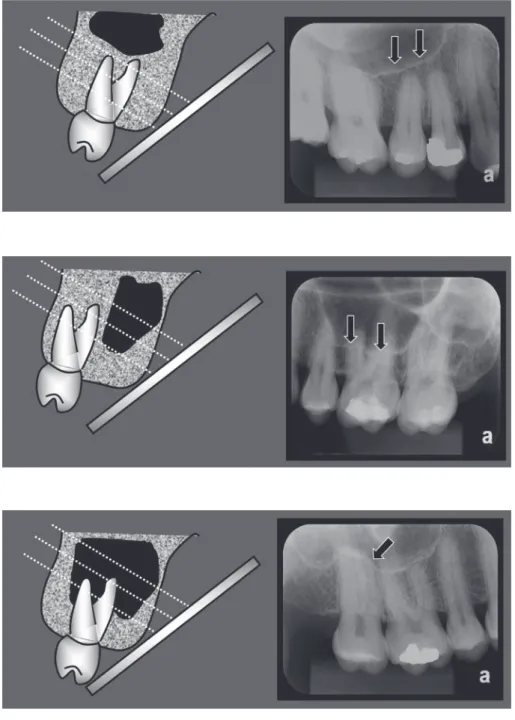
In adults, the maxillary sinuses have a pyramidal shape, extending from the root of the canine to the maxillary tu- berosity, and from the floor of the orbit to the apex region of the maxillary posterior teeth. On periapical radiogra- phy, the contour of the floor of a healthy maxillary sinus is seen as a slightly curved, thin, and radiopaque line(Fig.
1).16 When pneumatization of the maxillary sinus is pres- ent, two situations may occur. In the first, pneumatization occurs in the region near the tooth root, without actually coming in contact with it. As the radiographic image is two-dimensional, the image of the maxillary sinus floor projects itself over the roots of the posterior maxillary teeth; however, it should be noted that the contour of the maxillary sinus floor remains unaltered, that is, horizontal and slightly curved(Fig. 2). In the second, the pneumati- zation of the real maxillary sinus comes in contact with the dental roots. Thus, the maxillary sinus floor deviates from its linear and horizontal path in order to bypass the dental root of the posterior teeth, in turn taking on a sin- uous contour in the shape of a bell, with a format that is similar to the contour of the root apex, a phenomenon that we in this article term an ‘alveolar dome’(Fig. 3). In this scenario, on the periapical radiograph, one can observe that the radiopaque line of the contour of the maxillary sinus floor merges with the radiopaque line of the lamina dura that bypasses the dental apex, as if both were a sin- gle sinuous and radiopaque line in close contact with the root apex.
All of the images were evaluated by 2 dentists, special- ists in dental radiology and diagnostic imaging, after hav- ing been duly trained and calibrated. The interpretation of the digital images was performed directly with Kodak Dental Imaging software(Kodak Dental Systems, Roch- ester, NY, USA), allowing the use of all available resourc- es. This study used a computer that contained a GeForce 9500 GT graphics card(Nvidia Corporation, Santa Clara, CA, USA) and an LED LG Flatron E2241 monitor(LG Electronics, Greater Noida, Uttar Pradesh, India) with a resolution of 1920×1080 pixels, together with brightness and contrast levels of the monitor set to their pre-defined configurations.
BioEstat 5.0 software(Instituto de Desenvolvimento Sus- tentável Mamirauá, Belém, Pará, Brazil) was used to com-
pare the prevalence of alveolar domes among the max- illary teeth and, considering the molars, to compare the prevalence of alveolar domes among the different roots of the same tooth. The χ2 test was applied with a signifi- cance level of 5%.
results
The prevalence of alveolar domes was evaluated in 400 first pre-molars, 400 second pre-molars, 400 first mo- lars, 400 second molars, and 128 third molars. The re- sults demonstrated that the prevalence of alveolar domes
identified in the first pre-molars was 7.75%(31/400), which is statistically significantly lower when compared to the other maxillary posterior teeth(19.25% for second pre-molar [77/400], 30.5% for first molar [122/400], 32%
for second molar [128/400]), and 22.66% for third molars (29/128)(p<0.05)(Table 1). There was also no statisti- cally significant difference in the prevalence of alveolar domes between the first and second maxillary molars, and between the second pre-molar and third molars. Howev- er, for the second pre-molars and third molars, the prev- alence of alveolar domes was statistically lower when compared to the first and second molars and statistically
Fig. 1. Maxillary sinus without pneu- matization. The periapical radiograph shows the slightly curved, thin, delicate, and tenuous radiopaque line of the con- tour of the maxillary sinus floor(arrows).
Fig. 2. Maxillary sinus with pneuma- tization near the root of the maxillary molar. The contour of the maxillary sinus floor is projected over the roots of the maxillary molar; however, its format remains horizontal and slightly curved(arrows).
Fig. 3. Maxillary sinus with pneumatiza- tion involving the root of the maxillary molar. The radiopaque line of the con- tour of the maxillary sinus floor appears in the form of a bell, forming an alveolar dome(arrows).
higher when compared to the first pre-molars(p<0.05).
In the evaluation of the presence of alveolar domes among the roots of the first molars, it was observed that the palatal(P) root presented a lower prevalence of alveo- lar domes(11.25%, 45/400) when compared to the distob- uccal(DB)(28.25%, 113/400) and the mesiobuccal(MB) (29.75%, 119/400) roots(p<0.05). The buccal roots pre- sented no statistically significant differences among them (Table 2).
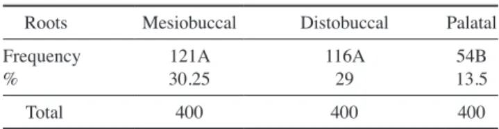
The same characteristics found for the roots of the first molars were also identified for the maxillary second mo- lars. In other words, no statistically significant difference was observed in the prevalence of alveolar domes among the buccal roots(30.25% for MB root [121/400], and 29%
for DB root [116/400]), which present a higher prevalence of alveolar domes when compared to the 13.5% for the P root(54/400)(p<0.05)(Table 3).
discussion
In this study, the prevalence of alveolar domes in max- illary sinus was examined using periapical radiographs.
Due to the anatomical proximity between the maxillary sinus and the root apices of the posterior teeth, various cases of maxillary sinusitis are of odontogenic origin or association with periapical and periodontal lesions, as well as tooth extractions.9,17 In addition, the endodontic treatment of pre-molars and molars can result in acci- dents, such as oral sinus communication, allowing for the displacement of infected tissues to the inner portion of the maxillary sinus, which can cause acute or chronic forms of sinusopathy.18
The distance between the maxillary sinus and the roots of maxillary posterior teeth were analyzed by Kilic et al.3 through 92 computed tomography(CT) images. These au- thors maintained that the roots of the first pre-molars had less contact with the maxillary sinus, whereas the buccal roots of the second molars had more contact but presented no statistically significant difference between them. These results are quite similar to those from the present study, which observed that the first pre-molars present a lower prevalence of alveolar domes, while the buccal roots, as compared to the palatal root, of the maxillary first and second molars present a greater prevalence of alveolar domes. This affirmation can be explained by the anatomy of the maxillary sinus, which shows a tendency towards a reduction in volume in the medial and posterior direc- tions.19
One study performed by Pagin et al.,2 conducted using CT images, verified that the root apices protruded into the maxillary sinus in 21.1% of the first pre-molars, 22.2%
of the second pre-molars, 20.3% of the first molars, 25%
of the second molars, and 11.1% of the third molars. In the present study, it was observed that the root apices protruded into the maxillary sinus in 7.75% of the first pre-molars, 19.25% of the second pre-molars, 30% of the first molars, 32% of the second molars, and 22.66% of the third molars. Upon comparing the two studies, a greater difference was found in the prevalence of the alveolar domes of the first pre-molars and third molars; however, this difference may well be related to the different types of exams used in each study.
As regards the roots of the maxillary first and second molars, many studies have demonstrated that the MB roots of these molars were most frequently associated with alveolar domes when compared to the DB and P roots, and that the P roots contained the lowest prevalence
Table 1. Prevalence and percentage of alveolar domes in the eval- uated teeth.
First
premolar Second
premolar First
molar Second
molar Third molar Prevalence
% 31C
7.75 77B
19.25 122A
30.5 128A
32 29B
22.66
Total 400 400 400 400 128
Frequency followed by different letters differ significantly between them (chi-square test; p<0.05)
Table 2. Prevalence and percentage of alveolar domes in the me- siobuccal, distobuccal, and palatal roots of the maxillary first mo- lars.
Roots Mesiobuccal Distobuccal Palatal Prevalence
% 119A
29.75 113A
28.25 45B
11.25
Total 400 400 400
Frequency followed by different letters differ significantly between them (chi-square test; p<0.05)
Table 3. Percentage of the appearance of alveolar domes in the mesiobuccal, distobuccal, and palatal roots of the maxillary second molars.
Roots Mesiobuccal Distobuccal Palatal Frequency
% 121A
30.25 116A
29 54B
13.5
Total 400 400 400
Frequency followed by different letters differ significantly between them (chi-square test; p<0.05)
of alveolar domes.12,13,20 The present study also observed a lower prevalence of alveolar domes in the P roots, when compared to the buccal roots(p<0.05). However, no sta- tistically significant difference was observed between the MB and DB roots(p>0.05). This divergence can be ex- plained by the sensitivity of the diagnostic method, given that the present study was conducted with digital peri- apical radiographs, while the other studies were conduct- ed using CT exams. The same was not true for the first pre-molar and the P root of the first and second molars, as they presented results that were similar to prior studies, that is, a lower prevalence of alveolar domes.8,13,20
No previous study using periapical radiographs has evaluated the anatomical relationship between the apices of the maxillary posterior teeth and the maxillary sinus floor. In this study, the results of the prevalence of alveo- lar domes, using two-dimensional periapical radiographs as an evaluation method, were similar to those found in works that used three-dimensional CT exams.6,8,13,19-22
However, periapical radiographs have the advantage of being an imaging method that is more commonly used by dentists, due to their cost, accessibility, and lower radia- tion dose.21 Once the periapical radiograph has identified an alveolar dome, the decision to recommend a CT exam should be based on the patient’s history and clinical ex- amination.23
In conclusion, the present study coined the term ‘alveo- lar dome,’ referring to the anatomical projection of the root into the floor of the maxillary sinus. In regard to prevalence, this study showed that the first and second molars presented a greater prevalence of alveolar domes, especially in the buccal roots, followed by the third mo- lars and second pre-molars. The first pre-molars present- ed a lower prevalence of alveolar domes. Although the periapical radiograph is a two-dimensional method, the results of this study showed that periapical radiographs can provide dentists with the auxiliary information nec- essary to identify alveolar domes, improving diagnosis, planning, and treatment.
references
1. Hauman CH, Chandler NP, Tong DC. Endodontic implica- tions of the maxillary sinus: a review. Int Endod J 2002; 35:
127-41.
2. Pagin O, Centurion BS, Rubira-Bullen IR, Alvares Capelozza AL. Maxillary sinus and posterior teeth: accessing close rela- tionship by cone-beam computed tomographic scanning in a Brazilian population. J Endod 2013; 39: 748-51.
3. Kilic C, Kamburoglu K, Yuksel SP, Ozen T. An assessment
of the relationship between the maxillary sinus floor and the maxillary posterior teeth root tips using dental cone-beam computerized tomography. Eur J Dent 2010; 4: 462-7.
4. de Oliveira AG, dos Santos Silveira O, Francio LA, de An- drade Marigo Grandinetti H, Manzi FR. Anatomic variations of paranasal sinuses - clinical case report. Surg Radiol Anat 2013; 35: 535-8.
5. Lana JP, Carneiro PM, Machado Vde C, de Souza PE, Manzi FR, Horta MC. Anatomic variations and lesions of the max- illary sinus detected in cone beam computed tomography for dental implants. Clin Oral Implants Res 2012; 23: 1398-403.
6. Sharan A, Madjar D. Correlation between maxillary sinus floor topography and related root position of posterior teeth using panoramic and cross-sectional computed tomography imaging. Oral Surg Oral Med Oral Pathol Oral Radiol Endod 2006; 102: 375-81.
7. Kretzschmar DP, Kretzschmar JL. Rhinosinusitis: review from a dental perspective. Oral Surg Oral Med Oral Pathol Oral Radiol Endod 2003; 96: 128-35.
8. Tan R, Spector S. Pediatric sinusitis. Curr Allergy Asthma Rep 2007; 7: 421-6.
9. Patel NA, Ferguson BJ. Odontogenic sinusitis: an ancient but under-appreciated cause of maxillary sinusitis. Curr Opin Otolaryngol Head Neck Surg 2012; 20: 24-8.
10. Pokorny A, Tataryn R. Clinical and radiologic findings in a case series of maxillary sinusitis of dental origin. Int Forum Allergy Rhinol 2013; 3: 973-9.
11. Didilescu A, Rusu M, Săndulescu M, Georgescu C, Ciulu- vică R. Morphometric analysis of the relationships between the maxillary first molar and maxillary sinus floor. Open J Stomatol 2012; 2: 352-7.
12. Eberhardt JA, Torabinejad M, Christiansen EL. A computed tomographic study of the distances between the maxillary si- nus floor and the apices of the maxillary posterior teeth. Oral Surg Oral Med Oral Pathol 1992; 73: 345-6.
13. Kwak HH, Park HD, Yoon HR, Kang MK, Koh KS, Kim HJ.
Topographic anatomy of the inferior wall of the maxillary si- nus in Koreans. Int J Oral Maxillofac Surg 2004; 33: 382-8.
14. American Dental Association, U.S. Department of Health and Human Services. Dental radiographic examinations: recom- mendations for patient selection and limiting radiation ex- posure. Revised: 2012 [Internet]. Chicago: American Dental Association; 2012 [cited 2016 Mar 1]. Available from: http://
www.ada.org/~/media/ADA/Member Center/FIles/Dental_
Radiographic_Examinations_2012.ashx
15. Low KM, Dula K, Bürgin W, von Arx T. Comparison of periapical radiography and limited cone-beam tomography in posterior maxillary teeth referred for apical surgery. J Endod 2008; 34: 557-62.
16. White SC, Pharoah MJ. Oral radiology; principles and inter- pretation. 6th ed. St. Louis: Mosby-Year Book Inc; 2009.
17. Shanbhag S, Karnik P, Shirke P, Shanbhag V. Association be- tween periapical lesions and maxillary sinus mucosal thicken- ing: a retrospective cone-beam computed tomographic study.
J Endod 2013; 39: 853-7.
18. Lu Y, Liu Z, Zhang L, Zhou X, Zheng Q, Duan X, et al. As- sociations between maxillary sinus mucosal thickening and apical periodontitis using cone-beam computed tomography
scanning: a retrospective study. J Endod 2012; 38: 1069-74.
19. Mossa-Basha M, Blitz AM. Imaging of the paranasal sinuses.
Semin Roentgenol 2013; 48: 14-34.
20. Lane JJ, O’Neal RB. The relationship between periodontitis and the maxillary sinus. J Periodontol 1984; 55: 477-81.
21. Butaric LN, McCarthy RC, Broadfield DC. A preliminary 3D computed tomography study of the human maxillary sinus and nasal cavity. Am J Phys Anthropol 2010; 143: 426-36.
22. Jung YH, Cho BH. Assessment of the relationship between the maxillary molars and adjacent structures using cone beam computed tomography. Imaging Sci Dent 2012; 42: 219-24.
23. Special Committee to Revise the Joint AAE/AAOMR Po- sition Statement on use of CBCT in Endodontics. AAE and AAOMR joint position statement: use of cone beam computed tomography in endodontics 2015 update. Oral Surg Oral Med Oral Pathol Oral Radiol 2015; 120: 508-12.