87 게재결정: 2008년 7월 8일
*교신저자: 이 숭, 502-157, 서남대학교 의과대학 내과학교실, Phone: 062- 370-7888, FAX: 062-371-3092, E-mail: [email protected]
Chonnam Medical Journal Vol. 44, No. 2, pp. 87∼92
복수가 동반된 간경변증 환자에서 폴리에틸렌 글리콜을 이용한 장 투과성 평가
서남대학교 의과대학 내과학교실, 1가정의학과교실
한문종ㆍ이 숭*ㆍ임재규ㆍ이현민ㆍ한민석ㆍ김우진ㆍ박인형ㆍ손승철1
Assessment of Intestinal Permeability Using Polyethylene Glycol in Liver Cirrhosis with Ascites
Moon-jong Han, Soong Lee*, Jae-kyu Lim, Hyun-min Lee, Min-seok Han, Woo-jin Kim, In-hyung Park and Seung-cheol Son1
Departments of Internal Medicine, 1Family Medicine, College of Medicine, Seonam University Hospital, Gwangju, Korea
Increased intestinal permeability and endotoxemia have been implicated in pathogenesis, progression and development of complications of liver cirrhosis (LC). No study has thus far been reported in Korea on the assessment of intestinal permeability using polyethylene glycol (PEG). The aim of this study was to determine whether intestinal permeability is altered in LC and correlated with clinical characteristics. 65 patients with LC and 26 healthy control subjects were studied. The intestinal permeability value is expressed as the percentage of retrieved PEG 400 and 3350 that had been orally administered in 8-hr urine samples as determined by high performance liquid chromatography. The causes of liver cirrhosis were alcohol (n=38), virus (HBV 17, HCV 9) (n= 26) and alcohol combined with HBV (n=1). Mean values for PEG 400 and 3350 retrieval were 46.5±3.22 and 0.24±0.03 in controls, 44.1±5.17 and 0.21±0.02 in LC without ascites, and 37.4±3.55 and 0.29±0.04 in LC with ascites, respectively. Intestinal permeability index (IPI) was significantly higher in LC with ascites than in healthy controls or LC without ascites (0.84±0.21 vs. 0.52±0.05 or 0.54±0.04, p<0.05). A sub-analysis relating intestinal permeability to the Child-Pugh class of LC showed significant differences between class A , B and C for PEG 3350 and IPI (p<0.05). According to sub-analysis relating IPI to the presence of encephalopathy, hypoalbuminemia of LC, there were significant differences (p<0.05), but not for patients as indicated by prolonged prothrombin time, esophageal varix or hyperbilirubinemia. The present study suggests that increased intestinal permeability is probably of importance in the pathophysiology and progression of liver cirrhosis with ascites.
Keywords: Permeability; Polyethylene glycol; Liver cirrhosis
서 론
인체 장관은 장내 세균의 저장소로서 화상, 출혈성 쇼크 등과 같은 심각한 임상 상황에서 중요한 역할을 하는 것으 로 알려져 있고,1 정상에서 점막 장벽 기능, 면역글로불린 분비, 대식 세포계 등과 같은 일련의 방어체계들이 독소나 장내 세균 전위를 억제하나 문맥압 항진증이 동반된 간 경 변증에서는 장관의 점막 이상이 존재하고, 간 손상과 장관의 기능적 활성도와 서로 연관 있음이 보고되고 있다.2,3 장 투과성 증가에 따른 장내세균의 전위, 내독소 유입 증 가는 만성 간 손상의 병인과 간 경변증에서 간성 뇌증과 세 균 감염 같은 심각한 합병증 발생의 기여 인자로서 연관되 어 있음이 보고되었으나,4 일부에서는 장 투과성 증가가 간 경변증 환자의 세균 감염 병태 생리에 중요하지 않음이 보 고되었다.5,6 이처럼 간 경변증에서 장 투과성은 정상 혹은 증가하는 것으로 다양하게 보고되고 있다.6 12 전신적인 내 독소 혈증은 싸이토카인의 분비를 통해 장 투과성을 증가시 키고, 숙주의 면역 손상과 장내 세균 전위를 촉진시켜 간 경 변증에서 감염성 합병증 즉, 자발성 세균성 복막염과 균혈증 을 초래하는 것으로 알려져 있으나,13,14 이런 비정상적 장 투과성 증가의 기전은 아직 정확히 알려져 있지 않으며 추 가적인 연구가 필요하다.8
복수가 동반된 간 경변증 환자의 폴리에틸렌 글리콜(poly- ethylene glycol, PEG)을 이용한 장 투과성 변화에 대한 연 구는 거의 없으며 국내에서는 아직 보고가 없는 실정이다.
이에 저자들은 복수를 동반한 간 경변증 환자에서 PEG를 이용하여 장 투과성 변화를 살펴보고 임상 양상과의 연관성 을 비교 분석하여 장 투과성의 역할을 규명하고자 하였다.
대상 및 방법
1. 연구 대상
32명의 복수가 있는 간 경변증 환자, 33명의 복수가 없는 간 경변증 환자, 그리고 26명의 건강 대조군을 대상으로 했 고 대조군은 알코올성, 바이러스성 간염의 기왕력이 없고 신 체 검사 및 혈액 검사상 정상이며 장 투과성이 증가할 만한 질환이 없는 경우로 하였다. 본 연구에서는 위장관 질환, 신 장 질환 및 당뇨 환자는 제외되었고, 장 투과성 검사에 영향 을 줄 수 있는 락툴로오스, 비스테로이드성 항염증제, 알코 올을 2주 전에 복용한 환자도 제외시켰다. 본 연구는 서남
대학교 건강과학연구소의 임상시험 위원회에서 승인 받았으 며 본 연구에 포함된 모든 대상자들의 동의서를 받았다.
간 경변증 환자는 초음파 및 상부소화관 내시경상 비 종 대, 식도 정맥류, 복수 등 전형적인 소견과 연장된 프로트롬 빈 시간, 저 알부민혈증 등의 검사실 결과를 보인 환자로 하 였다. 간 질환의 중증도는 Child-Pugh 분류에 따랐다. 자발 성 세균성 복막염의 진단은 뚜렷한 감염의 원인이 없으면서 복수에서 다형핵 세포수가 250 cells/mm3 이상일 때, 요로 감염은 소변에서 다형핵 세포수가 105/ml보다 많거나 소변 배양 검사상 양성일 때로 정의하였다. 전신 감염은 혈액 배 양 검사상 2번에서 양성일 때 진단하였고 폐렴은 객담 배양 검사상 양성이거나 전형적인 흉부방사선 소견, 폐결핵은 객 담 항산균 염색 검사상 양성과 전형적인 흉부방사선 소견에 근거하여 진단하였다.
2. 장 투과성의 측정
혈액 검사는 장 투과성 검사 1주 이내에 실시하였으며 장 투과성 검사는 PEG 400, 3350을 이용하였다. 시험 대상군 모두 검사 전날 저녁 8시 이후 금식시키고 시험 당일 아침 에 소변을 보게 한 후 아침 식전 1시간 전에 PEG 400 1 g, PEG 3350 10 g을 물 100 ml와 혼합하여 복용시켰다. 이후 8시간 소변을 모아 복용한 PEG 중 회수된 양을 백분율로 표시하였다.
소변에서 저분자량 표지자인 PEG 400과 고분자량 표지 자인 PEG 3350를 증발 광산란 효과를 이용한 고성능 액체 크로마토그래피(high performance liquid chromatography, HPLC) 방법을 이용하여 동시에 검출하였다. 모인 소변에서 2 ml를 채취하여 0.45μm syringe filter (Nylon membrane) 로 여과하여 4oC에서 보관하였다. 채취된 소변 1 ml는 곧바 로 냉각장치가 달린 Waters 717+autosampler에 놓여졌다.
소변에서 미세 불순물을 제거하기 위해 일회용 Security Guard kit (Phenomenex, Torrance, CA USA)를 거친 후 polystylene divinyl benzene (PS/DVB) 중합체 염주로 채 워진 5μm PLRP-S 100Å (150×4.6 mm, Polymer Labora- tories, Amherst, MA USA)를 통과시켰다. 그 후 친수성과 소수성 물질을 분리하기 위해 gradient mobile phase (acetonitrile/H2O)가 40~60분 동안 이용되었다. HPLC 전기용매는 불순물이 먼저 추출되고 후에 원하는 표지자인 PEG 400과 3350가 최대치 중복 없이 분리되도록 고안된 gradient controller program에 의해 분리되었다. 분리된 혼 합물은 증발 산란 광 검출기(PL-ELSD 2100 under con-
Fig. 1. HPLC chromatogram of urine of cirrhotic patient with ascites.
Table 1. Demographics and characteristics of the subjects Cirrhotics Cirrhotics Healthy with ascites without ascites controls
(n=32) (n=33) (n=26)
Age (yr±SE) 55.3±9.4 54.2±8.9 50.3±9.2
Sex (M/F) 28/4 26/7 17/9
Etiology
Alcohol 20 (62.5%) 18 (55.5%) Viral* 11 (34.3%) 15 (44.5%) Alcohol/viral† 1 (3.2%) 0 (0%)
Child class 1/20/11 27/6/0¶
(A/B/C) (3.2%/62.5%/34.3%) (81.8%/18.2%/0%) Child-Pugh score 8.8±0.44¶ 6.3±0.34 Serum albumin 2.8±0.11 3.5±0.12¶ (g/dl±SE)
Serum bilirubin 5.1±0.92∥ 2.0±0.43 (mg/dl±SE)
Prothrombin time
15.7±0.50§ 14.3±0.40 (s±SE)
AST (IU/L) 78.3±9.45 74.8±11.4
ALT (IU/L) 34.1±4.8 51.5±=8.9
Encephalopathy 11 (34.6%)§ 2 (7.4%) Esophageal varix 14 (42.3%) 11 (33.3%) Concurrent infection
Total patients 2 7
SBP 2 0
Bacteremia 0 1
UTI 0 4
Others‡ 0 2
*viral etiology-cirrhosis with ascites (HBV-7, HCV-4), cirrhosis without ascites (HBV-10, HCV-5); †viral etiology-HBV-1; ‡Others:
pneumonia, one; pulmonary tuberculosis, one; §Different letters denote significant difference by post hoc of Scheffe (p<.05); ∥ significantly different at p<.01; ¶significantly different at p<.001.
SBP, spontaneous bacterial peritonitis; UTI, urinary tract infection.
ditions of evaporation-85oC, nebulizer 85oC and gas flow 1.0; Polymer Laboratories)를 이용하여 분석 하였다(Fig. 1).
측정 곡선은 200~1,500 mg/L (PEG 400)과 10~200 mg/L (PEG 3350)의 범위에서 얻었으며 장 투과성은 PEG 농도와 총 소변량을 통해, 장 투과성 지수(IPI, in %)는 PEG 400에 비례한 PEG 3350의 장 투과성을 나타낸 PEG 회수율로 다음과 같이 계산하였다.
Total excretion of PEG (ppm)=urinary concentration×
volume of urine
Permeability (%)=(Total excretion of PEG)/(Ingestion of PEG)×100
Intestinal permeability index (%)=(Permeability of PEG 3350)/(Permeability of PEG 400)×100
3. 통계적 분석
자료는 변수들에 따라 평균값, 표준오차, 백분율로 작성 되었다. 정상 대조군, 복수를 동반한 간 경변증, 복수가 없 는 간 경변증 환자의 세 그룹 간 차이는 ANOVA test에 의해 분석하였고 의미가 있을 때 평균치 간의 차이를 조사 하기 위해 Scheffe post hoc comparisons를 이용하였다.
Independent samples t-test, Mann-Whitney test, Fisher’s exact test로 p value가 0.05 미만인 경우 통계적 유의성이 있는 것으로 단정하였다. 두 변수들 간의 관련성에 대해서는 Pearson’s correlation coefficient 또는 Kendall’s tub를 이 용했고 통계적 분석을 위해 SPSS 11.0 프로그램을 이용하 였다.
결 과
1. 환자의 특징
간 경변증 환자와 건강 대조군 간의 나이, 성별 간 차이는 없었고 간 경변증의 원인은 알코올(38예), 간염 바이러스 감염(HBV 17, HCV 9; 26예)과 알코올과 B형 간염 바이 러스 동반이 1예였다. 요소와 크레아티닌으로 평가된 신장 기능은 모든 대상군에서 정상이었다. 복수가 있는 간 경변증 환자 중 2명, 복수가 없는 간 경변증 환자 중 7명에서 동시 감염이 있었다. 나이, 성별, 원인, 중증도, 간 경변증의 합병 증 유무, 동시 감염에 대한 자세한 사항은 Table 1에 기술 하였다.
2. 건강 대조군 및 간 경변증 환자에서 장 투과성 변화 PEG 400과 3350 회수율의 평균값은 건강 대조군에서
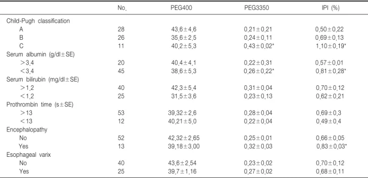
Table 3. Analysis relating intestinal permeability to the clinical and laboratory findings in the cirrhotic patients
No. PEG400 PEG3350 IPI (%)
Child-Pugh classification
A 28 43.6±4.6 0.21±0.21 0.50±0.22
B 26 35.6±2.5 0.24±0.11 0.69±0.13
C 11 40.2±5.3 0.43±0.02* 1.10±0.19*
Serum albumin (g/dl±SE)
>3.4 20 40.4±4.1 0.22±0.31 0.57±0.01
<3.4 45 38.6±5.3 0.26±0.22* 0.81±0.28*
Serum bilirubin (mg/dl±SE)
>1.2 40 42.3±5.4 0.31±0.04 0.70±0.12
<1.2 25 31.5±3.6 0.23±0.13 0.62±0.21
Prothrombin time (s±SE)
>13 53 39.32±2.6 0.28±0.04 0.69±0.3
<13 12 40.21±5.0 0.22±0.04 0.49±0.4
Encephalopathy
No 52 42.32±2.65 0.25±0.01 0.66±0.05
Yes 13 39.18±3.00 0.32±0.03 0.83±0.03*
Esophageal varix
No 40 43.6±2.54 0.23±0.02 0.70±0.12
Yes 25 39.7±1.16 0.27±0.02 0.68±0.11
*Different letters denote significant difference by post hoc of Scheffe (p<0.05); †significantly different at p<0.01; ‡significantly different at p<0.001. PEG, polyethylene glycol; IPI, intestinal permeability index.
Table 2. Intestinal permeability in the healthy control subjects and cirrhotic patients
Cirrhotics Cirrhotics Healthy with ascites without ascites controls
(n=32) (n=33) (n=26)
PEG400 37.4±3.55 44.1±5.17 46.5±3.22
PEG3350 0.29±0.04 0.21±0.02 0.24±0.03
IPI 0.84±0.21* 0.54±0.04 0.52±0.05
*Different letters denote significant difference by post hoc of Scheffe (p<0.05). PEG, polyethylene glycol; IPI, intestinal permeability index.
46.5±3.22와 0.24±0.03, 복수가 없는 간 경변증 환자군에 서 44.1±5.17와 0.21±0.02, 복수가 동반된 간 경변증 환자 군에서 37.4±3.55와 0.29±0.04로 복수를 동반한 간 경변증 환자군에서 두 군에 비해 PEG 3350 회수율이 높았으나 통 계적 유의성은 없었다. IPI의 평균값은 건강 대조군과 간 경 변증 환자군 간에 차이가 있었고, 건강 대조군과 복수가 없 는 간 경변증 환자군에 비해 복수가 있는 간 경변증 환자군 에서 유의하게 높았다(0.52±0.05 and 0.54±0.04 vs. 0.84±
0.21, p<0.05). 그러나 건강 대조군과 복수가 없는 간 경변 증 환자군 간에는 유의한 차이가 없었다(Table 2).
3. 간 경변증 환자에서 장 투과성과 임상 양상 및 검사실 소견 과의 유의성
장 투과성과 간 경변증의 중증도를 나타내는 Child-Pugh 분류의 비교에서 분류 C에서 A나 B에 비해 PEG 3350 회 수율(0.43±0.02 vs. 0.21±0.02, 0.24±0.11, p<0.05)과 IPI (1.10±0.19 vs. 0.50±0.22, 0.69±0.13, p<0.05)가 유 의하게 높았다. 또한 PEG 3350 회수율은 저 알부민혈증이 있는 경우 없는 군에 비해 유의하게 높았으며(0.26±0.22 vs. 0.22±0.31, p<0.05) IPI는 간성 뇌증과 저 알부민혈증 이 있는 군에서 없는 군에 비해 유의하게 높았고(0.81±0.28 vs. 0.57±0.01, 0.83±0.03 vs. 0.66±0.05, p<0.05) 프로트 롬빈 시간 연장, 식도 정맥류, 고빌리루빈혈증 유무 등에서 는 유의한 차이가 없었다(Table 3).
감염이 동반된 간 경변증 환자군과 동반되지 않은 군, 그 리고 알코올과 비알코올성 간 경변증 환자간 장 투과성 (PEG 400, 3350 회수율 및 IPI)에 있어서 유의한 차이는 없었으나 간 경변증 환자에서 Child-Pugh 점수와 장 투과 성 증가 간에는 양의 상관관계가 있었다(r=0.494, p<
0.005 for PEG 3350 and r=0.447, p<0.01 for IPI).
고 찰
장 투과성은 여러 병적인 상황에서 증가하는 것으로 알려 져 있고 알코올성 간 질환, 간 경변증 등에서 연관성이 보고 되고 있으며,5,15 중증 질환에서는 내독소와 장내 세균의 전 위가 증가하며 이는 간의 손상 정도를 증가시키고, 높은 빈 도로 감염과 패혈증을 초래한다고 알려져 있다.16
장투과성에 대한 다양한 연구가 시도되고 있으며 그 중 알코올성 간질환에서 51Cr-EDTA를 이용한 장 투과성 증가 가 보고된 후 여러 간 질환에서 당류 및 PEG 등을 이용한 장 투과성 연구가 이루어지고 있으나 간 경변증 환자에서 PEG를 이용한 연구는 국내에서는 아직 없는 실정이다. 투 과성 측정 물질로 PEG를 이용하는 장점은 첫째 이 중합체 는 항원과 같은 거대분자를 포함하는 넓은 범위의 분자크기 를 포함한다는 것이고, 둘째 크로마토그래프 측정은 음식과 소변 안에 있는 당의 존재에 영향을 받지 않고, 식이 조절도 필요치 않다는 것이다. 하지만 투과성 측정 물질의 모든 필 요조건이 충족되어도 장 투과성을 평가하기 위한 PEG 혼합 물의 전신적 사용은 작은 크기의 분자와 큰 크기의 분자를 결정할 때 다른 분석 방법을 사용해야 하는 본질적인 기술 적 복잡성의 문제가 있었으나 본 연구에서는 증발 광 산란 검출기를 이용한 HPLC 기술로 소변에서 PEG 400과 3350 의 분석을 할 수 있는 적출법 개발에 의해 이런 시간과 복잡 성 문제를 해결하였다.
PEG 회수율에 의한 장 투과성 측정에 관한 연구에서 알 코올성 간질환과 중증의 급성 췌장염 환자들의 PEG 회수율 증가와 혈중 내독소 농도 사이에 유의한 연관성이 있다고 보고되어 PEG가 이전 장 투과성 측정 물질들(분자량 300 Da-1200 Da)보다 내독소의 간 문맥과 소화관 림프계로의 이동 평가에 더 신뢰할 수 있는 물질이라는 것이 증명되었 다.7 분자량 200 Da와 1,200 Da 사이의 분자 크기를 갖는 PEG (PEG 400)는 장 투과성 변화와 관련된다고 생각되는 만성 염증성 질환, 류마티스성 관절염, 셀리악 병, 음식 알 레르기 등의 질환에 대한 연구에서 장 투과성 측정 물질로 사용되었다.17,18 그러나 Lipid A는 장내 세균의 주를 차지하 는 그램 음성 세균의 Lipopolysaccharide (LPS)의 내독소 주성분으로, 변화되지 않은 형태에서만 염증반응을 유발하 며 분자량은 1,900 Da이다.19 그러므로 51Cr-EDTA, PEG 400과 같은 분자량 1,200 Da 아래의 분자량을 가진 화합물 의 연구 결과는 내독소 전위 결과에 대한 신뢰성이 낮다.
최근 연구에서는 서로 다른 분자량을 갖는 PEG (PEG
400, 3350)가 화학적 구조의 독특한 특성과 연관하여 장 투 과성 연구에 이용되는데 이는 독성이 없고, 수용성이며 숙주 나 장내세균에 의해 대사되지 않는 특징이 있으며 혈액 내 로 이동 후 극성 PEG 분자는 소변으로 배설된다.20 균일한 화학적 성질, 적절한 분자 크기, 내독소와 유사한 선형, 체 인과 같은 모양 때문에 PEG는 장관을 통한 LPS 이동 평가 에 가장 적절한 측정 물질로 생각되고 있다.21 단당류 또는 이당류, 당 알코올, 방사성 핵종 복합체(51Cr-EDTA, 99mTc- DTPA), 단백질과 이들의 복합체 등 이전에 이용된 장 투 과성 측정 물질들은 이런 조건들을 충족시키지 못한다.22 분 자량이 다른 두 PEG 측정 물질을 동시에 사용하는 것은 쉽 게 확산되는 PEG 400에 비해 적게 투과되는 PEG 3350의 이동 값을 반영하는 지표로서 광범위한 장 투과성을 측정 가능하게 한다. 또한 측정 물질의 흡수 전 실험 결과에 영향 을 미칠 수 있는 위 배출과 소화액 분비에 의한 희석, 흡수 후 영향을 미칠 수 있는 전신적인 분포와 신장 청소율과 같 은 요인들이 두 분자에 똑같이 작용하므로 서로 다른 개인 간 직접적으로 비교될 수 있는 장점이 있다.23
측정 물질들은 분자량이 작은 경우 세포를 통한(trans- cellular) 경로로, 분자량이 큰 경우 세포 옆 공간을 통한 (paracellular pathway) 경로로 통과한다고 알려져 있으나,7 본 연구에서 모든 대상자들의 PEG 400과 3350 회수율의 유의한 차이가 없는 것으로 보아 내독소와 세균성 독소의 장관에서 간 문맥계로의 이동이 어느 경로를 통과하는지 명 확히 언급하기는 어렵다. 이에 관하여 PEG를 이용한 검사 동안 간 경변증 환자에서 장 점막의 형태적 또는 분자적 변 화에 대한 연구가 더 필요하다고 생각한다. 복수 내로 PEG 가 유출되어 낮은 소변 배설률을 초래하고 그에 따라 장 투 과성 검사 결과가 저평가될 가능성이 있으나 51Cr-EDTA를 이용한 9명의 간 경변증 환자의 장 투과성 검사에서
51Cr-EDTA의 복수 내로의 유출은 거의 없으며,24 복수 천 자가 소변 51Cr-EDTA 배설에 중요한 영향이 없는 것으로 보아 복수 자체가 결과에 영향을 미치지는 않는 것으로 보 고한 결과와 같이,5,6 본 연구에서도 6명의 간 경변증 환자의 복수에서 PEG가 검출되지 않아 복수 내로의 PEG 유출에 의한 낮은 소변 배설률의 가능성은 배제할 수 있었다.
장 투과성 증가가 자발성 세균염이나 균혈증 같은 감염성 합병증과 연관이 있다고 보고되고 있으나,25,26 본 연구에서 는 감염증이 있는 군과 없는 군 사이에 차이가 관찰되지 않 았다. 이는 감염이 동반된 환자수가 적고 각 감염 질환마다 차이가 있어 단순 비교보다는 질환의 발병 시기와 호전 시
기에 각각 측정 비교하는 것이 합리적이라고 생각하였다. 장 투과성 증가와 간 경변증의 중증도 사이의 관련성은 다양하 게 보고되고 있으나,7,10,11 본 연구에서는 Child-Pugh 분류 A와 B환자에 비해 C환자에서 PEG 3350 회수율과 IPI가 유의하게 높은 것이 관찰되었다. 이는 아마 타 보고와 검사 방법의 차이, 대상 환자군의 차이 때문일 것이다.
기존 연구들과 본 연구 결과의 단순 비교는 검사에 이용 된 측정물질의 구성과 사용된 분석 기술이 다를 수 있고 환 자 집단의 차이, 간 경변증의 중증도를 포함한 평가 방법의 차이가 있을 수 있어 다소 무리가 따른다고 볼 수 있다.
이상의 결과를 토대로 PEG를 이용한 장 투과성 연구는 임상적 유용성이 있을 것으로 사료되고 장 투과성 증가는 복수를 동반한 간 경변증의 병태생리와 진행에 아마도 중요 한 역할을 할 것으로 생각하며 저알부민혈증과 간성 뇌증 발 생에 일부 기여할 것으로 생각되었으나 추후 내독소, 싸이토 카인, 산화체 매개 등과 연관된 연구가 추가되어야 할 것이다.
References
1. Wilmore DW, Smith RJ, O'Dwyer ST, Jacobs DO, Ziegler TR, Wang XD. The gut: a central organ after stress. Surgery 1988;104:917-23.
2. Berg RD. Translocation and indigenous gut flora. In: Fuller (ed).
Probiotics. London:Champman Hall, 1992;55-85.
3. Manevska B. Enzymatic changes in the mucous membrane of the small intestine in tetrachlormethane-induced experimental liver cirrhosis.
Eksp Med Morfol 1976;15:107-11.
4. Kalaitzakis E, Johansson JE, Bjarnason I, Bjärnsson E. Intestinal permeability in cirrhotic with and without ascites. Scand J Gastro- enterol 2006;41:326-30.
5. Ramachandran A, Balasubramanian KA. Intestinal dysfunction in liver cirrhosis: its role in spontaneous bacterial peritonitis. J Gastroenterol Hepatol 2001;16:607-12.
6. Zuckerman MJ, Menzies IS, Ho H, Gregory GG, Casner NA, Crane RS, et al. Assessment of intestinal permeability and absorption in cirrhotic patients with ascites using combined sugar probes. Dig Dis Sci 2004;49:621-6.
7. Rahman SH, Ammori BJ, Larvin M, McMahon MJ. Increased nitric oxide excretion in patients with severe acute pancreatitis: evidence of an endotoxin mediated inflammatory response? Gut 2003;52:270-4.
8. DeMeo MT, Mutlu EA, Keshavarzian A, Tobin MC. Intestinal permeation and gastrointestinal disease. J Clin Gastroenterol 2002;34:
385-96.
9. Loret S, Nollevaux G, Remacle R, Klimek M, Barakat I, Deloyer P, et al. Analysis of PEG 400 and 4000 in urine for gut permeability assessment using solid phase extraction and gel permeation chroma- tography with refractometric detection. J Chromatogr B Analyt Technol Biomed Life Sci 2004;805:195-202.
10. Parlesak A, Schafer C, Schutz T, Bode JC, Bode C. Increased intestinal permeability to macromolecules and endotoxemia in patients
with chronic alcohol abuse in different stages of alcohol-induced liver disease. J Hepatol 2000;32:742-7.
11. Rasaratnam B, Connelly N, Chin-Dusting J. Nitric oxide and the hyperdynamic circulation in cirrhosis: is there a role for selective intestinal decontamination? Clin Sci (Lond) 2004;107:425-34.
12. Ramachandran A, Prabhu R, Thomas S, Reddy JB, Pulimood A, Balasubramanian KA. Intestinal mucosal alterations in experimental cirrhosis in the rat: role of oxygen free radicals. Hepatology 2002;
35:622-9.
13. Yasuda T, Takeyama Y, Ueda T, Shinzeki M, Sawa H, Nakajima T, et al. Breakdown of intestinal mucosa via accelerated apoptosis increases intestinal permeability in experimental severe acute pan- creatitis. J Surg Res 2006;135:18-26.
14. Fujii T, Seki T, Maruoka M, Tanaka J, Kawashima Y, Watanabe T, et al. Lactulose-L-rhamnose intestinal permeability test in patients with liver cirrhosis. Hepatol Res 2001;19:158-69.
15. Francés R, Chiva M, Sanchez E, González-Navajas JM, Llovet T, Zapater P, et al. Bacterial translocation is downregulated by anti- TNF-alpha monoclonal antibody administration in rats with cirrhosis and ascites. J Hepatol 2007;46:797-803.
16. Kasravi FB, Wang L, Wang XD, Molin G, Bengmark S, Jeppsson B.
Bacterial translocation in acute liver injury induced by D-galac- tosamine. Hepatology 1996;23:97-103.
17. Bjarnason I, MacPherson A, Hollander D. Intestinal permeability: an overview. Gastroenterology 1995;108:1566-81.
18. Hollander D. The intestinal permeability barrier. A hypothesis as to its regulation and involvement in Crohn's disease. Scand J Gastroenterol 1992;27:721-6.
19. Homma JY, Matsuura M, Kanegasaki S, Kawakubo Y, Kojima Y, Shibukawa N, et al. Structural requirements of lipid A responsible for the functions: a study with chemically synthesized lipid A and its analogues. J Biochem 1985;98:395-406.
20. Lin CY, Tsai IF, Ho YP, Huang CT, Lin YC, Lin CJ, et al.
Endotoxemia contributes to the immune paralysis in patients with cirrhosis. J Hepatol 2007;46:816-26.
21. Kiki I, Yilmaz O, Erdem F, Gundogdu M, Demircan B, Bilici M.
Tumour necrosis factor-alpha levels in hepatitis B virus-related chronic active hepatitis and liver cirrhosis and its relationship to Knodell and Child-Pugh scores. Int J Clin Pract 2006;60:1075-9.
22. Ataseven H, Bahcecioglu IH, Kuzu N, Yalniz M, Celebi S, Erensoy A, et al. The levels of ghrelin, leptin, TNF-alpha, and Il-6 in liver cirrhosis and hepatocellular carcinoma due to HBV and HDV Infection. Mediators Inflamm 2006;2006:78380.
23. Mohammed NA, Abd El-Aleem S, Appleton I, Maklouf MM, Said M, McMahon RF. Expression of nitric oxide synthase isoforms in human liver cirrhosis. J Pathol 2003;200:647-55.
24. Bac DJ, Swart RG, van den berg JWO, Wilson PJH. Small bowel wall function in patient with advanced liver cirrhosis and portal hyper- tension:studies on permeability and luminal bacterial overgrowth. Eur J Gastrohepatology 1993;5:383-7.
25. Zhang SC, Wang W, Ren WY, He BM, Zhou K, Zhu WN. Effect of cisapride on intestinal bacterial and endotoxin translocation in cirrhosis. World J Gastroenterol 2003;9:534-8.
26. Asai K, Buurman WA, Reutelingsperger CP, Schutte B, Kaminishi M.
Modular effects of estradiol on ethanol-induced apoptosis in human intestinal epithelial cells. Scand J Gastroenterol 2005;40:326-35.