Received on November 12, 2011. Revised on November 25, 2011. Accepted on December 6, 2011.
CC This is an open access article distributed under the terms of the Creative Commons Attribution Non-Commercial License (http://creativecommons.org/licenses/by-nc/3.0) which permits unrestricted non-commercial use, distribu- tion, and reproduction in any medium, provided the original work is properly cited.
*Corresponding Author. Tel: 82-2-880-4802; Fax: 82-2-873-2271 E-mail: cyun@snu.ac.kr Keywords: T cell receptor, nTreg, Intrathymic selection
T Cell Receptor Signaling That Regulates the Development of Intrathymic Natural Regulatory T Cells
Ki-Duk Song1, SuJin Hwang3 and Cheol-Heui Yun1,2*
1Center for Agricultural Biomaterials, 2Department of Agricultural Biotechnology and Research Institute for Agriculture and Life Sciences, Seoul National University, Seoul 151-921, Korea, 3Program on Genomics of Differentiation, Eunice Shriver National Institute of Child Health and Human Development, National Institutes of Health, Bethesda 20892, MD, USA
T cell receptor (TCR) signaling plays a critical role in T cell development, survival and differentiation. In the thymus, quantitative and/or qualitative differences in TCR signaling determine the fate of developing thymocytes and lead to positive and negative selection. Recently, it has been sug- gested that self-reactive T cells, escape from negative se- lection, should be suppressed in the periphery by regulatory T cells (Tregs) expressing Foxp3 transcription factor. Foxp3 is a master factor that is critical for not only development and survival but also suppressive activity of Treg. However, sig- nals that determine Treg fate are not completely understood.
The availability of mutant mice which harbor mutations in TCR signaling mediators will certainly allow to delineate sig- naling events that control intrathymic (natural) Treg (nTreg) development. Thus, we summarize the recent progress on the role of TCR signaling cascade components in nTreg de- velopment from the studies with murine model.
[Immune Network 2011;11(6):336-341]
INTRODUCTION
αβ TCR cells which express clonotypic T cell receptor chain α and β, develop in the thymus and egress out from the thymus after completion of differentiation into helper T cells or CD4+ T cells which express coreceptor CD4 and recognize antigen-MHC class II complex on antigen presenting cells, and cytotoxic T cells or CD8+ T cells which express cor- eceptor CD8 and recognize antigen-MHC class I complex.
Then, CD4+ and CD8+ T cells populate in periphery and un- dergo further differentiation. CD4+ T cells can differentiate into Th1, Th2, Th9, and Th17 effector cells depending on the micro-environmental milieu including cytokine production.
CD8+ T cells can destroy target cells by forming pores with perforin and secreting granzyme A/B (1). Potentially self-re- active T cells can develop because of the generation of ran- dom diversity in the T cell repertoire. Self-reactive T cells can be eliminated during the selection process in the thymus, namely central tolerance, and if they escape negative se- lection, they can be still suppressed by peripheral mecha- nisms including anergy and/or by CD4+CD25+ regulatory T cells (Tregs). Treg is a specialized subset of CD4+ T cells and able to suppress immune responses (2). Tregs are also generated in the thymus and constitutively express IL-2 re- ceptor α chain, CD25 and transcription factor Foxp3 (3). To note, it is not clear yet what signal triggers Foxp3 expression.
TCR signal has been suggested to be important to turn on the expression of Foxp3 in developing Treg precursors in the thymus (4). Although it is known that Treg generation relies on TCR, CD28 and IL-2 signaling during differentiation in the thymus, only a few studies have shown the role of proximal TCR signaling components in intrathymic or natural Treg (nTreg) generation.
OVERVIEW OF TCR SIGNALING
TCR signaling plays a critical role in development, survival
337
and effector cell differentiation and function of T cells. In the thymus, quantitative and qualitative differences in TCR signal- ing determine the fate of developing thymocytes that sub- sequently cause to positive and negative selection (5).
Interactions between TCR and peptide-MHC lead to the acti- vation of Lck, a member of Src family of tyrosine kinase in T cells, and then activated Lck phosphorylates two tyso- sine-residues on the ITAMs of CD3 and ζ chains. Then, ZAP-70, another tyrosine kinase, is recruited to phosphory- lated ITAMs. ITAM binding to ZAP70 relieves an auto- inhibitory conformation of the kinase and the phosphor- ylation of the specific tyrosine residues that are important for the activity of ZAP-70 (6). Activated ZAP-70 phosphorylates the tyrosine residues on the linker for activation of T cells (LAT), an adaptor molecule, which couples proximal TCR ac- tivation to downstream signaling pathways, e.g., PLC-γ1, Ca2+
flux, PKC activation, Ras/ERK activation (7-9). Then these molecules form signalosomes with other proteins containing the Src homology 2 (SH2) domains including PLC-γ1, Grb2, Gads, Grap, 3BP2, and Shb, and indirectly binds Sos, c-Cbl, Vav, SLP-76, and Itk (10,11). In human LAT, one of the four membrane-distal tyrosines, Y132 (Y136 in mouse LAT), asso- ciates with PLC-γ1, and further interaction of LAT with PLC- γ1 and Gads-SLP-76 complex is critical for PLC-γ1 activa- tion, which hydrolyzes phosphatidylinositol 4,5 bisphosphate (PIP2) to generate diacylglycerol (DAG) and inositol 1,4,5-tri- phosphate (IP3). While DAG activates PKC/NF-κB and RasGRP-Ras-Raf-ERK pathways, IP3 mediates Ca2+ flux from the endoplasmic reticulum, leading to the activation of claci- neurin, a calcium induced phosphatase, and dephosphor- ylation of NFAT family transcription factors. Finally, dephos- phorylated NFATs are transclocated into the nucleus for their transcriptional activities (12). On the other hand, three ty- rosines, Y171, Y191 and Y226, bind to Grb2 upon phospho- rylation. Grb2 recruits son of sevenless (Sos), a guanine nu- cleotide exchange factor (RasGEF), to the plasma membrane for activation of Ras molecule. Two tyrosines, Y171 and Y191 also bind to Gads, which constitutively interacts with SLP-76.
Tyrosine phosphorylation of the N-terminal, positions at 113 and 128 of SLP-76 results in the recruitment of the GEF Vav and the adapter protein Nck and these complexes regu- late directly actin cytoskeletal rearrangement following TCR ligation and JNK activation (8,9). Thus, TCR engagement trig- gers a series of signaling cascades that ultimately lead to T cell development, differentiation and effector function.
TCR SIGNALING AND CENTRAL TOLERANCE IN THE THYMUS
After successful completion of β-selection, where rearranged TCRβ chain associates preTα chain to form preTCR com- plex, preTCR signaling operates for proliferation and survival of double negative (DN) thymocytes (13). Then, DN thymo- cytes progress to double-positive (DP) thymocytes stage where thymocytes begin to express both CD4 and CD8 coreceptors. DP thymocytes also re-express RAG1/2 which is required for TCRα rearrangement, resulting αβ TCR com- plex expression on their surface (14). The development of DP thymocytes has three fates: death by neglect, positive se- lection or negative selection. During this thymic selection process, random rearrangement of TCRs is useless since these TCRs cannot bind to their cognate ligands (15). The failure of clonotypic αβ TCR expressed on DP thymocytes to en- gage cognate epitope-MHC could not generate signals re- quired to undergo positive selection and approximately 90%
of these cells die. Positive selection occurs when and if the TCR of the thymocytes engages an epitope-MHC complex with low affinity, and then generate signals for differentiation and survival. Positively selected DP thymocytes mature into CD4+ or CD8+ single-positive (SP) T cells. In a way, positive selection enriches for self-reactive cells too, making them a threat for autoimmunity in periphery. On the other hand, negative selection deletes potentially self-reactive thymocytes, thereby generating a repertoire of peripheral T cells that is largely self-tolerant. TCR of thymocytes engaging an epit- ope-MHC complex with high affinity leads to the apoptotic death of the cells (16).
NTREG DEVELOPMENT IN THE THYMUS
Development of nTregs seems to occur via a two-step process dependent on multiple intracellular pathways activated by a combination of TCR, CD28 and cytokine receptor mediated signals (17-19). The initial step in nTreg development de- pends on signals generated by TCR engaged with self-pep- tide-MHC class II complexes (20) and B7/CD28 interactions (21). Tregs emerge from a pool of DP thymocytes which ex- press TCRs with a relatively high affinity for self-antigens to CD4+ SP thymocytes which are CD25+GITRhiFoxp3− (22-24).
This requirement for the development of Tregs differs from the fate of conventional CD4+ T cells expressing higher affin-
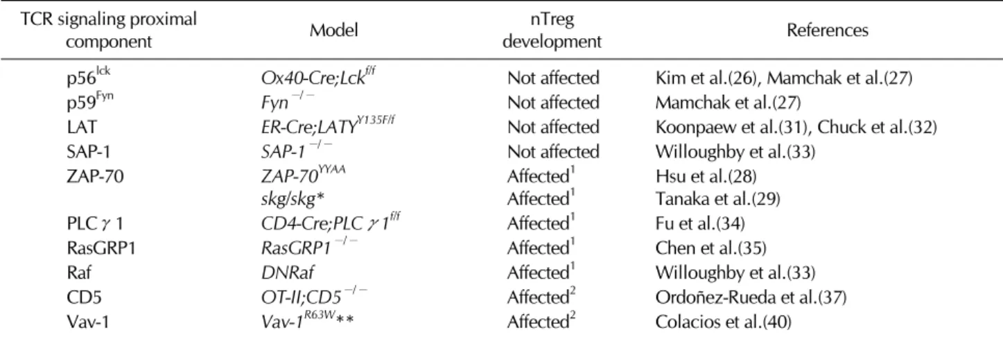
Table I. TCR proximal signaling molecules and intrathymicTreg development in mutant animal models TCR signaling proximal
component Model nTreg
development References
p56lck Ox40-Cre;Lckf/f Not affected Kim et al.(26), Mamchak et al.(27)
p59Fyn Fyn−/− Not affected Mamchak et al.(27)
LAT ER-Cre;LATYY135F/f Not affected Koonpaew et al.(31), Chuck et al.(32)
SAP-1 SAP-1−/− Not affected Willoughby et al.(33)
ZAP-70 ZAP-70YYAA
skg/skg* Affected1
Affected1 Hsu et al.(28) Tanaka et al.(29)
PLCγ1 CD4-Cre;PLCγ1f/f Affected1 Fu et al.(34)
RasGRP1 RasGRP1−/− Affected1 Chen et al.(35)
Raf DNRaf Affected1 Willoughby et al.(33)
CD5 OT-II;CD5−/− Affected2 Ordoñez-Rueda et al.(37)
Vav-1 Vav-1R63W** Affected2 Colacios et al.(40)
*A point mutation in ZAP-70 SH2 domain. **A point mutation (Arg63Trp) found in rat. 1Decreased Treg proportion or absolute number,
2increased Treg proportion or absolute number.
ity TCRs, which are eliminated by negative selection (25). The subsequent differentiation of Treg precursors into functional Tregs requires IL-2 and/or IL-15 signaling to induce and main- tain Foxp3 expression (24).
THE ROLE OF TCR SIGNALING COMPONENTS IN
NTREG DEVELOPMENT
The findings from mutant animal studies have contributed to understand the role of TCR signaling in nTreg development.
These animal models include p56Lck, p59Fyn, LAT, ZAP-70, CD5, PLC-γ1, RasGRP-1 and Vav-1 and findings are summar- ized in Table I.
p56Lck
p56Lck triggers TCR signaling in response to antigenic stim- ulation to regulate T cell development, survival and differ- entiation in the thymus. But, conditional ablation of p56Lck in DP and CD4-SP thymocytes has no impact on nTreg gen- eration (26) and inactivation of p59Fyn, another Src family member of tyrosine kinase, also did not affect nTreg develop- ment in the thymus (27). Furthermore, inactivation of Lck and Fyn in thymus did not impair nTreg development, suggesting that Lck and Fyn are not directly involved in the development of nTreg. Instead, inactivation of p56Lck seems to compromise Treg homeostasis, which is characterized by impaired turn- over, preferential redistribution to lymph nodes, and loss of suppressive function (26).
ZAP-70
Development of nTreg is impaired in mice with a mutation (skg mutation) or tyrosine to phenylalanine mutations of Y315 and Y319 of the ZAP-70 gene (ZAP-70YYAA) (28,29). In addi- tion, these mice are impaired in T cell development and hy- poresponsiveness on cells with TCR stimulation as well as de- fective on positive and negative selection. These two tyrosine residues appear to play an important role in recruitment of downstream molecules, LAT, and auto-inhibition of ZAP-70.
Furthermore, ZAP-70YYAA Tregs showed minimal suppressor activity (28), suggesting that these tyrosine residues are crit- ical for nTreg development as well as their function.
LAT
Knockin mutant mouse, where tyrosine 136, a PLC-γ1-bind- ing site in murine LAT, is replaced by phenylalanine (Y136F), develops lymphoproliferative disease (30) and has defective on Treg development (31). These studies suggested that the LAT-PLC-γ1 interaction plays a critical role in Foxp3 ex- pression and the development of nTreg cells. Elegant genetic approach by the same group has revealed that Treg develop- ment in LAT mutant mice, where wild-type LAT is used for selection and mutant LAT (Y136F) is replaced after tamoxifen treatment, is intact. However, their suppressor activity is com- promised and nonfunctional in periphery, indicating that LAT-PLC-γ1 interaction is essential for the suppressive activ- ity of Tregs not for nTreg development (32).
339
Raf/SAP-1
The study with mutant mice defective in Raf signaling, i.e., ERK effector SRF accessory protein 1 (SAP-1), and with domi- nant negative Raf-1 transgenic mice revealed the differential requirement for Treg development (33). To note, Treg devel- opment in SAP-deficient animal was intact. Dominant neg- ative-Raf transgenic mice, unlike SAP-1 knockout mice, are defective in Treg development. Nevertheless, neither ablation of SAP-1 nor inactivation of Raf signaling by DN-Raf affected the activity of Treg to suppress naïve T cells in vitro. Furthermore, SAP-1 deficient Tregs are functional to suppress colitits in vivo, suggesting that ERK signaling to SAP-1 is not required for the suppressive activity of Treg (33).
PLC-γ1
Investigation on the function of PLC-γ1 signaling in T cell including Treg development has been impeded by the fact that PLC-γ1 deficiency leads to embryonic lethal during em- bryonic development. Therefore, the generation and analysis of PLC-γ1 conditional knockout mice allowed the assess- ment of the role of PLC-γ1 signaling in conventional T cell and Treg development (34). It has been shown that PLC-γ1 ablation affects T cell development and impairs positive and negative selection, resulting in severe reduction of mature T cells in periphery. PLC-γ1 deficiency also leads to impaired TCR-stimulated proliferation and cytokine production, and the activation of multiple signaling mediators and transcriptional factors (e.g., ERK, JNK, AP-1, NFAT and NF-κB). In addi- tion, Treg development is impaired and their suppressor ac- tivity is compromised (34). But, it is not clear whether or not defective Treg development in PLC-γ1 mutant mice is due to impaired TCR-induced Foxp3 expression or defective IL-2/IL-2R signaling by impaired IL-2 production.
RasGRP-1
In RasGRP-1 deficient mice, Treg cell development in the thy- mus was impaired. But in the periphery frequencies of CD4+ Foxp3+ Tregs were significantly increased due to massive proliferation of RasGRP-1 deficient Tregs and increased apop- tosis of RasGRP-1 deficient Foxp3−CD4+ T cells. RasGRP-1 deficient Treg cells possessed a more activated cell surface phenotype and RasGRP-1 deficient Tregs are more sup- pressive than wild-type controls. This study strongly sug- gested that the intrathymic nTreg development relies on RasGRP-1 signaling pathway, but their peripheral expansion
and function do not (35).
CD5
CD5 is a negative regulator of TCR signaling and CD5 defi- ciency rendered thymocytes hyperresponsive to TCR stim- ulation, leading to increased proliferation, Ca2+ flux and ty- rosine phosphorylation of TCR-ζ, LAT, PLC-γ1 and Vav-1 (36). CD5 deficiency leads to increased nTreg generation in thymus and periphery, and importantly they are functional and suppress naïve T cells. Furthermore, CD5 deficient nTregs showed increased basal level of p-ERK compared with those of wild-type nTreg (37).
Vav-1
Vav-1 is a signal transducer downstream of TCR and is critical for T cell development and activation (38,39). In a genetic study with two rat strains, which have different susceptibility to autoimmune disease, a non-synonymous mutation was identified in the 1st exon of Vav-1, responsible for the sub- stitution of an arginine by tryptophan at position 63. This mu- tation is associated with increased proportion and absolute numbers of Tregs. This mutation resulted in constitutive acti- vation of Vav-1 protein and increased activity of guanine nu- cleotide exchange factor (40).
In summary, even considering critical role of Src family kin- ase and adaptors in T cell development in general, p56Lck, p59Fyn and LAT appear to be dispensable for intrathymic nTreg development but are required for maintenance or sup- pressive function of Treg in the periphery (28,29,34). ZAP-70, PLC-γ1, RasGRP1 and Raf positively regulate nTreg develop- ment in the thymus, since ablation of these mediators impairs nTreg generation in the thymus (30,31,35-37). However, whether IL-2 production or IL-2 signaling is affected in these mutant mice is yet to be defined. Especially, IL-2 production in PLC-γ1 conditional knockout mice is decreased, suggest- ing that both diminished TCR signal and impaired IL-2 signal may lead to impaired nTreg development.
CONCLUSION
During the last few years, we have seen much progress in our understanding of nTreg generation by TCR signaling.
New studies from animal model analysis support the role of TCR signaling in intrathymic Treg development and revealed specific requirement for this process, which is distinct from
conventional T cell development. But the challenge remains how to dissect the impact of TCR signaling on negative se- lection versus Treg generation since these two processes are closely related, yet distinct. We anticipate that more mouse models will be developed and analyzed to understand how TCR signaling pathways regulate nTreg generation in near future.
ACKNOWLEDGEMENTS
This work was supported by the research grant of the Next- Generation BioGreen 21 Program (No. PJ81272011, PJ008196 and PJ0077932011), Rural Development Administration, Repu- blic of Korea.
CONFLICTS OF INTEREST
The authors have no financial conflict of interest.
REFERENCES
1. Zúñiga-Pflücker JC, Jones LA, Chin LT, Kruisbeek AM: CD4 and CD8 act as co-receptors during thymic selection of the T cell repertoire. Semin Immunol. 3;167-175, 1991.
2. Sakaguchi S, Fukuma K, Kuribayashi K, Masuda T: Organ-specific autoimmune diseases induced in mice by elimination of T cell subset. I. Evidence for the active participation of T cells in natural self-tolerance; deficit of a T cell subset as a possible cause of autoimmune disease. J Exp Med 161;72-87, 1985.
3. Kim JM, Rasmussen JP, Rudenky AY: Regulatory T cells prevent catastrophic autoimmunity throughout the life span. Nature Immunol 8;191-197, 2007.
4. Sauer S, Bruno L, Hertweck A, Finlay D, Leleu M, Spivakov M, Knight ZA, Cobb BS, Cantrell D, O'Connor E, Shokat KM, Fisher AG, Merkenschlager M: T cell receptor signaling controls Foxp3 expression via PI3K, Akt, and mTOR. Proc Natl Acad Sci USA 105;7797-7802, 2008.
5. Starr TK, Jameson SC, Hogquist KA: Positive and negative se- lection of T cells. Annu Rev Immunol 21;139-176, 2002.
6. Au-Yeung BB, Deindl S, Hsu LY, Palacios EH, Levin SE, Kuriyan J, Weiss A: The structure, regulation, and function of ZAP-70.
Immunol Rev 228;41-57, 2009.
7. Samelson LE: Signal transduction mediated by the T cell antigen receptor: the role of adapter proteins. Annu Rev Immunol 20;371-394, 2002.
8. Sommers CL, Samelson LE, Love PE: LAT: a T lymphocyte adapter protein that couples the antigen receptor to downstream signaling pathways. Bioessays 26;61-67, 2004.
9. Fuller DM, Zhu M, Ou-Yang CW, Sullivan SA, Zhang W: A tale of two TRAPs: LAT and LAB in the regulation of lymphocyte de- velopment, activation, and autoimmunity. Immunol Res 49;97-108, 2011.
10. Wange RL: LAT, the linker for activation of T cells: a bridge be-
tween T cell-specific and general signaling pathways. Sci STIKE 2000 63;re1, 2000.
11. Samelson LE: Signal transduction mediated by the T cell antigen receptor: the role of adapter proteins. Annu Rev Immunol 20;
371-394, 2002.
12. Ho SN, Thomas DJ, Timmerman LA, Li X, Francke U, Crabtree GR: NFATc3, a lymphoid-specific NFATc family member that is calcium-regulated and exhibits distinct DNA binding specificity.
J Biol Chem 270;19898-19907, 1995.
13. Michie AM, Zúñiga-Pflücker JC: Regulation of thymocyte differ- entiation: pre-TCR signals and beta-selection. Semin Immunol 14;311-323, 2002.
14. Ellmeier W, Sawada S, Littman DR: The regulation of CD4 and CD8 coreceptor gene expression during T cell development.
Annu Rev Immunol 17;523-554, 1999.
15. Werlen G, Hausmann B, Naeher D, Palmer E: Signaling life and death in the thymus: timing is everything. Science 299;1859-1863, 2003.
16. Gascoigne NR, Palmer E: Signaling in thymic selection. Curr Opin Immunol 23;207-212, 2011.
17. Burchill MA, Yang J, Vang KB, Moon JJ, Chu HH, Lio CW, Vegoe AL, Hsieh CS, Jenkins MK, Farrar MA: Linked T cell receptor and cytokine signaling govern the development of the regulatory T cell repertoire. Immunity 28;112-121, 2008.
18. Lio CW, Hsieh CS: A two-step process for thymic regulatory T cell development. Immunity 28;100-111, 2008.
19. Cheng G, Yu A, Malek TR: T-cell tolerance and the multi-func- tional role of IL-2R signaling in T-regulatory cells. Immunol Rev 241;63-76, 2011.
20. Bensinger SJ, Bandeira A, Jordan MS, Caton AJ, Laufer TM: Major histocompatibility complex class II-positive cortical epithelium mediates the selection of CD4+25+ immunoregulatory T cells. J Exp Med 194;427–438, 2001.
21. Tai X, Cowan M, Feigenbaum L, Singer A: CD28 costimulation of developing thymocytes induces Foxp3 expression and regu- latory T cell differentiation independently of interleukin 2. Nat Immunol 6;152-162, 2005.
22. Fontenot JD, Dooley JL, Farr AG, Rudensky AY: Developmental regulation of Foxp3 expression during ontogeny. J Exp Med 202;901-916, 2005.
23. Wan YY, Flavell R: Identifying Foxp3-expressing suppressor cells with a bicistronic reporter. Proc Natl Acad Sci USA 102;5126-5131, 2005.
24. Lio CW, Hsieh CS: Becoming self-aware: the thymic education of regulatory T cells. Curr Opin Immunol 23;213-219, 2011.
25. Robey E, Fowlkes BJ: Selective events in T cell development.
Annu Rev Immunol 12;675–705, 1994.
26. Kim JK, Klinger M, Benjamin J, Xiao Y, Erle DJ, Littman DR, Killeen N: Impact of the TCR signal on regulatory T cell homeo- stasis, function, and trafficking. PLoS One 4;e6580, 2009.
27. Mamchak AA, Thien CB, Dagger SA, Lyandres J, Jiang S, Langdon WY, DeFranco AL: Unaltered negative selection and Treg devel- opment of self-reactive thymocytes in TCR transgenic Fyn-defi- cient mice. Eur J Immunol 40;539-547, 2010.
28. Hsu LY, Tan YX, Xiao Z, Malissen M, Weiss A: A hypomorphic allele of ZAP-70 reveals a distinct thymic threshold for auto- immune disease versus autoimmune reactivity. J Exp Med 206;2527-2541, 2009.
341
29. Tanaka S, Maeda S, Hashimoto M, Fujimori C, Ito Y, Teradaira S, Hirota K, Yoshitomi H, Katakai T, Shimizu A, Nomura T, Sakaguchi N, Sakaguchi S: Graded attenuation of TCR signaling elicits distinct autoimmune diseases by altering thymic T cell se- lection and regulatory T cell function. J Immunol 185;2295-2305, 2010.
30. Sommers CL, Park CS, Lee J, Feng C, Fuller CL, Grinberg A, Hildebrand JA, Lacaná E, Menon RK, Shores EW, Samelson LE, Love PE: A LAT mutation that inhibits T cell development yet in- duces lymphoproliferation. Science 296;2040-2043, 2002.
31. Koonpaew S, Shen S, Flowers L, Zhang W: LAT-mediated signal- ing in CD4+CD25+ regulatory T cell development. J Exp Med 203;119-129, 2006.
32. Chuck MI, Zhu M, Shen S, Zhang W: The role of the LAT- PLC-gamma1 interaction in T regulatory cell function. J Immunol 184;2476-2486, 2010.
33. Willoughby JE, Costello PS, Nicolas RH, Robinson NJ, Stamp G, Powrie F, Treisman R: Raf signaling but not the ERK effector SAP-1 is required for regulatory T cell development. J Immunol 179;6836-6844, 2007.
34. Fu G, Chen Y, Yu M, Podd A, Schuman J, He Y, Di L, Yassai M, Haribhai D, North PE, Gorski J, Williams CB, Wang D, Wen
R: Phospholipase Cγ1 is essential for T cell development, activa- tion, and tolerance. J Exp Med 207;309-318, 2010.
35. Chen X, Priatel JJ, Chow MT, Teh HS: Preferential development of CD4 and CD8 T regulatory cells in RasGRP1-deficient mice.
J Immunol 180;5973-5982, 2008.
36. Tarakhovsky A, Kanner SB, Hombach J, Ledbetter JA, Müller W, Killeen N, Rajewsky K: A role for CD5 in TCR-mediated signal transduction and thymocyte selection. Science 269;535-537, 1995.
37. Ordoñez-Rueda D, Lozano F, Sarukhan A, Raman C, Garcia- Zepeda EA, Soldevila G: Increased numbers of thymic and pe- ripheral CD4+ CD25+Foxp3+ cells in the absence of CD5 signal- ing. Eur J Immunol 39;2233-2247, 2009.
38. Turner M, Billadeau DD: VAV proteins as signal integrators for multi-subunit immune-recognition receptors. Nat Rev Immunol 2;476-486, 2002.
39. Tybulewicz VL: Vav-family proteins in T-cell signalling. Curr Opin Immunol 17;267-274, 2005.
40. Colacios C, Casemayou A, Dejean AS, Gaits-Iacovoni F, Pedros C, Bernard I, Lagrange D, Deckert M, Lamouroux L, Jagodic M, Olsson T, Liblau RS, Fournié GJ, Saoudi A: The p.Arg63Trp poly- morphism controls Vav1 functions and Foxp3 regulatory T cell development. J Exp Med 208;2183-2191, 2011.