Received on December 3, 2013. Revised on January 14, 2014. Accepted on January 15, 2014.
CC This is an open access article distributed under the terms of the Creative Commons Attribution Non-Commercial License (http://creativecommons.org/licenses/by-nc/3.0) which permits unrestricted non-commercial use, distribu- tion, and reproduction in any medium, provided the original work is properly cited.
*Corresponding Author. Je-Min Choi, Department of Life Science, Hanyang University, 222, Wangsimni-ro, Seongdong-gu, Seoul, Korea. Tel: 82-2-2220-4765; Fax: 82-2-2299-3495; E-mail: jeminchoi@hanyang.ac.kr
#These authors contributed equally to this work.
Keywords: Follicular helper T cells, Germinal Center, Follicular regulatory T cells, Cytokines, Autoimmunity
Abbreviations: TFH, Follicular helper T cell; TFR, Follicular regulatory T cell; Bcl-6, B cell lymphoma-6; GC, Germinal Center; PD-1, Programmed cell death protein 1; SAP, SLAM - associated protein; BTLA4, B- and T-lymphocyte attenuator 4; ROR, RAR-related orphan receptor; PSGL-1, P-selectin glycoprotein ligand-1; CCR7, C-C chemokine receptor type 7;
SLE, Systemic Lupus Erythematosus; RA, Rheumatoid Arthritis
Insights into the Role of Follicular Helper T Cells in Autoimmunity
Hong-Jai Park1,2#, Do-Hyun Kim1,2#, Sang-Ho Lim1,2, Won-Ju Kim1,2, Jeehee Youn3, Youn-Soo Choi4 and Je-Min Choi1,2*
1Department of Life Science, 2Research Institute for Natural Sciences, Hanyang University, Seoul 133-791, Korea, 3Department of Anatomy & Cell Biology, College of Medicine, Hanyang University, Seoul 133-791, Korea, 4Division of Vaccine Discovery, La Jolla Institute for Allergy and Immunology, La Jolla, CA 92037, USA
Follicular helper T (TFH) cells are recently highlighted as their crucial role for humoral immunity to infection as well as their abnormal control to induce autoimmune disease. During an infection, naïve T cells are differentiating into TFH cells which mediate memory B cells and long-lived plasma cells in germi- nal center (GC). TFH cells are characterized by their ex- pression of master regulator, Bcl-6, and chemokine receptor, CXCR5, which are essential for the migration of T cells into the B cell follicle. Within the follicle, crosstalk occurs be- tween B cells and TFH cells, leading to class switch recombi- nation and affinity maturation. Various signaling molecules, including cytokines, surface molecules, and transcription fac- tors are involved in TFH cell differentiation. IL-6 and IL-21 cy- tokine- mediated STAT signaling pathways, including STAT1 and STAT3, are crucial for inducing Bcl-6 expression and TFH
cell differentiation. TFH cells express important surface mole- cules such as ICOS, PD-1, IL-21, BTLA, SAP and CD40L for mediating the interaction between T and B cells. Recently, two types of microRNA (miRNA) were found to be involved in the regulation of TFH cells. The miR-17-92 cluster induces Bcl-6 and TFH cell differentiation, whereas miR-10a neg- atively regulates Bcl-6 expression in T cells. In addition, fol- licular regulatory T (TFR) cells are studied as thymus-derived CXCR5+PD-1+Foxp3+ Treg cells that play a significant role in limiting the GC response. Regulation of TFH cell differ- entiation and the GC reaction via miRNA and TFR cells could be important regulatory mechanisms for maintaining immune
tolerance and preventing autoimmune diseases such as sys- temic lupus erythematosus (SLE) and rheumatoid arthritis (RA). Here, we review recent studies on the various factors that affect TFH cell differentiation, and the role of TFH cells in autoimmune diseases.
[Immune Network 2014;14(1):21-29]
INTRODUCTION
CD4 helper T cells play a significant role in regulating adap- tive immune responses against foreign antigens. Once acti- vated by the antigen, they differentiate into various types of T cells, including Th1, Th2, Th17, Th9, and Treg cells, depend on environmental cytokines to control antigen-specific im- mune responses. IL-6 and IL-21 contribute to follicular helper T (TFH) cell differentiation when naive T cells are stimulated with T cell Receptor (TcR) and co-stimulatory molecules such as ICOS and CD28 (1). TFH cells are a distinct subset of T cells by expressing Bcl-6 and are localized to B cell follicle in lymphoid organs with critical roles in the mediation of hu- moral adaptive immunity (2,3).
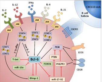
Various cytokines, surface molecules, and transcription fac- tors are reported to be involved in TFH cell differentiation (Fig. 1). IL-6 and IL-21 are critical cytokines for TFH cell differ-
Figure 1. Molecular mechanisms of Bcl-6 expression in T cells. Bcl-6, the master regulator of TFH cell differentiation is controlled by a complex signaling pathway. Co-stimulatory molecules such as CD28 and ICOS activate PI3K to induce Bcl-6 expression. PTEN, PHLPP2 inhibit Bcl-6 expression through interfering PI3K signaling and Foxo1 directly inhibits Bcl-6 expression. Various cytokines, such as IL-6, IL-21, IL-12, and IFN-γ induce Bcl-6 expression through JAK-STAT signaling pathway while high level of IL-2 in combination with IL-12 induces T-bet to inhibit Bcl-6. Blimp-1 and Bcl-6 is reciprocally regulating each other to make a decision of effector T cell fate between TFH and non-TFH effector cells. Some miRNA such as miR-17-92 induces Bcl-6 expression by interfering phosphatases, which inhibit PI3K signaling pathway while miR-10a directly inhibits Bcl-6 expression.
Figure 2. Germinal center reaction controlled by TFH and TFR cells.
Naïve T cells following stimulation with TcR and co-stimulatory molecules with IL-6 and IL-21 by dendritic cells can differentiate into TFH cells and migrate to the CXCL13-rich B cell follicle region. In the B cell follicle, TFH cells interact with B cells via TcR and co-stimulatory molecules such as ICOS and CD40L. Upon interaction between TFH cells and B cells, IL-4 and IL-21 from TFH cells allow B cells to differentiate into memory B cells or plasma cells, which are involved in long-lasting antibody production. TFR cells derived from nTreg precursor cells from the thymus by expressing Bcl-6 and CXCR5 migrate to B cell follicle and inhibit both TFH cell and B cell function.
entiation (4). Surface molecules, including ICOS, CD40L, PD-1, BTLA, and SAP are also important for TFH cell differ- entiation and their functions (5). Inhibiting the interaction be- tween CD40 and CD40L, or deficiency of ICOS or its ligand causes defects in formation of the germinal center (GC) (6) and TFH cell differentiation (7,8). In addition, SAP contributes to TFH cell differentiation by maintaining stable T and B cell interaction (6,9). Cytokine- and co-stimulatory molecule-me- diated signaling pathways are essential for expression of the transcription factor B cell lymphoma-6 (Bcl-6), which is the master regulator of TFH cell differentiation and is inhibited by the antagonizing transcription factor Blimp-1. Expression of Bcl-6 and Blimp-1 is reciprocally regulated during T cell dif- ferentiation (1).
Bcl-6-deficient T cells failed to differentiate into TFH cells and the GC responses are hardly developed, demonstrating the absolute requirement for Bcl-6 (2,3). TFH cell differ- entiation program involves a dramatic change in surface ex- pression of chemokine receptors. Reciprocal up-regulation of CXC-chemokine receptor 5 (CXCR5) and down-regulation of
CCR7 enables TFH cells to migrate into B cell follicles by re- sponding to CXCL13, the ligand of CXCR5 (10-12). Inside of B cell follicles, TFH cells provide B cell help signals by ex- pressing co-stimulatory molecules and secreting cytokines such as IL-4 and IL-21, which are essential for germinal center B cells to undergo class switch recombination, somatic hy- per-mutation, affinity maturation, and differentiation of plas- ma cells and memory B cells in the GC (13-15).
Recently, it was reported that some microRNAs (miRNAs) have a regulatory role in TFH cell differentiation and the GC reaction. The miR-17-92 cluster acts as a positive regulator of TFH cell differentiation via suppression of phosphatases that inhibits ICOS-mediated PI3K signaling pathways (16). In addi- tion, the miR-17-92 cluster represses the expression of RORα, which induces inappropriate gene expression during TFH cell differentiation (17). By contrast, miR-10a directly inhibits Bcl-6 expression (18), which strongly indicating that miRNAs are involved in dynamic regulation of TFH differentiation.
If the GC reaction mediated by TFH cells is dysregulated or if autoreactive T and B cells are activated, high levels of autoantibody can be accumulated through abnormal GC for- mation, which contributes to the development of autoimmune
diseases (19). Thus, TFH cells should be tightly regulated to prevent autoimmunity by limiting germinal center reactions to self antigen (20). Recently, follicular regulatory T (TFR) cells expressing CXCR5 were demonstrated to limit the GC reaction and reduce antibody production by migrating into B cell fol- licles (21). The regulation of germinal center reaction by TFH
and TFR cells for normal immunity is summarized as figure (Fig. 2).
In this review, we discuss the function of cytokines, tran- scription factors, and signaling pathways related to the differ- entiation or characteristics of TFH cells. Additionally, we dis- cuss the role of the GC reaction related to TFH and TFR cells in the maintenance of immune homeostasis and provide both a better understanding of the importance of TFH cells in auto- immunity and their clinical relevance in human autoimmune diseases.
SIGNALING PATHWAYS REQUIRED FOR TFH CELL DIFFERENTIATION
ICOS, PI3K, and Foxo1
It has been reported that a strong interaction between the TcR and major histocompatibility complex (MHC) class II mole- cules triggers TFH cell differentiation, which indicates that a strong TcR signal is essential for TFH cell differentiation (22).
In addition, among surface co-stimulatory molecules being expressed by TFH cells, ICOS is induced when CD4 T cells become activated by recognizing antigen through TcRs, which then interact with ICOS-L that is expressed on B cells (7,11,23,24). Its binding to the ligand ICOS-L triggers activation signals in a similar way to other members of CD28 family co-stimulatory receptors (25,26). ICOS plays a significant role in increasing T cell proliferation and the production of cyto- kines, including IL-21 and IL-4 (11,27,28).
ICOS-mediated PI3K activation is crucial for TFH cell differ- entiation, as a point mutation on the cytoplasmic tail of ICOS, where PI3K binds to and activates, led to a severely impaired TFH cell differentiation of CD4 T cells (28). In contrast, over- expression of ICOS is sufficient to maintain TFH cells in CD28-deficient mice (7). Among PI3K subunit p110γ ap- pears to convey ICOS-mediated TFH cell differentiation signal- ing pathway, as p110γ deficiency resulted in a defective TFH
cell differentiation, further strongly indicating that ICOS and PI3K are important for either differentiation or survival of TFH
cells. These results imply that ICOS-mediated PI3K signaling is crucial for the differentiation of TFH cells (25). Moreover,
Heping et al. reported that ICOS signaling is critical for mo- tility of TFH cells into the B cell follicle in a Bcl-6 independent manner (29).
PI3K signaling pathways following TcR and co-stimulation regulate the phosphorylation of Foxo1 to relocate it from the nucleus to the cytoplasm (30,31). A recent study revealed that Foxo1 negatively regulates T cell activation and contributes to T cell tolerance (32). Foxo1-deficient CD4 T cells contrib- ute to the development of autoimmune phenotypes including increased autoantibody production with reduced Foxp3+ reg- ulatory T cell development and function, and augmented gen- eration of TFH cells and GC formation. In addition, the pres- ence of Foxo-binding elements has been identified in the pro- moter region of Bcl-6 (33), which suggests that Foxo1 might act as a transcriptional repressor of Bcl-6 and, if so, Foxo1 might negatively regulate TFH differentiation. Thus, ICOS and PI3K signaling is crucial for TFH differentiation and Foxo1 could be a regulator of GC reaction.
IL-21, IL-6, and STATs
IL-6 and IL-21 are well-known pro-inflammatory cytokines with important roles in Bcl-6 expression and TFH cell differ- entiation (4). IL-21 induces B cell proliferation and class switch recombination and IL-21R is required for antibody re- sponse and GC formation (34). The IL-6-mediated STAT3 acti- vation is also important for IL-21 expression in human and mouse naïve CD4 T cells upon TcR stimulation (35,36).
STAT3 is phosphorylated by JAK upon IL-6 stimulation, and activated STAT3 was shown to bind to Bcl-6 in T cells (33).
IL-6 is an important factor for Bcl-6 induction in CD4 T cells during dendritic cell priming stage of CD4 T cell activation (37). However, other signaling pathways could compensate for IL-6 dependent TFH differentiation pathway, as TFH cells are normally found at the peak of the immune response to infection and immunization (38,39). IL-6 signaling is required for IL-21 expression via c-Maf (40-42). Once being produced, IL-21 further increases its own production through a positive feedback mechanism (43).
Augmented IL-21 was reported to induce the expression of the master regulator for TFH cell differentiation, Bcl-6 (44), while controversies exist whether IL-21 is a critical factor for Bcl-6 induction in CD4 T cells (37-39). At a downstream level, Choi et al. showed that IL-6-mediated STAT1 signaling can also prime TFH cells by compensating for STAT3 and inducing Bcl-6 expression. Another recent study demonstrated that IL-12-mediated STAT4 signaling can induce expression of
both Bcl-6 and T-bet, and T-bet inhibits the function of Bcl-6 (45). The balance between T-bet and Bcl-6 expression might be regulated by IL-2 concentration (33). Furthermore, IFN-γ was accounted to lead to abnormal TFH cell differentiation in the sanroque mouse model (46). Given that IFN-γ induced Bcl-6 via pSTAT1 which binds to an IRE in an exon of Bcl-6 (47), IFN-γ could function as a positive regulator by directly inducing Bcl-6 expression in CD4 T cells. This supported by recent study by Lee et al., which demonstrated that T cell specific deletion of IFN-γR resulted in decreased TFH cell dif- ferentiation in sanroque mice (46). Further studies are needed to clarify how this complex cytokine network regulates Bcl-6 expression and TFH cell differentiation.
Bcl-6 and Blimp-1
The zinc-finger-containing transcriptional repressor Bcl-6 was originally described as a key molecule in GC formation and B cell response (48,49). Bcl-6-deficient mice cannot develop somatic hyper-mutation in B cell, result impaired GC for- mation (50,51). In addition, B cells from these mice do not undergo affinity maturation, somatic hyper-mutation, and class switch recombination of immunoglobulin (49). Recently, Bcl-6 was identified as a crucial factor for TFH cell differ- entiation (3). Bcl-6-deficient mice show impaired TFH cell dif- ferentiation (2) and non-TFH CD4 T cells do not express in- creased levels of Bcl-6 (2,52). Bcl-6 directly inhibits a number of transcription factors, including T-bet and RORγt, which are key modulators of differentiation of Th1 and Th17 cells, respectively (3). Bcl-6 also inhibits expression of CCR7 and PSGL-1, which negatively regulate the migration of T cells in- to B cell follicles (39,53). Moreover, Bcl-6 regulates the ex- pression of various TFH cell-related molecules, including ICOS, PD-1, PTLA, CD200, and SAP (23). Turner et al. identified the mouse form of Blimp-1, which is induced by cyto- kine-mediated B cell differentiation (54). Recent studies re- ported that the transcription factor Blimp-1 has an antago- nistic role of Bcl-6 (1,52,55) and inhibits TFH cell differ- entiation (1). Blimp-1 is highly expressed in non-TFH effector T cells such as Th1, Th2, and Th17 cells (1,52), whereas Bcl-6 is highly expressed only in TFH cells. Moreover, constitutive expression of Blimp-1 inhibited TFH cell formation (1) and Blimp-1 is important for terminal differentiation of both CD4+ and CD8+ T cells, which is characterized by high levels of effector molecule secretion and low proliferative potential (52). IL-2 mediated STAT5 signaling in activated CD4+ T cells induces expression of Blimp-1, which suppresses Bcl-6 and
TFH cell differentiation (56). High level of IL-2, especially in effector Th1 cells, induces T-bet, which also inhibits Bcl-6 ex- pression and TFH cell differentiation (33). Th1 cells might have the flexibility to regulate the expression of T-bet and Bcl-6 depending on environmental conditions (33). IL-6- and IL-21-mediated STAT3 signaling can also induce Blimp-1 or Bcl-6 (57) through the participation of additional transcription factors (5). To summarize, effector T cell fate seems to rely on the expression of Bcl-6 or Blimp-1 and they are recip- rocally inhibit each other via complex signaling pathway, eventually act as a decision maker between TFH cell and other effector T cell differentiation.
REGULATION OF TFH CELL DIFFERENTIATION VIA TFR CELL AND miRNA
Follicular regulatory T cells
Foxp3-expressing regulatory T (Treg) cells contribute to the maintenance of immune tolerance by suppressing the dysre- gulated immune response to self-antigens (58). Scurfy mice without Foxp3+ T cells demonstrate severe systemic auto- immune phenotype. In addition, CD4 T cells isolated from scurfy mouse are hyper-responsive to TcR stimulation (59,60).
It has been recently reported that the mice with CXCR5- deficient Treg cells have more GC with augmented im- munoglobulin production owing to the limited capability of these cells to migrate into B cell follicular region. This sug- gests that CXCR5 expression of Treg cells is crucial for regu- lation of the GC reaction (61). In addition, Treg cells express- ing Bcl-6 and CXCR5, which originate from CXCR5- natural Treg cell precursors, are found in GC (21). In the absence of CXCR5+Bcl-6+ Treg cells, the GC reaction was not controlled efficiently leading to enhanced immunoglobulin production and increased B cell population in GCs. This result implies that Treg cells expressing CXCR5 have important roles in regu- lation of the GC reaction. Treg cells in GC are called follicular regulatory T (TFR) cells, which share characteristics of both TFH and Treg cells since Bcl-6, CD28 and SAP also affect devel- opment of TFR cells. 5∼25% of TFH cells expressing CXCR5 and PD-1 are also Foxp3+ TFR cells and are located in the B cell follicle region (62). Recent study demonstrated that lack of the PD-1-PD-L1 pathway induced increase of TFR cells and its suppressive ability, suggesting the regulatory role of PD-1 in the differentiation of TFR cells (63). Co-transfer ex- periments with thymus-derived Foxp3+ CD4 T cells and Foxp3− CD4 T cells into recipient demonstrated that Foxp3+
CD4 T cells become TFR cells in mice immunized with antigen suggesting TFR cells are derived from Treg cell precursors. A recent study demonstrated Ag-specific antibody production was increased in the mice with Bcl-6-deficient Treg cells (21).
Furthermore, increased levels of high affinity antibody, plas- ma cells, and memory B cells are found in the mice demon- strating that TFR cells expressing Bcl-6 control the GC reaction including plasma cell production and affinity maturation. By contrast, Blimp-1 down- regulates the number of TFR cells, suggesting that Bcl-6 and Blimp-1 also reciprocally regulate differentiation of TFH and TFR cells to control the GC reaction (1). TFR cells therefore seem to play a crucial role in the main- tenance of immune tolerance, preventing autoimmune re- sponse by inhibiting the GC reaction and antibody production.
micro RNAs
miRNAs are functional single stranded RNAs (ssRNAs), which are encoded endogenously, and are involved in immune cell development and differentiation (64,65). Recent study re- ported that the miR-17-92 cluster was regulated by Bcl-6 in CD4 T cells (3). T cells overexpressing Bcl-6 demonstrated diminished expression of the miR-17-92 cluster, as do TFH
cells, which, suppresses the expression of CXCR5. However, several studies have shown that the miRNA-17-92 cluster in- duces TFH cell differentiation (16,17). T cell specific miR-17-92 transgenic mice demonstrate spontaneous Bcl-6 expression, TFH cell differentiation, and GC formation (16). In contrast, miR-17-92-deficient mice show impaired TFH cell differ- entiation during acute and chronic virus infection. The miR-17-92 cluster induces TFH cell differentiation through sup- pression of PTEN and PHLPP2 expression, which regulate the ICOS-PI3K pathway. The miR-17-92 cluster also directly in- hibits expression of RORα, which is involved in gene ex- pression of non-TFH effector T cell differentiation (17). In ad- dition, miR-10a, which is specifically expressed in Treg cells by TGF-β and retinoic acid, directly suppresses Bcl-6 ex- pression (18). Some induced-Treg (iTreg) cells migrate to GC in Peyer’s patch and have TFH-like phenotypes. miR-10a ex- pression is down-regulated in these iTreg cells and over- expression of miR-10a significantly inhibits the conversion of iTreg into TFH-like cells. More studies on the role of TFR cells and miRNA in TFH differentiation are needed to improve our understanding on dynamic regulation of germinal center reaction.
TFH CELLS IN AUTOIMMUNE DISEASES Systemic lupus erythematosus
Systemic lupus erythematosus (SLE) is an autoimmune disease with a complex phenotype that includes systemic inflam- mation, fever, fatigue, and chills (66). Diagnosis of SLE is very difficult because its phenotype overlaps with other diseases. Recent studies have suggested that the pathogenesis of SLE is profoundly related to TFH cells (44,67,68). Sponta- neous GC formation and autoantibody production have been reported in many mouse models of SLE (44,67), suggesting that TFH cells might be associated with pathogenesis of SLE.
Indeed, recent studies demonstrated that TFH cell differ- entiation is spontaneously induced in these mouse models (44,67,68). Also, dysregulated TFH cell activity contributes to the pathogenesis of SLE through aberrant GC formation and massive production of autoantibodies, such as anti-dsDNA and ANA. TFH cells induce these phenomena via cytokines and co-stimulatory molecules which stimulate B cells (69,70).
Autoimmune phenotypes were alleviated when TFH cell differ- entiation was inhibited in sanroque mice, which have in- creased GC formation and TFH cell differentiation (70).
Linterman et al. crossed sanroque mice with IL-21- or SAP-de- ficient mice, or mice heterozygous for Bcl-6 to examine the role of Bcl-6 in development of the lupus-like phenotype (71). They found that the deficiencies of Bcl-6 or SAP amelio- rate the lupus-like phenotype in sanroque mice IL-21 in- dependently. However, lupus-like autoimmune phenotypes were reduced in another study when IL-21 signaling is not present in BXSB-Yaa mice, another mouse model of human SLE (72), recapitulating the complexity of pathogenesis of SLE in human. Remarkably, IL-21 expression was up-regulated in SLE patients than in healthy controls (73), and elevated pro- duction of TFH relating factors such as CXCL13, BAFF were reported in human SLE patients (74). These results suggest that abnormal TFH cell differentiation strongly related to SLE pathogenesis.
Rheumatoid arthritis
Rheumatoid arthritis (RA) is an autoimmune disorder, which is recently studied that it is associated with dysregulated TFH
cell differentiation. Deborah et al. found that blockade of IL-21 signaling by IL-21R-Fc fusion protein treatment reduces disease severity in mouse and rat RA models (75). Further- more, IL-21 blockade in animal models results in decreased IL-6 expression. A recent study by Victoratos et al. found that
TFH cells have a critical role in the maintenance of follicular dendritic cell (FDC)-mediated GC formation and autoantibody production in KRN/B mice that spontaneously develop RA (76,77). In addition, Jang et al. reported that IL-21 re- ceptor-deficient KBx/N mice have less severe RA with re- duced TFH cell population in draining lymph node (43). An IL-21R-Fc fusion protein that inhibits IL-21 signaling can delay disease onset and progression. Platt et al. also found in- creased TFH cells and antibody production in an OVA-induced RA mouse model (78). In this study, they showed that abata- cept, a fusion protein composed of the Fc region of IgG and the extracellular domain of CTLA-4 has a role in regulation of TFH cell differentiation in OVA-induced RA mouse models.
In human RA patients, up-regulated IL-21 level was reported with increased TFH-like cells that enhanced IL-21R expression (79). This increased TFH-like cells population correlated with enhanced 28-joint count disease activity score and anti-CCP antibody, which indicate disease severity.
Synthetically, TFH cells seem to be involved in the patho- genesis of human autoimmune diseases such as SLE, RA, etc., therefore, regulation of TFH cell differentiation could be an important strategy for the suppression of autoimmune diseases.
CONCLUSION
Recently, characterization of TFH cells and germinal center re- action has been highlighted in immunology field that TFH cells have crucial roles in B cell response in adaptive immunity.
IL-6 and IL-21 signaling induce expression of CXCR5, which enables the migration of T cells into B cell follicles with ex- pressing Bcl-6, a master transcription factor for TFH cell differentiation. These characteristics of TFH cells distinguish them from other helper T cells. TFH cells can induce affinity maturation, somatic hyper-mutation which mediate memory B cells and long-lived plasma cells with increased germinal centers. However, abnormally activated TFH cell function give rise to an immune reaction against auto-antigens, and sub- sequently could trigger autoimmune diseases such as SLE, RA, etc. There are several ways including miRNAs, TFR cells, and IL-21 blockade to potentially correct abnormal germinal cen- ter reaction by negatively regulating aberrant TFH cell differ- entiation. Through better understanding of current knowledge of TFH cell mediated dynamic germinal center reaction, we hope to discover novel therapeutic approaches by targeting TFH cells in human autoimmune diseases.
ACKNOWLEDGEMENTS
This study is supported by Basic Science Research Program through National Research Foundation of Korea grants (NRF- 2011-0012859 and NRF-2013R1A 1A2A 10060048).
CONFLICT OF INTEREST
The authors have no financial conflicts of interest to declare.
REFERENCES
1. Johnston, R. J., A. C. Poholek, D. DiToro, I. Yusuf, D. Eto, B. Barnett, A. L. Dent, J. Craft, and S. Crotty. 2009. Bcl6 and Blimp-1 are reciprocal and antagonistic regulators of T follicular helper cell differentiation. Science 325: 1006-1010.
2. Nurieva, R. I., Y. Chung, G. J. Martinez, X. O. Yang, S.
Tanaka, T. D. Matskevitch, Y. H. Wang, and C. Dong. 2009.
Bcl6 mediates the development of T follicular helper cells.
Science 325: 1001-1005.
3. Yu, D., S. Rao, L. M. Tsai, S. K. Lee, Y. He, E. L. Sutcliffe, M. Srivastava, M. Linterman, L. Zheng, N. Simpson, J. I.
Ellyard, I. A. Parish, C. S. Ma, Q. J. Li, C. R. Parish, C. R.
Mackay, and C. G. Vinuesa. 2009. The transcriptional re- pressor Bcl-6 directs T follicular helper cell lineage commitment. Immunity 31: 457-468.
4. Nurieva, R. I., Y. Chung, D. Hwang, X. O. Yang, H. S. Kang, L. Ma, Y. H. Wang, S. S. Watowich, A. M. Jetten, Q. Tian, and C. Dong. 2008. Generation of T follicular helper cells is mediated by interleukin-21 but independent of T helper 1, 2, or 17 cell lineages. Immunity 29: 138-149.
5. Crotty, S. 2011. Follicular helper CD4 T cells (TFH). Annu.
Rev. Immunol. 29: 621-663.
6. Qi, H., J. L. Cannons, F. Klauschen, P. L. Schwartzberg, and R. N. Germain. 2008. SAP-controlled T-B cell interactions un- derlie germinal centre formation. Nature 455: 764-769.
7. Dong, C., U. A. Temann, and R. A. Flavell. 2001. Cutting edge: critical role of inducible costimulator in germinal center reactions. J. Immunol. 166: 3659-3662.
8. Iwai, H., M. Abe, S. Hirose, F. Tsushima, K. Tezuka, H.
Akiba, H. Yagita, K. Okumura, H. Kohsaka, N. Miyasaka, and M. Azuma. 2003. Involvement of inducible cos- timulator-B7 homologous protein costimulatory pathway in murine lupus nephritis. J. Immunol. 171: 2848-2854.
9. Lu, K. T., Y. Kanno, J. L. Cannons, R. Handon, P. Bible, A. G. Elkahloun, S. M. Anderson, L. Wei, H. Sun, J. J.
O'Shea, and P. L. Schwartzberg. 2011. Functional and epi- genetic studies reveal multistep differentiation and plasticity of in vitro-generated and in vivo-derived follicular T helper cells. Immunity 35: 622-632.
10. Breitfeld, D., L. Ohl, E. Kremmer, J. Ellwart, F. Sallusto, M.
Lipp, and R. Förster. 2000. Follicular B helper T cells express CXC chemokine receptor 5, localize to B cell follicles, and support immunoglobulin production. J. Exp. Med. 192:
1545-1552.
11. Schaerli, P., K. Willimann, A. B. Lang, M. Lipp, P. Loetscher, and B. Moser. 2000. CXC chemokine receptor 5 expression defines follicular homing T cells with B cell helper function.
J. Exp. Med. 192: 1553-1562.
12. Balkwill, F. 2004. Cancer and the chemokine network. Nat.
Rev. Cancer 4: 540-550.
13. Jacob, J., G. Kelsoe, K. Rajewsky, and U. Weiss. 1991.
Intraclonal generation of antibody mutants in germinal centres. Nature 354: 389-392.
14. Berek, C., A. Berger, and M. Apel. 1991. Maturation of the immune response in germinal centers. Cell 67: 1121-1129.
15. Liu, Y. J., F. Malisan, O. de Bouteiller, C. Guret, S. Lebecque, J. Banchereau, F. C. Mills, E. E. Max, and H. Martinez- Valdez. 1996. Within germinal centers, isotype switching of immunoglobulin genes occurs after the onset of somatic mutation. Immunity 4: 241-250.
16. Kang, S. G., W. H. Liu, P. Lu, H. Y. Jin, H. W. Lim, J.
Shepherd, D. Fremgen, E. Verdin, M. B. Oldstone, H. Qi, J. R. Teijaro, and C. Xiao. 2013. MicroRNAs of the miR-17 approximately 92 family are critical regulators of T(FH) differentiation. Nat. Immunol. 14: 849-857.
17. Baumjohann, D., R. Kageyama, J. M. Clingan, M. M. Morar, S. Patel, D. de Kouchkovsky, O. Bannard, J. A. Bluestone, M. Matloubian, K. M. Ansel, and L. T. Jeker. 2013. The microRNA cluster miR-17~92 promotes TFH cell differentiation and represses subset-inappropriate gene expression. Nat.
Immunol. 14: 840-848.
18. Takahashi, H., T. Kanno, S. Nakayamada, K. Hirahara, G.
Sciumè, S. A. Muljo, S. Kuchen, R. Casellas, L. Wei, Y.
Kanno, and J. J. O'Shea. 2012. TGF-beta and retinoic acid induce the microRNA miR-10a, which targets Bcl-6 and con- strains the plasticity of helper T cells. Nat. Immunol. 13:
587-595.
19. Vinuesa, C. G. and M. C. Cook. 2001. The molecular basis of lymphoid architecture and B cell responses: implications for immunodeficiency and immunopathology. Curr. Mol.
Med. 1: 689-725.
20. King, C., S. G. Tangye, and C. R. Mackay. 2008. T follicular helper (TFH) cells in normal and dysregulated immune responses. Annu. Rev. Immunol. 26: 741-766.
21. Chung, Y., S. Tanaka, F. Chu, R. I. Nurieva, G. J. Martinez, S. Rawal, Y. H. Wang, H. Lim, J. M. Reynolds, X. H. Zhou, H. M. Fan, Z. M. Liu, S. S. Neelapu, and C. Dong. 2011.
Follicular regulatory T cells expressing Foxp3 and Bcl-6 sup- press germinal center reactions. Nat. Med. 17: 983-988.
22. Fazilleau, N., L. J. McHeyzer-Williams, H. Rosen, M. G.
McHeyzer-Williams. 2009. The function of follicular helper T cells is regulated by the strength of T cell antigen receptor binding. Nat. Immunol. 10: 375-384.
23. Chtanova, T., S. G. Tangye, R. Newton, N. Frank, M. R.
Hodge, M. S. Rolph, and C. R. Mackay. 2004. T follicular helper cells express a distinctive transcriptional profile, re- flecting their role as non-Th1/Th2 effector cells that provide help for B cells. J. Immunol. 173: 68-78.
24. Choi, Y. S., R. Kageyama, D. Eto, T. C. Escobar, R. J.
Johnston, L. Monticelli, C. Lao, and S. Crotty. 2011. ICOS re- ceptor instructs T follicular helper cell versus effector cell dif- ferentiation via induction of the transcriptional repressor Bcl6.
Immunity 34: 932-946.
25. McAdam, A. J., T. T. Chang, A. E. Lumelsky, E. A.
Greenfield, V. A. Boussiotis, J. S. Duke-Cohan, T. Chernova, N. Malenkovich, C. Jabs, V. K. Kuchroo, V. Ling, M. Collins, A. H. Sharpe, and G. J. Freeman. 2000. Mouse inducible cos- timulatory molecule (ICOS) expression is enhanced by CD28 costimulation and regulates differentiation of CD4+ T cells.
J. Immunol. 165: 5035-5040.
26. Hutloff, A., A. M. Dittrich, K. C. Beier, B. Eljaschewitsch, R.
Kraft, I. Anagnostopoulos, and R. A. Kroczek. 1999. ICOS is an inducible T-cell co-stimulator structurally and functionally related to CD28. Nature 397: 263-266.
27. Vogelzang, A., H. M. McGuire, D. Yu, J. Sprent, C. R.
Mackay, and C. King. 2008. A fundamental role for inter- leukin-21 in the generation of T follicular helper cells.
Immunity 29: 127-137.
28. Gigoux, M., J. Shang, Y. Pak, M. Xu, J. Choe, T. W. Mak, and W. K. Suh. 2009. Inducible costimulator promotes helper T-cell differentiation through phosphoinositide 3-kinase.
Proc. Natl. Acad. Sci. USA 106: 20371-20376.
29. Xu, H., X. Li, D. Liu, J. Li, X. Zhang, X. Chen, S. Hou, L.
Peng, C. Xu, W. Liu, L. Zhang, and H. Qi. 2013. Follicular T-helper cell recruitment governed by bystander B cells and ICOS-driven motility. Nature 496: 523-527.
30. Brunet, A., A. Bonni, M. J. Zigmond, M. Z. Lin, P. Juo, L.
S. Hu, M. J. Anderson, K. C. Arden, J. Blenis, and M. E.
Greenberg. 1999. Akt promotes cell survival by phosphorylat- ing and inhibiting a Forkhead transcription factor. Cell 96:
857-868.
31. Yuan, T. L. and L. C. Cantley. 2008. PI3K pathway alterations in cancer: variations on a theme. Oncogene 27: 5497-5510.
32. Kerdiles, Y. M., E. L. Stone, D. R. Beisner, M. A. McGargill, I. L. Ch'en, C. Stockmann, C. D. Katayama, and S. M.
Hedrick. 2010. Foxo transcription factors control regulatory T cell development and function. Immunity 33: 890-904.
33. Oestreich, K. J., S. E. Mohn, and A. S. Weinmann. 2012.
Molecular mechanisms that control the expression and activity of Bcl-6 in TH1 cells to regulate flexibility with a TFH-like gene profile. Nat. Immunol. 13: 405-411.
34. Spolski, R. and W. J. Leonard. 2008. Interleukin-21: basic bi- ology and implications for cancer and autoimmunity. Annu.
Rev. Immunol. 26: 57-79.
35. Diehl, S. A., H. Schmidlin, M. Nagasawa, B. Blom, and H.
Spits. 2012. IL-6 Triggers IL-21 production by human CD4(+) T cells to drive STAT3-dependent plasma cell differentiation in B cells. Immunol. Cell Biol. 90: 802-811.
36. Eddahri, F., S. Denanglaire, F. Bureau, R. Spolski, W. J.
Leonard, O. Leo, and F. Andris. 2009. Interleukin-6/STAT3 signaling regulates the ability of naive T cells to acquire B-cell help capacities. Blood 113: 2426-2433.
37. Choi, Y. S., D. Eto, J. A. Yang, C. Lao, and S. Crotty. 2013.
Cutting edge: STAT1 is required for IL-6-mediated Bcl6 in- duction for early follicular helper cell differentiation. J.
Immunol. 190: 3049-3053.
38. Eto, D., C. Lao, D. DiToro, B. Barnett, T. C. Escobar, R.
Kageyama, I. Yusuf, and S. Crotty. 2011. IL-21 and IL-6 are critical for different aspects of B cell immunity and re- dundantly induce optimal follicular helper CD4 T cell (Tfh)
differentiation. PLoS One 6: e17739.
39. Poholek, A. C., K. Hansen, S. G. Hernandez, D. Eto, A.
Chandele, J. S. Weinstein, X. Dong, J. M. Odegard, S. M.
Kaech, A. L. Dent, S. Crotty, and J. Craft. 2010. In vivo regu- lation of Bcl6 and T follicular helper cell development. J.
Immunol. 185: 313-326.
40. Takeda, K., T. Kaisho, N. Yoshida, J. Takeda, T. Kishimoto, and S. Akira. 1998. Stat3 activation is responsible for IL-6-de- pendent T cell proliferation through preventing apoptosis:
generation and characterization of T cell-specific Stat3-defi- cient mice. J. Immunol. 161: 4652-4660.
41. Nurieva, R., X. O. Yang, G. Martinez, Y. Zhang, A. D.
Panopoulos, L. Ma, K. Schluns, Q. Tian, S. S. Watowich, A.
M. Jetten, and C. Dong. 2007. Essential autocrine regulation by IL-21 in the generation of inflammatory T cells. Nature 448: 480-483.
42. Yang, Y., J. Ochando, A. Yopp, J. S. Bromberg, and Y. Ding.
2005. IL-6 plays a unique role in initiating c-Maf expression during early stage of CD4 T cell activation. J. Immunol. 174:
2720-2729.
43. Jang, E., S. H. Cho, H. Park, D. J. Paik, J. M. Kim, and J.
Youn. 2009. A positive feedback loop of IL-21 signaling pro- voked by homeostatic CD4+CD25− T cell expansion is essen- tial for the development of arthritis in autoimmune K/BxN mice. J. Immunol. 182: 4649-4656.
44. Ozaki, K., R. Spolski, R. Ettinger, H. P. Kim, G. Wang, C.
F. Qi, P. Hwu, D. J. Shaffer, S. Akilesh, D. C. Roopenian, H. C. Morse, 3rd, P. E. Lipsky, and W. J. Leonard. 2004.
Regulation of B cell differentiation and plasma cell generation by IL-21, a novel inducer of Blimp-1 and Bcl-6. J. Immunol.
173: 5361-5371.
45. Nakayamada, S., Y. Kanno, H. Takahashi, D. Jankovic, K.
T. Lu, T. A. Johnson, H. W. Sun, G. Vahedi, O. Hakim, R.
Handon, P. L. Schwartzberg, G. L. Hager, and J. J. O'Shea.
2011. Early Th1 cell differentiation is marked by a Tfh cell-like transition. Immunity 35: 919-931.
46. Lee, S. K., D. G. Silva, J. L. Martin, A. Pratama, X. Hu, P.
P. Chang, G. Walters, and C. G. Vinuesa. 2012. Interfer- on-gamma excess leads to pathogenic accumulation of fol- licular helper T cells and germinal centers. Immunity 37:
880-892.
47. Zhou, G. and S. J. Ono. 2005. Induction of BCL-6 gene ex- pression by interferon-gamma and identification of an IRE in exon I. Exp. Mol. Pathol. 78: 25-35.
48. Dent, A. L., A. L. Shaffer, X. Yu, D. Allman, and L. M.
Staudt. 1997. Control of inflammation, cytokine expression, and germinal center formation by BCL-6. Science 276:
589-592.
49. Klein, U. and R. Dalla-Favera. 2008. Germinal centres: role in B-cell physiology and malignancy. Nat. Rev. Immunol. 8:
22-33.
50. Ye, B. H., G. Cattoretti, Q. Shen, J. Zhang, N. Hawe, R. de Waard, C. Leung, M. Nouri-Shirazi, A. Orazi, R. S. Chaganti, P. Rothman, A. M. Stall, P. P. Pandolfi, and R. Dalla-Favera.
1997. The BCL-6 proto-oncogene controls germinal-centre formation and Th2-type inflammation. Nat. Genet. 16: 161- 170.
51. Toyama, H., S. Okada, M. Hatano, Y. Takahashi, N. Takeda,
H. Ichii, T. Takemori, Y. Kuroda, and T. Tokuhisa. 2002.
Memory B cells without somatic hypermutation are generated from Bcl6-deficient B cells. Immunity 17: 329-339.
52. Crotty, S., R. J. Johnston, and S. P. Schoenberger. 2010.
Effectors and memories: Bcl-6 and Blimp-1 in T and B lym- phocyte differentiation. Nat. Immunol. 11: 114-120.
53. Haynes, N. M., C. D. Allen, R. Lesley, K. M. Ansel, N.
Killeen, and J. G. Cyster. 2007. Role of CXCR5 and CCR7 in follicular Th cell positioning and appearance of a pro- grammed cell death gene-1high germinal center-associated subpopulation. J. Immunol. 179: 5099-5108.
54. Turner, C. A. Jr., D. H. Mack, and M. M. Davis. 1994.
Blimp-1, a novel zinc finger-containing protein that can drive the maturation of B lymphocytes into immunoglobulin-secret- ing cells. Cell 77: 297-306.
55. Martins, G. and K. Calame. 2008. Regulation and functions of Blimp-1 in T and B lymphocytes. Annu. Rev. Immunol.
26: 133-169.
56. Johnston, R. J., Y. S. Choi, J. A. Diamond, J. A. Yang, and S. Crotty. 2012. STAT5 is a potent negative regulator of TFH cell differentiation. J. Exp. Med. 209: 243-250.
57. Kwon, H., D. Thierry-Mieg, J. Thierry-Mieg, H. P. Kim, J.
Oh, C. Tunyaplin, S. Carotta, C. E. Donovan, M. L. Goldman, P. Tailor, K. Ozato, D. E. Levy, S. L. Nutt, K. Calame, and W. J. Leonard. 2009. Analysis of interleukin-21-induced Prdm1 gene regulation reveals functional cooperation of STAT3 and IRF4 transcription factors. Immunity 31: 941-952.
58. Sakaguchi, S. 2004. Naturally arising CD4+ regulatory t cells for immunologic self-tolerance and negative control of im- mune responses. Annu. Rev. Immunol. 22: 531-562.
59. Kanangat, S., P. Blair, R. Reddy, M. Daheshia, V. Godfrey, B. T. Rouse, and E. Wilkinson. 1996. Disease in the scurfy (sf) mouse is associated with overexpression of cytokine genes. Eur. J. Immunol. 26: 161-165.
60. Clark, L. B., M. W. Appleby, M. E. Brunkow, J. E. Wilkinson, S. F. Ziegler, and F. Ramsdell. 1999. Cellular and molecular characterization of the scurfy mouse mutant. J. Immunol. 162:
2546-2554.
61. Wollenberg, I., A. Agua-Doce, A. Hernández, C. Almeida, V.
G. Oliveira, J. Faro, and L. Graca. 2011. Regulation of the germinal center reaction by Foxp3+ follicular regulatory T cells. J. Immunol. 187: 4553-4560.
62. Linterman, M. A., W. Pierson, S. K. Lee, A. Kallies, S.
Kawamoto, T. F. Rayner, M. Srivastava, D. P. Divekar, L.
Beaton, J. J. Hogan, S. Fagarasan, A. Liston, K.G. Smith, and C. G. Vinuesa. 2011. Foxp3+ follicular regulatory T cells con- trol the germinal center response. Nat. Med. 17: 975-982.
63. Sage, P. T., L. M. Francisco, C. V. Carman, A. H. Sharpe.
2013. The receptor PD-1 controls follicular regulatory T cells in the lymph nodes and blood. Nat. Immunol. 14: 152-161.
64. Cobb, B. S., T. B. Nesterova, E. Thompson, A. Hertweck, E. O'Connor, J. Godwin, C. B. Wilson, N. Brockdorff, A. G.
Fisher, S. T. Smale, and M. Merkenschlager. 2005. T cell line- age choice and differentiation in the absence of the RNase III enzyme Dicer. J. Exp. Med. 201: 1367-1373.
65. Muljo, S. A., K. M. Ansel, C. Kanellopoulou, D. M.
Livingston, A. Rao, and K. Rajewsky. 2005. Aberrant T cell differentiation in the absence of Dicer. J. Exp. Med. 202:
261-269.
66. Doria, A., M. Zen, M. Canova, S. Bettio, N. Bassi, L. Nalotto, M. Rampudda, A. Ghirardello, and L. Iaccarino. 2010. SLE diagnosis and treatment: when early is early. Autoimmun.
Rev. 10: 55-60.
67. Luzina, I. G., S. P. Atamas, C. E. Storrer, L. C. daSilva, G.
Kelsoe, J. C. Papadimitriou, and B. S. Handwerger. 2001.
Spontaneous formation of germinal centers in autoimmune mice. J. Leukoc. Biol. 70: 578-584.
68. Simpson, N., P. A. Gatenby, A. Wilson, S. Malik, D. A.
Fulcher, S. G. Tangye, H. Manku, T. J. Vyse, G. Roncador, G. A. Huttley, C. C. Goodnow, C. G. Vinuesa, and M. C.
Cook. 2010. Expansion of circulating T cells resembling fol- licular helper T cells is a fixed phenotype that identifies a subset of severe systemic lupus erythematosus. Arthritis Rheum. 62: 234-244.
69. Daikh, D. I., B. K. Finck, P. S. Linsley, D. Hollenbaugh, and D. Wofsy. 1997. Long-term inhibition of murine lupus by brief simultaneous blockade of the B7/CD28 and CD40/gp39 costimulation pathways. J. Immunol. 159: 3104-3108.
70. Vinuesa, C. G., M. C. Cook, C. Angelucci, V.
Athanasopoulos, L. Rui, K. M. Hill, D. Yu, H. Domaschenz, B. Whittle, T. Lambe, I. S. Roberts, R. R. Copley, J. I. Bell, R. J. Cornall, and C. C. Goodnow. 2005. A RING-type ubiq- uitin ligase family member required to repress follicular help- er T cells and autoimmunity. Nature 435: 452-458.
71. Linterman, M. A., R. J. Rigby, R. K. Wong, D. Yu, R. Brink, J. L. Cannons, P. L. Schwartzberg, M. C. Cook, G. D.
Walters, and C. G. Vinuesa. 2009. Follicular helper T cells are required for systemic autoimmunity. J. Exp. Med. 206:
561-576.
72. Bubier, J. A., T. J. Sproule, O. Foreman, R. Spolski, D. J.
Shaffer, H. C. Morse 3rd, W. J. Leonard, and D. C.
Roopenian. 2009. A critical role for IL-21 receptor signaling in the pathogenesis of systemic lupus erythematosus in BXSB-Yaa mice. Proc. Natl. Acad. Sci. USA 106: 1518-1523.
73. Dolff, S., W. H. Abdulahad, J. Westra, B. Doornbos-van der Meer, P. C. Limburg, C. G. Kallenberg, and M. Bijl. 2011.
Increase in IL-21 producing T-cells in patients with systemic lupus erythematosus. Arthritis Res. Ther. 13: R157.
74. Wong, C. K., P. T. Wong, L. S. Tam, E. K. Li, D. P. Chen, and C. W. Lam. 2010. Elevated production of B cell chemo- kine CXCL13 is correlated with systemic lupus erythematosus disease activity. J. Clin. Immunol. 30: 45-52.
75. Young, D. A., M. Hegen, H. L. Ma, M. J. Whitters, L. M.
Albert, L. Lowe, M. Senices, P. W. Wu, B. Sibley, Y.
Leathurby, T. P. Brown, C. Nickerson-Nutter, J. C. Keith Jr, and M. Collins. 2007. Blockade of the interleukin-21/inter- leukin-21 receptor pathway ameliorates disease in animal models of rheumatoid arthritis. Arthritis Rheum. 56: 1152- 1163.
76. Kouskoff, V., A. S. Korganow, V. Duchatelle, C. Degott, C.
Benoist, and D. Mathis. 1996. Organ-specific disease pro- voked by systemic autoimmunity. Cell 87: 811-822.
77. Victoratos, P. and G. Kollias. 2009. Induction of autoanti- body-mediated spontaneous arthritis critically depends on fol- licular dendritic cells. Immunity 30: 130-142.
78. Platt, A. M., V. B. Gibson, A. Patakas, R. A. Benson, S. G.
Nadler, J. M. Brewer, I. B. McInnes, and P. Garside. 2010.
Abatacept limits breach of self-tolerance in a murine model of arthritis via effects on the generation of T follicular helper cells. J. Immunol. 185: 1558-1567.
79. Liu, R., Q. Wu, D. Su, N. Che, H. Chen, L. Geng, J. Chen, W. Chen, X. Li, and L. Sun. 2012. A regulatory effect of IL-21 on T follicular helper-like cell and B cell in rheumatoid arthritis. Arthritis Res. Ther. 14: R255.