서론
외상성 장애, 종양제거, 심한 골흡수 등으로 인한 골결손 부가 발생하고 있으며, 골결손부의 심미적, 기능적 회복, 안
정 및 치유를 증진시키기 위해 자가골, 동종골, 이종골, 합 성골 등의 골이식재를 이용한 재건술이 시행되고 있다. 이 상적인 골이식재는 생체 적합성이 뛰어나고, 이식한 후 골 형성을 유도 또는 전도하며, 시간이 지나면 결국은 완전히 흡수되고, 소실된 골을 영구적으로 대체할 수 있어야 한다.
이러한 골이식재 중 자가골이 골유도성과 골형성능이 뛰어 나 가장 예지성이 높은 골 이식재로 평가되지만, 자가골 채 득을 위한 부가적인 수술이 필요하고 이로 인한 합병증과 회복기간의 증가, 많은 골이 필요한 경우 충분한 양을 얻을 수 없으며 채취에 부가적인 수술 시간이 더 소요되는 등의 단점이 있다1-3). 이러한 문제점을 극복하고 자가골을 대체 하기 위한 다양한 종류의 골이식재의 개발과 연구가 지속되
수종의 탈단백 우골 이식재의 특성 비교 분석
여신일
1, 박성환
1, 노우창
1, 박진우
1, 이재목
1, 서조영
1,2*1. 경북대학교 치의학전문대학원 치주과학교실 2. 경조직 바이오치아 재생연구소
A comparative analysis of basic characteristics of several deproteinized bovine bone substitutes
Shin-il Yeo
1, Sung-Hwan Park
1, Woo-Chang Noh
1, Jin-Woo Park
1, Jae-Mok Lee
1, Jo-Young Suh
1,2*1. Department of Periodontology, School of Dentistry
2. Institute for Hard tissue and Bio-tooth Regeneration(IHBR), School of Dentistry, Kyungpook National University
ABSTRACT
Purpose: Deproteinized bovine bone substitutes are commonly used in dental regenerative surgery for treatment of alveolar defects. In this study, three different bovine bone minerals - OCS-B (NIBEC, Seoul, Korea), Bio-Oss (Geistlich - Pharma, Switzerland), Osteograft/N - 300 (OGN, Dentsply Friadent Ceramed. TN, USA) - were investigated to analyze the basic characteristics of commercially available bone substitutes.
Methods: Their physicochemical properties were evaluated by scanning electron microscopy, energy dispersive X-ray spectrometer (EDS), surface area analysis, and Kjeldahl protein analysis. Cell proliferation and alkaline phosphatase (ALP) activity of human osteosarcoma cells on different bovine bone minerals were evaluated.
Results: Three kinds of bone substitutes displayed different surface properties. Ca/P ratio of OCS - B shown to be lower than other two bovine bone minerals in EDS analysis. Bio-Oss had wider surface area and lower amount of residual protein than OCS - B and OGN. In addition Bio - Oss was proved to have lower cell proliferation and ALP activity due to lots of residual micro particles, compared with OCS - B and OGN.
Conclusions: Based on the results of this study, three bovine bone minerals that produced by similar methods appear to have different property and characteristics. It is suggested that detailed studies and quality management is needed in operations for dental use and its biological effects on new bone formation. (J Korean Acad Periodontol 2009;39:149-156)
KEY WORDS: bovine bone substitute; physicochemical property; cell proliferation; alkaline phosphatase activity.
* Correspondence: Jo-Young Suh, Ph.D. D.D.S.
Department of Periodontology, School of Dentistry, IHBR, Kyungpook National University, 188-1 2 Samduk-dong, Jung-gu, Daegu, 700-412, Korea
E-mail: [email protected], Tel: 82-53-600-7521, Fax: 82-53-427-3263
* This work was supported by the Korea Science and Engineering Foundation (KOSEF) grant funded by the Korean government (MOST) (No. M10646020002-08N4602–00210).
Received: Apr. 30, 2009; Accepted: Jun. 1, 2009
고 있다. 최근까지 많이 사용되고 있는 동종골 이식재는 사 체에서 채취하므로 환자의 불편함이나 양의 부족 등의 문제 는 극복할 수 있으나 조작이 쉽지 않고, 약 1백만에서 8백 만 분에 1의 확률로 질병 감염의 가능성이 있고, 면역학적 거부현상을 감소시키기 위한 과정에서 골형성 능력이 감소 되고 표본별로 골유도 능력에 상당한 차이를 보이는 단점이 있다4-8). 동종골 이식재의 이러한 여러 가지 문제점 때문에 다양한 골이식재에 대한 연구가 지속적으로 이루어지고 있 으며, 최근에는 우골에서 단백질 성분을 제거한 이종골 이 식재 및 합성골 이식재인 tricalcium-phosphate와 hy- droxyapatite (HA)와 같은 bioceramic 제재 등에 대한 연 구와 개발이 행해지고 있다9-11). 그러나 합성골은 이식 시 임상적으로 긍정적 결과를 보이기는 하나, 이들은 골전도체 로서 숙주골이 자라 들어 올 수 있는 공간만 유지해주는 비 계로서 역할을 주로 하는 것으로 알려져 있다12-14).
반면 이종골 이식재인 탈단백 우골은 사람과 같은 칼슘/
인 비율을 가지고 있고, 비교적 낮은 온도인 300℃에서 화 학적인 과정을 거쳐 골의 유기성분을 제거하기 때문에 합성 골과 달리 골 본래의 다공성 구조를 그대로 유지하는 특징 을 갖는다15,16). 또한 유기성분이 제거되고 남은 우골의 무기 질은 인간의 골과 매우 유사한 화학적인 조성과 형태 및 초 미세구조를 보인다17,18). 탈단백 우골은 1960년대에는 대중 적으로 사용되었으나, 이종이식재로서 골형성 인자 등의 단 백질 성분이 존재하는 경우 시술 후 자가면역 질환이 발생 하는 등의 문제점으로 사용이 감소하였다19). 하지만 유기물 질을 제거하는 기술이 나날이 발전하고 최근에는 탈단백 우 골을 사용한 골재건술의 성공적인 임상증례의 보고가 증가 되고 있다20-22). 여러 가지의 골이식재를 비교한 문헌에 따 르면 타이식재에 비해 탈단백 우골이 5배 이상의 높은 표면 적을 보이고, 수많은 결정체 공간과 미세터널을 갖기 때문 에 혈관과 골의 내부성장을 증가시킨다고 보고되고 있다23). 또한, 탈단백 우골은 생체친화성이 우수하며, 골형성의 척 도가 되는 골전도성이 높은 것으로 보고되었다24). 탈단백 우골의 흡수성에 대해서는 아직 많은 논란이 계속되고 있는 데, 탈단백 우골이 신생골에 의해 대체되면서 빠르게 흡수 된다는 보고25)가 있는가 하면 흡수소와가 드물고 아주 느리 게 흡수된다는 의견이 있고26,27), 전혀 흡수가 일어나지 않 는다는 주장도 있다28). 지금까지의 여러 임상보고들을 종합 해 보면, 탈단백 우골이 다른 골이식재와 비교하였을 때 부 적절한 흡수율과 잔존 단백질로 인한 문제점 등을 제외하면,
치조골 재생을 위한 시술 시 성공적인 임상결과를 나타냄을 알 수 있다. 하지만 많은 수의 임상보고나 연구논문에 비해 현재 판매되고 있는 여러 가지 탈단백 우골 제품의 기본적 특성과 장·단점에 대한 자료는 많지 않은 실정이다.
그러므로 본 연구에서는 탈단백 우골인 OCS-B와 Osteograft/N-300, Bio-Oss의 형태학적, 화학적 특성과 세포증식률을 평가하여 상호 비교하고자 하였다.
재료 및 방법
1. 실험재료
본 실험에 사용된 세 종류의 탈단백 우골은 다음과 같다.
1) Bio-Oss(Geistlich, Wolhusen, 스위스); 250~1000㎛
입자 크기, 망상골 이식재
2) Osteograft/N-300(OGN; Dentsply Friadent Ceramed, TN, 미국); 250~420 ㎛ 입자 크기, 피질골 이식재
3) OCS-B(NIBEC, 서울, 한국); 200~1000 ㎛ 입자 크기, 망상골 이식재
2. 골이식재의 형태학적, 화학적 특성 평가
탈단백 우골의 표면 미세구조를 알아보기 위하여 주사전 자현미경[Scanning electron microscope (SEM); S-4200, Hitachi, 일본]을 사용하여 관찰하였다. 칼슘과 인을 포함한 구성 성분의 정성과 정량 분석을 위해 Energy dispersive X-ray spectrometer (EDS)를 사용하여 칼슘과 인의 성분 비를 구하였다. 시료의 표면적을 측정하기 위해 질소가스를 흡착시켜 흡착 등온선으로부터 단위 질량당 표면적을 나타 내는 Brunauer-Emmett-Teller (BET)법을 이용한 비표면 적/기공도분석기 (High Speed Gas Sorption Analyzer;
Nova 2000-version 7.02; Quantachrome, 미국)를 사용 하였다. 잔존 단백질량 측정은 질소 분석의 국제적인 표준 분석법으로 알려진 Kjeldahl법을 이용하여 측정하였다. 탈 단백 우골 500 ㎎당 진한 황산 20㎖와 분해촉매제를 넣어 고온에서 가열하여 분해한 후 일련의 과정을 거쳐 Kjeldahl 단백질/질소 분석기(Vapodest; Gerhardt, 독일)를 사용하 여 시행하였다.
3. 골이식재에 대한 세포증식 평가
Human osteosarcoma (HOS) cell line을 이용하여 각각 의 탈단백 우골에 대한 세포증식률을 평가하기 위해 24 well plate에 2×10⁴개의 HOS 세포를 접종하여 1, 4, 7일간 배양한 후 hemocytometer를 이용하여 증식된 세포수를 측 정하였다. 양성대조군으로 탈단백 우골을 넣지 않고 HOS 세포만을 배양하여 사용하였다.
4. 골이식재에 대한 Alkaline phosphatase(ALP) 활성도 측정
배양된 세포를 well당 25㎎의 이식재가 함유된 12 well plate에 5×10⁴개의 세포를 접종하고 골화세포 분화에 필요 한 Ascorbic acid (50㎍/㎖), β-GP (10mM), dex- amethasone (100 mM/㎖)이 포함된 배양액에서 4, 7, 10 일간 배양하였다. ALP 활성도는 FLUOstar OPTIMA (BMG, 독일)로 측정하였으며, 기질로는 p-nitrophenyl-
phosphate를 사용하였다. 총 단백질량은 BCA 단백질 정량 시약 (Pierce, 미국)으로 측정하였으며, 흡광도 값과 총단백 질량의 비율로 최종 ALP 활성도를 산출하였다.
5. 통계분석
모든 자료는 평균±표준편차로 표기하였고 각각의 탈단 백 우골에 대한 세포 독성, 증식 및 ALP 활성도의 유의성 을 알아보기 위하여 one-way analysis of variance (ANOVA)를 이용하여 통계분석을 시행하였고, Dunnett t-test 와 Tukey test로 사후검정을 시행하였다(p< 0.05).
결과
1. 골이식재에 대한 주사전자현미경 관찰소견 세 종류 이식재의 주사전자현미경을 이용한 미세구조 관 찰에서 이식재 표면은 서로 다른 형태학적 특성을 나타내었
a b c
d e f
g h i
Figure. 1. Scanning electron micrographs of OCS-B (a, b, c), Bio-Oss (d, e, f) and OGN (g, h, i) at magnifications of X100 (a, d, g), X1000 (b, e, h) and X3000 (c, f, i). Each bone substitute displayed characteristic surface structure.
다(Fig. 1). 550~650℃에서 고온 처리된 OCS-B는 Bio- Oss와 OGN에 비해 다소 균일한 표면형태를 보여주었다.
Bio-Oss는 고배율에서 OCS-B와 OGN과는 전혀 다른 마이 크론 단위의 빗살무늬 형태의 표면구조를 보여주었고 많은 미세분말이 잔존하였다.
2. 골이식재에 대한 성분분석 및 비표면적 측정 결과
EDS 분석을 통한 이식재 표면 구성성분의 정성, 정량분 석을 위하여 시료당 3회 분석 후의 평균값을 측정한 결과 칼슘과 인의 원소비율 (Ca/P)은 OCS-B는 1.42, Bio-Oss 는 1.62, OGN은 1.62로 나타났으며, 탄소, 산소, 칼슘, 인 이 많은 비율을 차지하였고, 그 외 나트륨과 마그네슘이 미 량 존재하였다. 이 중 OCS-B의 칼슘과 인의 원소비율이 1.42로 일반적인 골 미네랄의 결과보다 조금 낮은 수치를 나타내었다(Table 1).
비표면적 측정을 위한 BET 분석결과 이식재의 비표면적 은 Bio-Oss는 77.5 m2/g, OGN은 41.0 m2/g로 나타났으 며, OCS-B는 63.6 m2/g로 측정되었다.
3. 골이식재 내 잔존 단백질량 측정
Kjeldahl법을 이용한 잔존 단백질량의 측정에서 Bio-Oss 의 조 단백질량(% of crude protein)은 0.118%, OGN은
0.140%, OCS-B는 0.053%로 OCS-B가 세 가지 중 가장 낮은 수치를 나타내었다(Table 2).
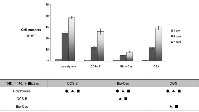
4. 골이식재에 대한 세포증식 평가
각 이식재에 대한 세포 증식을 평가한 결과 1, 4, 7일의 배양기간 동안 증식세포 수는 시간이 경과함에 따라 증가하 는 양상을 보였다(Fig. 2). 각 군간 비교에서 polystyrene은 1, 4, 7일 모두 다른 군에 비해 통계학적으로 유의한 증가 양상을 보였다(p<0.05). Bio-Oss는 4, 7일째 OCS-B와 OGN 모두에서 통계학적으로 유의한 낮은 값을 보였다 (p<0.05). 그러므로 Bio-Oss는 실험군 중 가장 낮은 세포 증식을 보였으며 OCS-B와 OGN의 두 군 사이에서는 별다 른 차이가 없었다.
5. ALP 활성도 측정
골화세포 분화의 지표가 되는 ALP 활성도를 측정한 결과 polystyrene은 7일째 증가하고 10일째 감소하였다. OCS-B 는 배양기간 동안 ALP 활성도가 증가하는 양상을 보였으며, Bio-Oss와 OGN은 7일째 감소하고 10일째 증가되었다.
Bio-Oss는 7일째 polystyrene군보다 통계학적으로 유의한 ALP 활성도의 감소를 보였다(p<0.05) (Fig. 3).
Table 1. EDS Results of the Bone Substitutes (Atomic %; Mean ± SD)
Elements OCS-B Bio-Oss OGN
C 21.162 ± 3.49 22.80 ± 4.41 20.21 ± 6.43
O 56.62 ± 8.97 60.43 ± 2.64 61.84 ± 7.23
Na 0.20 ± 0.19 0.14 ± 0.20 0.33 ± 0.04
Mg 0.34 ± 0.20 0.21 ± 0.20 0.65 ± 0.09
P 8.84 ± 2.59 6.47 ± 2.17 6.48 ± 0.33
Ca 12.84 ± 5.41 9.94 ± 2.15 10.49 ± 1.45
Ca/P 1.42 ± 0.16 1.62 ± 0.43 1.62 ± 0.20
Table 2. Results of Kjeldahl Analysis of the Bone Substitutes(% of crude protein)
OCS-B Bio-Oss OGN
% of crude protein 0.053 0.118 0.140
75 60 45 30 15 0
1 day 4 days 7 days
polystyrene OCS-B Bio-Oss OGN
Cell numbers
(1×10⁴)
1(●), 4(▲), 7(■)days OCS-B Bio-Oss OGN
Polystyrene ●, ▲, ■ ●, ▲, ■ ●, ▲, ■
OCS-B ▲, ■
Bio-Oss ▲, ■
● : statistically significant difference between each groups at 1day (p<0.05)
▲ : statistically significant difference between each groups at 4days (p<0.05)
■ : statistically significant difference between each groups at 7days (p<0.05)
Figure. 2. Cell proliferation measured at 1, 4 and 7 days of culture (n = 4 per each group). Polystyrene was significantly in- creased in optical density compared to OCS-B, Bio-Oss and OGN at 1, 4 and 7 days (p < 0.05). OCS-B was significantly increased in optical density compared to Bio-Oss at 4 and 7 days(p<0.05). Bio-Oss was significantly decreased in optical density compared to OGN at 4 and 7 days (p < 0.05).
1.2
1.0
0.8
0.6
0.4
0.2
0.0
4 days 7 days 10 days
polystyrene OCS-B Bio-Oss OGN ALP aclivity
(nmol/min/mg)
* statistically significant difference compared to polystyrene(p<0.05)
Figure 3. ALP activity measured after 4, 7 and 10 days at wavelength 410 nm by FLUO star OPTIMA (n = 5 per each group).
Bio-Oss was significantly decreased in ALP activity compared to polystyrene at 7 days ( p < 0.05).
고찰
치과 매식 재료들은 생체적합성과 생체기능성을 지녀야 하며, 이러한 기능을 수행하기 위해서는 기계적 성질뿐만 아니라 체내에 독성이나 자극, 이물 반응이 없어야 하며, 골 전도 효과와 신생골 형성을 유발하는 골유도 효과도 있어야 한다29). 이러한 효과를 지닌 가장 좋은 이식재는 자가골이 지만, 채취를 위한 부가적인 수술과 채취량의 한계 등으로 이 자가골을 대신하여 사용할 수 있는 여러 가지 골이식재 들이 개발되어 왔으며, 이에 대한 연구와 성공적인 임상 결 과에 대해 보고되어져 왔다30,31). 이 중 이종골 이식재인 탈 단백 우골은 골재건 수술에서 비교적 성공적인 결과가 많은 연구를 통해 보고 되어지고 있다32,33). 그러나 많은 수의 임 상보고나 연구논문에 비해 현재 판매되고 있는 여러 가지 탈단백 우골 제품의 기본적 특성과 장·단점에 대한 자료는 많지 않은 실정이다. 그러므로 이 연구에서는 골이식재로 판매되는 국산 탈단백 우골인 OCS-B (나이벡(주), 서울, 한국)와 두 가지의 수입 탈단백 우골인 Osteograft/ N-300 (OGN; Dentsply Friadent Ceramed, TN, 미국), Bio-Oss (Geistlich-Pharma, 스위스)의 형태학적, 화학적 특성과 세포증식률을 평가하여 상호 비교하고자 하였다.
이식재의 표면적 측정을 위해 시행된 BET분석결과 Bio-Oss는 다른 문헌34)에서 보고된 79.7 m2/g과 유사한 수 치인 77.5 m2/g를 나타내어 가장 높은 표면적을 보였고, Bio-OSS와 동일하게 우골에서 유래한 이종골 이식재인 OCS-B의 표면적은 63.6 m2/g으로 Bio-Oss와 비교시 조금 낮은 수치를 나타내었다. 이에 반해 OGN은 41.0 m2/g의 표면적을 나타내었고, 앞의 두 가지 이식재들보다 다소 낮 은 수치를 나타내었다. Bio-Oss와 OCS-B가 동일하게 우골 에서 유래한 이종골 이식재이지만, 다소 표면적의 차이를 보이는 이유는 두 이식재간 미소공의 구조적 차이와 연관이 있을 것으로 여겨지며, 두 이식재간의 소결온도가 다른 점 역시 표면구조에 영향을 주었으리라 사료된다. 1,100℃의 온도에서 열처리를 하여 제작되는 것으로 알려진 OGN은 고 온의 열처리 과정을 통해서 표면의 미세한 구조가 좀 더 치 밀한 양상을 보일 것으로 사료되며, 실제 SEM 관찰에서 이 러한 소견이 관찰되었고 비표면적 역시 낮게 측정되었다.
EDS 분석을 통한 시료의 성분분석 결과 칼슘과 인의 원 소비율(Ca/P)은 OCS-B는 1.42, Bio-Oss는 1.62, OGN은 1.62로 나타났으며 이 중 OCS-B의 칼슘과 인의 원소비율이
1.42로 일반적인 골 미네랄의 결과와 다른 연구35)에서 나온 1.68보다 조금 낮은 수치를 나타내었고 Ca/P 비율이 제품 간 차이가 나는 이유는 골이식재 제작에 사용된 우골의 종 (species), 나이, 골 유형 등에 의존할 것으로 사료된다.
세포증식 평가에서 Bio-Oss는 실험군 중 가장 낮은 세포 증식을 보였으며 1, 4, 7일의 배양기간 동안 뚜렷한 세포 수 증식을 나타낸 다른 두 가지 이식재에서와 달리 그 증가양상 이 완만하였고, OCS-B와 OGN의 두 군 사이에서는 별다른 차이가 없었다. 골화세포분화의 지표가 되는 ALP 활성도를 측정한 결과에서도 Bio-Oss는 다른 두 가지 이식재보다 다 소 낮은 ALP 활성도를 나타내었다. 이는 Bio-Oss 표면의 무수한 잔존 미세분말의 존재가 적절한 세포부착과 증식을 저해한 결과인 것으로 보인다. 아울러 많은 동물실험과 임상 결과에서 좋은 평가를 받아오던 골대체재인 Bio-Oss가 세포 단위의 실험에서 좋지 않은 결과를 보인 것에 대해 차후 더 많은 실험과 분석이 요구될 것으로 생각된다.
현재 우골을 골이식 대체재로 사용하는데 있어서 중요시 되는 점들은 합성골과 달리 질병전염의 가능성이 있는지 여 부와 우골의 생체 내 흡수에 따라 신생골이 대체되어 지지 체로서의 역할을 하는가이다. 우골의 잔존 단백질에 대한 보고는 확실하게 밝혀지지 않았으며 상반된 보고가 존재한 다36). 우골을 이용한 이종골 이식재는 화학적인 처리방법과 열처리과정을 통해 원재료 내 존재하는 단백질을 변성, 제 거하고 다른 유기성분을 제거하는 단계를 통해 제조된다.
Bio-Oss의 경우 강알칼리 처리 후, 300℃에서 최소 15시간 동안 열처리 과정과 이후 유기용매를 이용한 부가적인 처리 과정 후 감마선 멸균을 시행하여 제조되는 것으로 알려져 있다. OCS-B는 소의 대퇴부 부위 뼈를 잘게 절단하여 유기 용매 중에서 탈지처리 후 강알칼리 처리를 통해 1차적으로 단백질을 제거하고, 열처리를 통한 2차 탈단백 과정을 거쳐 제조되는 것으로 알려져 있다35). 이에 반해 OGN은 1,100℃
에 이르는 초고온으로 열처리하여 제작되는 것으로 알려져 있으며, 이러한 열처리 과정이 화학용매의 사용과 관계없이 광우병 병원체를 불활성시키는데 가장 효과적인 것으로 보 고되었다37).
조직 내 존재하는 잔존 단백질량을 측정하는데 여러 가지 방법이 사용될 수 있으나, 본 실험에서는 질소 함량을 이용 한 단백질 분석법으로 널리 이용되는 Kjeldahl법38)을 사용 하여 세 가지 종류 이식재 내의 잔존 단백질량을 측정하였 다. Kjeldahl법을 이용한 잔존 단백질량의 측정은 유기질소
를 중화 적정법에 의하여 총질소를 정량하여 질소계수를 이 용하여 산출하는데 순수한 단백질뿐만 아니라 아미노산, purine 염기, amide 화합물 등도 함께 정량된다. 하지만 골 조직의 구성 성분의 종류와 비율을 고려하면 본 실험에서 측정된 조 단백질의 수치인 0.05% (OCS-B)와 0.1% (Bio- Oss, OGN)대의 수치에 포함되는 기타 구성 성분에 대해서 는 향후 추가적인 분석이 고려되며, 다른 종류의 분석방법 을 통해서 좀 더 구체적인 단백질의 정성, 정량이 필요할 것 으로 사료된다.
현재 우골을 이용하여 제작되는 이종골 이식재들은 면역 원성 등의 단백질 유래 부작용을 줄이기 위해 고온 열처리 와 강알칼리를 이용한 화학처리 등으로 잔존 단백질을 최대 한 제거하려고 노력하고 있고, 원재료 역시 광우병에 비교 적 안전한 것으로 알려진 것들을 사용하고 있는 실정이지만, 감염의 위험에서 절대적으로 안전하다고는 말할 수 없다.
유사한 방법으로 제조되는 국산 탈단백 우골인 OCS-B와 수입품인 탈단백 우골들과의 비교에서 표면의 형태학적, 화 학적 특성은 세 종류 모두 각기 다른 성상과 특징을 나타내 었으며, 세포 적합성에서는 OCS-B의 경우 수입품인 골이 식재와 비슷하거나 좀 더 나은 결과를 보여주고 있지만 향 후 동물실험과 임상실험을 거쳐 그 효과를 검증해야 할 것 이다. 환자에게 보다 안전하고 예후가 좋은 골대체제를 선 택하는데 있어 필요한 생체재료들의 비교 연구가 더욱 활발 해져야 할 것이며, 제조사들 또한 안전하고 엄격한 공정 관 리가 필요할 것으로 사료된다.
참고문헌
1. Borstlap W A, Heidbuchel KL, Freihofer HP, Kuijpers- Jagtman AM. Early secondary bone grafting of alveolar cleft defects: A comparison between chin and rib grafts. J Craniomaxillofac Surg 1990;18:210-205.
2. Tayapongsak P, W imsatt JA, LaBanc JP, Dolwick M F.
Morbidity from anterior ilium bone harvest : A com- parative study of lateral versus medial surgical approach.
Oral Surg Oral Med Oral Pathol 1994;78:296-300.
3. Clavero J, Lundgren S. Ramus or chin grafts for maxillary sinus inlay and local onlay augmentation: comparison of donor site morbidity and complications. Clin Implant Dent Relat Res 2003;5:154-160.
4. Emmings FG. Chemically modified osseous material for the restoration of bone defects. J Periodontol 1974;45:385-390.
5. Froum SJ, Thaler R, Scopp IW, Stahl SS. Osseous autografts. I. Clinical responses to bone blend or hip mar- row grafts. J Periodontol 1975;46:515-521.
6. Mellonig JT. Autogenous and allogeneic bone grafts in pe- riodontal therapy. Crit Rev Oral Biol Med 1992;3:333-352.
7. Rosenberg E, Rose LF. Biologic and clinical considerations for autografts and allografts in periodontal regeneration therapy. Dent Clin North Am 1998;42:467-490.
8. Wolff LF. Guided tissue regeneration in periodontal therapy. Northwest Dent 2000;79:23-28.
9. Schmitt JM, Buck DC, Joh SP, Lynch SE, Hollinger JO.
Comparison of porous bone mineral and biologically active glass in critical-sized defects. J Periodontol 1997;68:1043-1053.
10. Mangano C, Bartolucci E, Mazzocco C. A new porous by- droxyapatite for promotion of bone regeneration in maxil- lary sinus augmentation: clinical and histologic study in humans. Clin Oral Implants Res 2003;18:23-30.
11. Wiltfang J, Schlegel KA, Schultze-Mosgau S et al. Sinus floor augmentation with β-tricalciumphosphate(β-TCP):
dose platelet-rich plasma promote its osseo-intergration and degradation. Clin Oral Implants Res 2003;14:213-218.
12. Barnett JD, Mellonig JT, Gray JL, Towle HJ. Comparison of freeze-dried bone allograft and porous hydroxyapatite in human periodontal defects. J Periodontol 1989;60:231-237.
13. Mellonig JT. Freeze-dried bone allografts in periodontal re- constructive surgery. Dent Clin North Am 1991;35:505-520.
14. Schllohorn RG, McClain PK. Combined osseous composite grafting, root conditioning and guided tissue regeneration.
Int J Periodontics Restorative Dent 1998;8:8-30.
15. Gross J. Bone grafting material for dental application: A practical guide. Compendium 1997;18:1013-1036.
16. Slotte C, Lundgren D. Augmentation of calvarial tissue us- ing non-permeable silicone domes and bovine bone mineral.
An experimental study in the rat. Clin Oral Implants Res 1999;10:468-476.
17. de Bruijn JD, Klein CP, de Groot K, van Blitterswijk CA.
The ultrastructure of the bone-hydroxyapatite interface in vitro. J Biomed Mater Res 1992;26:1365-1382.
18. Rosen BV, Hobbs LW, Spector M. The ultrastructure of anorganic bovine bone and selected synthetic hydrox- yapatite used Bone:present and future. In: Babbush CA, ed- itor, Dental implants: the art and science/edited by Babbush
CA. W.B. Sanduers Company. Philadelphia 2001, p.70.
20. Yildirim M, Spiekermann H, Handt S, Edelhoff D.
Maxillary sinus augmentation with the xenograft Bio-Oss and autogenous intraoral bone for qualitative improvement of the implant site: a histologic and histomorphometric clinical study in humans. Int J Oral Maxillofac Implants 2001;16:23-33.
21. Mellonig JT. Human histologic evaluation of a bo- vine-derived bone xenograft in the treatment of periodontal osseous defects. Int J Periodontics Restorative Dent 2000;20:
19-29.
22. Harris RJ. Human histologic evaluation of a bone graft combined with GTR in the treatment of osseous dehiscence defects: a case report. Int J Periodontics Restorative Dent 2000;20:510-519.
23. Wetzel AC, Stich H, Caffesse RG. Bone apposition onto oral implants in the sinus area filled with different frafting materials. Clin Oral Implants Res 1995;6:155-163.
24. Klinge B, Alberius P, Isaksson S, Jonsson J. Osseous re- sponse to implanted natural bone mineral and synthetic hy- droxylapatite ceramic in the repair of experimental skull bone defects. J Oral Maxillofac Surg 1992;50:241-249.
25. Wheeler SL, Holmes RE, Clahoun CJ. Six-year clinical and histologicstudy of sinus-lift grafts. Int J Oral Maxillofac Implants 1996;11:26-34.
26. Berglundh T, Lindhe J. Healing around implants placed in bone defects treated with Bio-Oss. An experimental study in the dog. Clin Oral Implants Res 1997;8:117-124.
27. Skoglund A, Hising P, Young C. A clinical and histologic examination in humans of the osseous response to im- planted natural bone mineral. Int J Oral Maxillofac Implants 1997;12:194-199.
28. Valentini P, Bbensur D, Densari D, Graziani JN, Hammerle C. Histological evaluation and implantation procedure. A human case report. Clin Oral Implants Res 1998;9:59-64.
29. Eisenbarth E, Velten D, Muller M, Thull R, Breme J.
Biocompatibility of beta-stabilizing elements of titanium alloys. Biomaterials 2004;25:5705-5713.
30. Piattelli M, Favero GF, Scarano A, Orsini G, Piattelli A.
Bone reactions to anorganic bovine bone used insiuns lift- ing procedure: a histologic long-term report of 20 cases in man. Int J Oral Maxillofac Implants 1999;14:835-840.
31. Norton MR, Odell EW, Thompson ID, Cook RJ. Efficacy of bovine bone mineral for alveolar augmentation: a human histologic study. Clin Oral Implants Res 2003;14:775-783.
32. Buser D, Bragger U, Land NP, Nyman S. Regeneration and enlargement of jaw bone using guided tissue regeneration.
Clin Oral Implants Res 1990;1:22-32.
33. Hammerle CHF, Olah AJ, Schmid J et al. The biological effect of deproteinized bovine bone on bone neoformation on the rabbit skull. Clin Oral Implants Res 1997;8:198-207.
34. Park JW. Evaluation of deproteinized bovine bone mineral as a bone graft substitute : A comparative analysis of basic characteristics of three commercially available bone substitutes. J Korean Acad periodontol 2005;35:863-875.
35. Park JB, Han SH, Kim KH et al. Evaluation on the bone regenerative capacity of deproteinized bovine bone-derived bone graft material(OCS-B). The Journal of the Korean Dental Association 2006;44:359-366.
36. Storgard-Jensen S, Aaboe M, Pinholt EM et al. Tissue re- action and material characteristics of four bone substitutes.
Int J Oral Maxillofac Implants 1996;11:55-66.
37. Sogal A, Tofe AJ. Risk assessment of bovine spongiform encephalopathy transmission through bone graft material derived from bovine bone used for dental applications. J Periodontal 1999;70:1053-1063.
38. Brooks SPJ, Lampi BJ, Sarwar G, Botting HG. A compar- ison of methods for determining total body protein. Anal Biochem 1995;226:26-30.