www.krspine.org
Necessity of Whole Spine Standing Lateral Radiograph in Adults over 50 Years Old Who Have Degenerative Lumbar Disease
- Comparison with Supine Lumbar Lateral Radiograph -
Whoan Jeang Kim, M.D., Kun Young Park, M.D., Hwan Il Sung, M.D., Jae Yun Ku. M.D., Won Jo Kwon, M.D., Chang Kyu Park, M.D., Won Sik Choy, M.D.
J Korean Soc Spine Surg 2014 Jun;21(2):76-83.
Originally published online June 30, 2014;
http://dx.doi.org/10.4184/jkss.2014.21.2.76
Korean Society of Spine Surgery
Department of Orthopedic Surgery, Inha University School of Medicine
#7-206, 3rd ST. Sinheung-Dong, Jung-Gu, Incheon, 400-711, Korea Tel: 82-32-890-3044 Fax: 82-32-890-3467
©Copyright 2014 Korean Society of Spine Surgery pISSN 2093-4378 eISSN 2093-4386
The online version of this article, along with updated information and services, is located on the World Wide Web at:
http://www.krspine.org/DOIx.php?id=10.4184/jkss.2014.21.2.76
This is an Open Access article distributed under the terms of the Creative Commons Attribution Non-Commercial License (http://
creativecommons.org/licenses/by-nc/3.0) which permits unrestricted non-commercial use, distribution, and reproduction in any medium, provided the original work is properly cited.
Journal of Korean Society of
Spine Surgery
Necessity of Whole Spine Standing Lateral
Radiograph in Adults over 50 Years Old Who Have Degenerative Lumbar Disease
- Comparison with Supine Lumbar Lateral Radiograph -
Whoan Jeang Kim, M.D., Kun Young Park, M.D., Hwan Il Sung, M.D., Jae Yun Ku. M.D., Won Jo Kwon, M.D., Chang Kyu Park, M.D., Won Sik Choy, M.D.
Department of Orthopedic Surgery, Eulji University College of Medicine, Daejeon, Korea Study Design: Cross-sectional study.
Objectives: Sagittal imbalance cannot be predicted depending on the degree of lumbar lordosis. Thus, we tried to evaluate the necessity of whole spine standing lateral radiograph through comparison of the spinal and pelvic parameter between supine lumbar lateral radiograph and whole spine standing lateral radiograph.
Summary of the Literature Review: No studies in the literature compare supine lumbar lateral radiograph and whole spine standing lateral radiograph.
Materials and Methods: We randomly selected 50 males and 50 females among the patients over the age of 50 who visited our hospital for outpatient due to degenerative lumbar disease. Lumbar lordosis (sLL/wLL), sacral slope (sSS/wSS), and pelvic tilt (sPT/
wPT) were measured and compared respectively by supine lumbar lateral radiograph and whole spine standing lateral radiograph. We categorized as group AI (sLL<30˚) and group AII (sLL≥30˚) by supine lumbar lateral radiograph and analyzed them. We also categorized as group BI (SVA≤5 cm) and group BII (SVA>5 cm) by whole spine standing lateral radiograph and analyzed them.
Results: There were no statistical difference in lumbar lordosis (sLL/wLL: 35.1˚/37.7˚) and pelvic parameter (sSS/wSS: 32˚/31.7˚, sPT/
wPT: 24.3˚/24.2˚. sPI/wPI: 56.3˚/58.2˚) between supine lumbar lateral radiograph and whole spine standing lateral radiograph, and there were also no statistical difference between two groups (group AI & AII) in SVA, lumbar lordosis and pelvic parameter. Pelvic parameter compared by supine lumbar lateral radiograph and whole spine standing lateral radiograph based on sagittal balance was no significant difference, but lumbar lordosis appeared statistical difference.
Conclusion: Sagittal imbalance appears quite a lot in patients with degenerative lumbar disease and supine lateral radiograph can’t reflect the whole sagittal imbalance. So, whole spine standing lateral radiograph should be performed routinely to analyze the sagittal alignment.
Key Words: Degenerative lumbar disease, Supine lumbar lateral radiograph, Whole spine standing lateral radiograph, Sagittal balance
Received: December 28, 2012 Revised: February 13, 2013 Accepted: June 2, 2014 Published Online: June 30, 2014
Corresponding author: Whoan Jeang Kim, M.D.
Department of Orthopedic Surgery, Eulji University Hospital, 95, Dunsanseo- ro, Seo-gu, Daejeon 302-799, Korea
TEL: +82-42-611-3267 , FAX: +82-42-259-1289 E-mail: [email protected]
“This is an Open Access article distributed under the terms of the Creative Commons Attribution Non-Commercial License (http://
creativecommons.org/licenses/by-nc/3.0/) which permits unrestricted non-commercial use, distribution, and reproduction in any medium, provided the original work is properly cited.”
서 론
사회가 고령화되어 가면서 퇴행성 요추질환으로 내원하는 환 자들이 늘어나고 있으며 삶의 질을 향상시키기 위해 수술적 치 료가 필요한 경우도 많아지게 되었다.1,2) 50대 이후의 성인들의 경우 퇴행성 질환이 호발하며, 이로 인해 다양한 수술적 치료의 대상이 되었고 그동안 시상면 정렬에 관해서도 많은 연구들이 있어왔다. 외래로 내원하는 고령의 퇴행성 요추 질환 환자들을 진단하는데 있어 방사선 검사를 일반적으로 시행하는데, 앙와 위 요추부 방사선 전후면 사진, 측면 사진, 측면 굴곡 및 신전 사 진을 촬영 하지만, 요추부 전후면, 측면(중립, 굴곡 및 신전) 사진
Necessity of Whole Spine Standing Lateral Radiograp Journal of Korean Society of Spine Surgery
www.krspine.org
77
만으로는 척추가 균형을 유지하고 있는지, 혹은 시상면 불균형상태에 있는지 파악할 수 없기 때문에 기립 전신 척추 전후면 및 측면 사진이 필요하다.
그 동안 여러 저자들이 정상인에서의 시상면 정렬과 시상면 불균형의 임상적 중요성에 대해서 보고하였으며3-9) 척추와 골반 의 시상면상 주요 지표들을 파악하고 서로의 상관관계를 아는 것은 여러 가지 척추 질환의 진단과 치료에 있어서 매우 중요하
다.10-17) 예를 들어, 퇴행성 요추 질환에 대한 수술적 치료가 시상
면 정렬에 관한 이해 없이 척추관 협착증이나 전방 전위증 등 국 소적인 문제에만 초점을 맞추어 치료를 하게 되면 인접 분절의 문제, 시상면 불균형 같은 합병증이 발생할 수 있다.11,14,18)
이에 저자들은 왕와위 사진상 척추가 균형을 유지하고 있는 것처럼 보이는 환자에서도 일괄적으로 기립 전신 측면 사진 촬 영이 필요한지에 대한 의문에서 본 연구를 시작하였으며 외래 환자들을 대상으로 앙와위 요추부 측면 사진과 기립 전신 척추 측면사진에서의 척추지표와 골반지표를 비교하여 그 차이점과 기립 전신 척추 측면 사진이 가지는 의의 및 필요성을 알아보고 자 하였다.
연구 대상 및 방법
1. 연구 대상
2012년 3월부터 2012년 8월까지 본원에서 요통으로 외래를 통해 내원한 50세 이상의 고령 환자에서 과거력상, 시상면 정
렬에 영향을 미칠 수 있는 요인인 척추 골절, 척추 변형 및 종 양, 척추 수술, 고관절 및 슬관절 수술, 척추 측만 및 후만증, 전 방 전위증 등이 있었던 환자들은 모두 제외한 퇴행성 요추질환 환자를 대상으로 남자 50명과 여자 50명을 무작위로 선정하여 횡단 연구를 시행하였다. 방사선학적 검사는 앙와위 전후방 사 진 및 측면 사진(고관절 포함)외에도 기립 전신 척추 측면 사진 을 촬영하였고, 의학 영상 정보 시스템(PACS, Picture Archiving Communications System)을 이용하여 측정하였다. 기립 전신 척추 측면 사진 촬영 시 Faro등19)의 연구에서처럼 상지는 주 관절을 굴곡하고 수부를 쇄골 위에 위치시키는 fits-on clavicle position과 Kim 등20)의 연구에서처럼 양팔을 끌어안은 자세 (cross-arm position)를 시행하였으며, 하지의 자세는 고관절과 슬관절을 신전하여 기립 자세를 취한 후 촬영하였다(Fig. 1).
2. 분류
대상자 모두에서 앙와위 요추부 측면 사진과 기립 전신 척추 측면 사진에서 요추 전만각(lumbar lordosis), 천추 경사(sacral slope), 골반 경사(pelvic tilt), 골반 지수(pelvic incidence)를 각각 측정하여 비교 분석 하였다.
정상적인 시상 만곡의 각도는 범위가 커 성별에 따라, 연령에 따라, 또한 보고자에 따라 약간씩 차이가 있는데, 대체로 요추 전만각의 정상 범위는 30~80도이다.21-26) 따라서 일반적으로 알려져 있는 정상범위에서 최소한의 전만각인 30도를 기준으 로, 앙와위 요추부 측면 사진 상에서 측정한 요추 전만각이 30
A B C D
Fig.1. Fits-on-clavicle position (A, B) or cross-arm position (C, D) is recommended during taking radiographs.
도 미만인 군(AI)과 30도 이상인 군(AII)으로 분류하여 척추 및 골반 지표를 비교 분석 하였다. 또한 Schwab 등27)에 의해 시상 수직축(SVA)이 5cm이하일 때 환자들에게서 만족스런 척추 정 렬을 보인다고 하여, 본 연구에서도 5 cm을 기준으로 BI(SVA≤
5 cm), BII(SVA>5 cm) 군으로 분류하여 척추 및 골반 지표를 비 교하였다.
3. 방사선학적 검사
방사선학적 시상면상 척추 지표로 요추 전만각은 제 1요추 상 연에서 제 1천추 상연까지로 Cobb각을 측정하였고, 후만의 경 우 양의 값으로, 전만의 경우 음의 값으로 표시하였다. 시상 수직 축은 제 7경추 중심에서 내린 수선(C7 plumb line)에서 제 1천추 후상연까지의 거리를 측정하여 수선이 전방에 위치하는 경우 양 의 값으로 후방에 위치하는 경우 음의 값으로 표시하였다. 골반 지표는 천추 경사, 골반 경사, 골반 지수를 측정 하였다. 천추 경 사는 제 1천추의 상연과 수평선과의 각을, 골반 경사는 제 1천추 상연의 중심에서 고관절 축을 잇는 선과 고관절 중심을 지나는 수직선과의 각으로 측정하였다(Fig. 2). 골반 지수는 천추 경사와 골반 경사의 합으로 하였다.
4. 통계
연구 대상자의의 수가 적어 Power analysis를 시행한 결과 집
단수가 30명 이상이면 표본오차 0.253, 신뢰수준 1.96으로 나타 나 다음과 같이 분석하였다. 모든 환자에서 요추 전만각, 천추 경 사, 골반 경사각을 측정하여 Paired T-test를 이용하여 비교하였 고, AI 군과 AII 군에서 기립 전신 척추 측면 사진에서의 척추지 표와 골반지표를 Independent T-test를 이용하였다. 각 지표들간 의 연관성 분석은 Pearson’s correlation를 이용하였다. 통계의 분 석은 SPSS(Statistical Package for the Social Sciences 13.0) 소프트 웨어를 이용하여 시행하였으며 p-value가 0.05미만시 통계적으 로 유의한 것으로 간주하였다. 20년 이상의 정형외과 전문의 경 험을 갖고 있는 한 명의 관찰자 및 전공의로 재직 중인 다른 한 명의 관찰자가 최소 일주 이상의 간격을 두고 2회씩 계측을 시 행하였고, 관찰자 내 신뢰성 및 관찰자 간 신뢰성은 집단 내 상 관 계수(Intraclass Correlation Coefficients, ICCs)를 이용하여 평 가하였으며 상관 계수가 0.8~1.00일 때 신뢰도가 높은 것(strong reliability)으로 하였다.28)
결 과
1. 대상자 결과
전체 평균 연령은 67.7세(범위, 50~87세)이었고, 남자는 67.0 세(범위, 50~87세), 여자는 69.2세(범위, 53~86세)이었다. 전 체 대상자들의 신장은 평균 166.9±5.4 cm이었고, 체중은 평균 67.0±9.6 kg이었으며, 신체 질량 지수(body mass index; BMI) 는 평균 24.0±3.1이었다.
관찰자 내 및 관찰자 간 신뢰도는 각기 0.967, 0.882로 모두 집 단 내 상관 계수(ICCs)는 0.8이상이었다.
2. 방사선학적 결과
1) 앙와위 요추부 측면 사진과 기립 전신 척추 측면 사진과의 비교 앙와위 요추부 측면 사진과 기립 전신 척추 측면 사진에서 요 추 전만각은 각각 평균 35.1도(±14.4도), 37.7도(±16.8도), 천 추 경사는 32도(±9.6도), 31.7도(±9.8도), 골반 경사는 24.3도 (±10.2도), 24.2도(±11.1도)로 측정 되었으나 모두 통계적으 로 유의한 차이는 없었다(Table 1). 앙와위 요추부 측면 사진 상 에서 측정한 척추 및 골반 지표를 시상 수직축과의 상관관계를 알기 위하여 지표간 Pearson 상관성을 확인하였으나 Pearson Fig. 2. Spinal & Pelvic parameters.
LL: lumbar lordosis, SS: Sacral slope, SVA: Sagittal vertical axis, PI: Pelvic incidence, PT: Pelvic tilt.
Table 1. Supine lumbar lateral radiograph vs whole spine lateral radiograph
Supine Lumbar Lateral Radiograph Whole spine Lateral Radiograph p-value
LL 35.1˚(±14.4˚) 37.7˚(±16.8˚) 0.170
SS 32˚(±9.6˚) 31.7˚(±9.8˚) 0.085
PT 24.3˚(±10.2˚) 24.2˚(±11.1˚) 0.132
Necessity of Whole Spine Standing Lateral Radiograp Journal of Korean Society of Spine Surgery
www.krspine.org
79
correlation>0.05로 통계학적으로 유의하지 않았다(Table 2).2) 앙와위 요추 전만각(sLL)을 기준으로 분류한 AI과 AII 군 간의 비교 앙와위 요추부 측면 사진에서 측정한 요추 전만각을 30도 미 만(AI: 36예)과 30도 이상(AII: 64예)인 군으로 분류한 뒤, 각 군 내에서 앙와위 요추부 측면 사진과 기립 전신 척추 측면 사진상 에서 측정한 요추 전만각을 비교 분석하였으나 통계학적으로 유 의한 차이를 보이지 않았다(Table 3). 각 군의 기립 전신 척추 측 면 사진 상에서 측정한 시상 수직축이 6.7 cm(±6.3), 5.4 cm(±
9.7)로 측정되었으나 두 군간에 통계학적으로 유의한 차이가 없 었다(p=0.240).
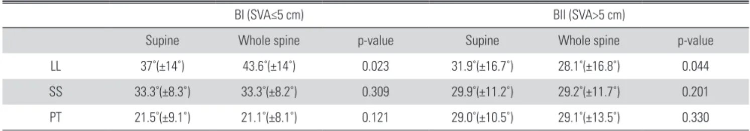
3) 시상 수직축(SVA)을 기준으로 분류한 BI과 BII 군 간의 비교 100예 중 62예가 BI(SVA≤5 cm), 38예가 BII(SVA>5 cm)였다.
BI 군에서 앙와위 요추부 측면 사진과 기립 전신 척추 측면 사 진에서 요추 전만각은 각각 37도(±14도), 43.6도(±14도)로 통 계적으로 유의하게 증가하였으나 천추 경사, 골반경사는 모두 통계적으로 유의한 차이는 없었다. BII 군에서도 요추 전만각이 31.9도(±16.7도), 28.1도(±16.8도)로 통계적으로 유의하게 감 소하였으나 천추 경사, 골반경사는 모두 통계적으로 유의한 차 이는 없었다. BI과 BII 군간에 기립 전신 척추 측면 사진에서 측 정한 척추 및 골반 지표를 비교하였을 때 모두 통계학적으로 유 의한 차이가 있었다(Table 4).
고 찰
그 동안 여러 저자들이 정상인에서의 시상면 정렬과 시상면 불균형의 임상적 중요성에 대해서 보고하였으며 척추와 골반의 시상면상 주요 지표들을 파악하고 서로의 상관관계를 아는 것은 여러 가지 척추 질환의 진단과 치료에 있어서 매우 중요하다.
일반적으로 기립(standing)과 좌위(sitting)시 시행한 측정한 요 추 전만각은 기립(standing)시가 좌위(sitting)시보다 현저히 증 가한다고 Endo 등이 보고하였으며,28) Philippot 등에 의하면 골 반 지표를 자세에 따라 비교 분석 하여 좌위(sitting)시에는 기립 (standing)과 앙와위(supine)와 비교하여 차이가 있었으나 기립 (standing)과 앙와위(supine) 간에는 차이가 없다고 보고한 바 있 다.29) 또한 시상면 정렬상태는 양팔의 자세나 하지의 자세에 따 라서도 변할 수 있어 그 동안 시상면 불균형 환자에서 표준화된 자세에 대하여 많은 연구가 있었다.4,7,19,30-32) 그러나 현재까지 앙 와위 요추부 측면 사진과 기립 전신 측면 사진과의 비교에 관한 연구는 없었다.
저자들의 경우에도 자세에 따른 시상면 불균형이나 척추 및 골반 지표들의 변화가 있을 것이라고 생각하였기 때문에 기립 전신 척추 측면 사진 촬영 시 표준화가 필요하였다. 그래서 척추 에 대하여 충분한 시야 확보를 제공하면서 견관절에서 최소한의 굴곡 모멘트를 발생시켜 시상면 정렬의 변화를 최소화 할 수 있 는 fits-on-clavicle 자세나 cross-arm 자세를 통하여 기립 전신 척추 측면 사진을 촬영하고 있다. 또한 시상면 불균형이 있는 환 자에서 전방으로 굽어지는 자세를 보상하기 위해 몸의 무게 중 심을 조금이라도 정상 위치로 환원시키려는 여러 가지 보상 작 용이 발생할 수 있기 때문에 촬영 시 하체의 자세도 고관절과 슬 관절을 최대한 신전한 상태에서 촬영하고 있다.
Table 2. Spine & pelvic parameter(˚) vs SVA(cm)
p-value
sLL* and SVA 0.612
sSS† and SVA 0.820
sPT‡ and SVA 0.510
*sLL lumbar lordosis on supine lumbar lateral radiograph.
†sSS sacral slope on whole spine standing lateral radiograph.
‡sPT pelvic tilt on whole spine standing lateral radiograph.
Table 3. Lumbar lordosis(˚) & SVA(cm) in AI and AII
sLL* wLL† p-value
AI (<30˚) 20.0˚(±7.6˚) 23.5˚(±13.5) 0.520 AII (≥30˚) 43.5˚(±9.6˚) 45.7˚(±12.7) 0.174
*sLL lumbar lordosis on supine lumbar lateral radiograph.
†wLL lumbar lordosis on whole spine standing lateral radiograph.
Table 4. BI vs BII
BI (SVA≤5 cm) BII (SVA>5 cm)
Supine Whole spine p-value Supine Whole spine p-value
LL 37˚(±14˚) 43.6˚(±14˚) 0.023 31.9˚(±16.7˚) 28.1˚(±16.8˚) 0.044
SS 33.3˚(±8.3˚) 33.3˚(±8.2˚) 0.309 29.9˚(±11.2˚) 29.2˚(±11.7˚) 0.201
PT 21.5˚(±9.1˚) 21.1˚(±8.1˚) 0.121 29.0˚(±10.5˚) 29.1˚(±13.5˚) 0.330
본 연구에서, 기립 전신 측면 사진에서의 요추 전만각이 앙와 위 측면 사진 촬영 시와 차이가 있을 것으로 예상하였으나, 앙와 위 요추부 측면 사진과 기립 전신 척추 측면 사진에서의 요추 전 만각이 각각 35.1도, 37.7도로 약간 증가하는 경향이 있지만 통 계학적으로 유의한 차이는 없었다.
요추 전만의 정도에 따라서 분류한 AI, AII 군 내에서도 앙와 위 요추부 측면 사진과 기립 전신 척추 측면 사진에서의 요추 전 만각이 통계학적으로 유의한 차이가 없었다. 또한, 앙와위 요추 부 측면 사진과 기립 전신 측면 사진에서 측정한 천추 경사 32도 (±9.6도), 31.7도(±9.8도), 골반 경사 24.3도(±10.2도), 24.2도 (±11.1도)로 통계학적으로 유의한 차이는 없었다.
시상면 균형 유무에 따른 요추 전만각과 골반 지표 또한 유사 한 결과를 보이는지 확인하기 위하여 시상 수직축(SVA)에 따라 분류한 BI 군(SVA≤5 cm)과 BII 군(SVA>5 cm)내에서 앙와위 방 사선 측면 사진과 기립 전신 척추 측면 사진에서 측정한 각 골반 지표는 통계학적으로 유의한 차이가 없었다. 유일하게 요추 전 만각만이 기립 전신 척추 측면 사진과 앙와위 방사선 측면 사진 과 비교하여 BI 군에서는 37도(±14도)에서 43.6도(±14도)로 증가하였고, BII 군에서는 31.9도(±16.7도)에서 28.1도(±16.8 도)로 감소하여 두 군에서 통계학적으로 유의한 차이가 있었다.
특히, 시상면 불균형이 있는 환자에서 앙와위 측면 사진에서보 다 기립 전신 측면 사진에서 요추 전만각이 감소하는 흥미로운 결과를 얻었다.
전체 환자를 대상으로 앞에 언급된 요추 전만각이 앙와위와 기립 시, 차이가 없다는 결과와 상이한 결과가 나오게 된 것은
정상인에서 앙와위와 기립시 요추 전만의 차이가 있는지에 대한 더 많은 연구가 필요할 것으로 사료된다.
본 연구에서 앙와위 방사선 측면 사진에서 측정한 요추 전만 각 및 골반 지표 기립 전신 척추 측면 사진에서 측정된 시상 수 직축과의 상관 관계를 알아보았으나 Pearson correlation>0.05 로 통계학적으로 관계는 없었다. 또한, 앙와위 방사선 측면 사진 의 전체 평균 요추 전만각은 35.1도로 정상 범위 내에 있으나, 시 상 수직축이 5 cm 이상인 BII 군이 38예나 되는 것으로 보아 앙 와위 방사선 측면 사진에서 측정한 요추 전만각 및 골반 지표만 으로는 시상면 불균형 유무를 판단할 수 없다. 예를 들면, 그림 3과 4에서 보듯이 두 환자에서 단순 방사선 측면 사진 상 측정 한 요추 전만각은 각각 29.5도, 26.4도로 비슷하였는데, 기립 전 신 척추 측면 사진 상 측정한 요추 전만각은 51도, 2.5도로 큰 차 이를 보여주고 있으며, 시상 수직축(SVA) 또한 0.2 cm, 20.7 cm 으로 단순 방사선 측면 사진에서 발견할 수 없었던 전혀 다른 척 추 상태를 보여주고 있다(Fig. 3, 4). 하지만, 천추 경사가 요추 전 만과 밀접한 관련이 있고 골반 지수 또한 천추 경사와 관계가 있
어7,33) 앙와위 및 기립시 요추 전만각이 변한다면 이로 인해 골반
지표값들도 유의한 상관관계를 가질 것으로 예상하였지만 앙와 위 방사선 측면 사진과 기립 전신 척추 측면 사진 상에서 측정한 이 값들은 통계학적으로 차이는 없었다. 시상면 균형 여부에 따 라 앙와위 측면 사진과 기립 전신 척추 측면 사진 상에서 비교한 골반 지표도 차이가 없었지만, 요추 전만각은 유의한 차이를 보 였다.
이처럼 퇴행성 요추 질환환자에 있어 시상면 정렬의 파악을
A B
Fig. 3. A 53-year-old man’s supine lumbar lateral radiograph (A) and whole spine lateral radiograph with SVA ≤ 5 cm (B).
B A
Fig. 4. A 75-year-old woman’s supine lumbar lateral radiograph (A) and whole spine lateral radiograph with SVA > 5 cm (B).
Necessity of Whole Spine Standing Lateral Radiograp Journal of Korean Society of Spine Surgery
www.krspine.org
81
위해선 앙와위 측면 사진에서 뿐 아니라 기립 전신 척추 사진은반드시 필요할 것으로 사료되며 더불어 일반적인 기립 측면 사 진의 촬영에 있어서도 기립 전신 측면 사진과 같은 자세로 촬영 시 추후 추시에 있어 매우 요긴한 자료로 쓰일 것으로 사료된다.
결 론
퇴행성 요추 질환이 있는 환자에서 시상면 불균형이 적지 않 게 나타나며 앙와위 요추부 측면사진에서의 전만 정도에 따라서 는 시상면 불균형을 파악할 수 없고 앙와위 측면 사진과 기립 전 신 척추 측면 사진에서 비교한 골반 지표는 차이가 없었으나 요 추 전만각은 시상면 균형 유무에 따라서 유의한 차이를 보여 시 상면 상태를 알기 위해서는 기립 전신 척추 측면 사진을 일괄적 으로 촬영해야 될 것으로 사료된다. 하지만, 본 연구는 연구 기간 이 짧고 연구 대상이 적으며 정상인에서 두 집단과의 차이를 비 교하지 않아 추후 이에 관해 더 많은 연구가 필요할 것으로 사료 된다.
REFERANCES
1. Benz RJ, Ibrahim ZG, Afshar P, Garfin SR. Predicting com- plications in elderly patients undergoing lumbar decompres- sion. Clin Orthop Relat Res. 2001:116-21.
2. Deyo RA, Ciol MA, Cherkin DC, Loeser JD, Bigos SJ.
Lumbar spine fusion. A cohort study of complications, re- operations, and resource use in the Medicare population.
Spine (Phila Pa 1976). 1993;18:1463-70.
3. Gelb DE, Lenke LG, Bridwell KH, Blanke K, McEnery KW.
An analysis of sagittal spinal alignment in 100 asymptomatic middle and older aged volunteers. Spine (Phila Pa 1976).
1995;20:1351-8.
4. Vedantam R, Lenke LG, Keeney JA, Bridwell KH. Com- parison of standing sagittal spinal alignment in asymp- tomatic adolescents and adults. Spine (Phila Pa 1976).
1998;23:211-5.
5. Propst-Proctor SL, Bleck EE. Radiographic determination of lordosis and kyphosis in normal and scoliotic children. J Pediatr Orthop. 1983;3:344-6.
6. Lenke LG, Bridwell KH, Blanke K, Baldus C, Weston J.
Radiographic results of arthrodesis with Cotrel-Dubousset instrumentation for the treatment of adolescent idiopathic scoliosis. A five to ten-year follow-up study. J Bone Joint Surg Am. 1998;80:807-14.
7. Stagnara P, De Mauroy JC, Dran G, et al. Reciprocal an- gulation of vertebral bodies in a sagittal plane: approach to references for the evaluation of kyphosis and lordosis. Spine (Phila Pa 1976). 1982;7:335-42.
8. Voutsinas SA, MacEwen GD. Sagittal profiles of the spine.
Clin Orthop Relat Res. 1986:235-42.
9. Bernhardt M, Bridwell KH. Segmental analysis of the sag- ittal plane alignment of the normal thoracic and lumbar spines and thoracolumbar junction. Spine (Phila Pa 1976).
1989;14:717-21.
10. Barrey C, Jund J, Noseda O, Roussouly P. Sagittal bal- ance of the pelvis-spine complex and lumbar degenerative diseases. A comparative study about 85 cases. Eur Spine J.
2007;16:1459-67.
11. Kawakami M, Tamaki T, Ando M, Yamada H, Hashizume H, Yoshida M. Lumbar sagittal balance influences the clini- cal outcome after decompression and posterolateral spinal fusion for degenerative lumbar spondylolisthesis. Spine (Phila Pa 1976). 2002;27:59-64.
12. Korovessis P, Dimas A, Iliopoulos P, Lambiris E. Correla- tive analysis of lateral vertebral radiographic variables and medical outcomes study short-form health survey: a com- parative study in asymptomatic volunteers versus patients with low back pain. J Spinal Disord Tech. 2002;15:384-90.
13. Kumar MN, Baklanov A, Chopin D. Correlation between sagittal plane changes and adjacent segment degeneration following lumbar spine fusion. Eur Spine J. 2001;10:314-9.
14. Videbaek TS, Bunger CE, Henriksen M, Neils E, Chris- tensen FB. Sagittal spinal balance after lumbar spinal fusion:
the impact of anterior column support results from a ran- domized clinical trial with an eight- to thirteen-year radio- graphic follow-up. Spine (Phila Pa 1976). 2011;36:183-91.
15. Roussouly P, Nnadi C. Sagittal plane deformity: an over- view of interpretation and management. Eur Spine J.
2010;19:1824-36.
16. Roussouly P, Nnadi C. Sagittal plane deformity: an over- view of interpretation and management. European Spine Journal. 2010;19:1824-36.
17. Barrey C, Jund J, Noseda O, Roussouly P. Sagittal balance of the pelvis-spine complex and lumbar degenerative dis- eases. A comparative study about 85 cases. European Spine Journal. 2007;16:1459-67.
18. Kim YJ, Bridwell KH, Lenke LG, Glattes CR, Rhim S, Cheh
G. Proximal junctional kyphosis in adult spinal deformity after segmental posterior spinal instrumentation and fu- sion: minimum five-year follow-up. Spine (Phila Pa 1976).
2008;33:2179-84.
19. Faro FD, Marks MC, Pawelek J, Newton PO. Evaluation of a functional position for lateral radiograph acquisition in adolescent idiopathic scoliosis. Spine (Phila Pa 1976).
2004;29:2284-9.
20. Kim MS, Chung SW, Hwang CJ, Lee CK, Chang BS. A radiographic analysis of sagittal spinal alignment for the standardization of standing lateral position. J Korean Or- thop Assoc. 2005;40:861-8.
21. Aebi M. The adult scoliosis. European Spine Journal.
2005;14:925-48.
22. Moon MS, Lee KS, Lim CI. A clinical study of degenerative lumbar scoliosis. Proc 5th Conf Lumbar Fusion and Stabili- zation. Tokyo, Springer-Verlag. 1993:98-112.
23. Glassman SD, Berven S, Bridwell K, Horton W, Dimar JR.
Correlation of radiographic parameters and clinical symp- toms in adult scoliosis. Spine (Phila Pa 1976). 2005;30:682- 8.
24. Pritchett JW, Bortel DT. Degenerative symptomatic lumbar scoliosis. Spine (Phila Pa 1976). 1993;18:700-3.
25. Kim JH, Suk SS, Chung ER, et al. Epidemiologic study of lumbar scoliosis with plain abdominal x-ray. J Kor Soc Spine Surg. 2004;11:246-52.
26. Bridwell KH, Dewald RL. The textbook of spinal surgery.
3rd ed. Philadelphia:Wolters Kluwer/Lippincott Williams &
Wilkins Health. 2011.
27. Schwab F, Patel A, Ungar B, Farcy JP, Lafage V. Adult
spinal deformity-postoperative standing imbalance: how much can you tolerate? An overview of key parameters in assessing alignment and planning corrective surgery. Spine (Phila Pa 1976). 2010;35:2224-31.
28. Prieto L, Lamarca R, Casado A. [Assessment of the reliabil- ity of clinical findings: the intraclass correlation coefficient].
Med Clin (Barc). 1998;110:142-5.
29. Philippot R, Wegrzyn J, Farizon F, Fessy MH. Pelvic balance in sagittal and Lewinnek reference planes in the standing, supine and sitting positions. Orthop Traumatol Surg Res.
2009;95:70-6.
30. Suzuki H, Endo K, Mizuochi J, Kobayashi H, Tanaka H, Yamamoto K. Clasped position for measurement of sagittal spinal alignment. European Spine Journal. 2010;19:782-6.
31. Jackson RP, Kanemura T, Kawakami N, Hales C. Lum- bopelvic lordosis and pelvic balance on repeated standing lateral radiographs of adult volunteers and untreated pa- tients with constant low back pain. Spine (Phila Pa 1976).
2000;25:575-86.
32. Jackson RP, McManus AC. Radiographic analysis of sag- ittal plane alignment and balance in standing volunteers and patients with low back pain matched for age, sex, and size. A prospective controlled clinical study. Spine (Phila Pa 1976). 1994;19:1611-8.
33. Legaye J, Duval-Beaupere G, Hecquet J, Marty C. Pel- vic incidence: a fundamental pelvic parameter for three- dimensional regulation of spinal sagittal curves. Eur Spine J.
1998;7:99-103.
Necessity of Whole Spine Standing Lateral Radiograp Journal of Korean Society of Spine Surgery
www.krspine.org
83
50세 이상의 퇴행성 요추 질환 환자에서 기립 전신 척추 측면 사진의 필요성 - 앙와위 요추부 측면 사진과의 비교 -
김환정 • 박건영 • 성환일 • 구제윤 • 권원조 • 박창규 • 최원식 을지대학교 의과대학 정형외과학교실
연구 계획: 횡단 연구
목적: 요추 전만각 정도에 따라 시상면 불균형을 예측할 수 없어, 앙와위 요추부 측면 사진과 기립 전신 척추 측면 사진에서의 척추 지표와 골반 지표를 비교하여 기립 전신 척추 측면 사진의 필요성을 알아보고자 하였다.
선행문헌의 요약: 앙와위 요추부 측면 사진과 기립 전신 척추 측면 사진을 비교한 연구는 없었다.
대상 및 방법: 퇴행성 요추 질환으로 외래로 내원한 50세 이상 환자에서 남자 50명과 여자 50명을 무작위로 선정하였다. 앙와위 요추부 측면 사진과 기 립 전신 척추 측면 사진에서 요추 전만각(sLL/wLL), 천추 경사(sSS/wSS), 골반 경사(sPT/wPT)를 각각 측정하여 비교 분석 하였다. 앙와위 요추부 측면 사진 상에서 요추 전만각이 30도 미만인 Group AI과, 30도 이상인 Group AII으로 분류하여 비교분석 하였다. 또한 기립 전신 척추 측면 사진에서 시상 수직축이 제 1천추의 후상연 모서리를 기준점으로 전방 5 cm 미만인 Group BI과 5 cm 이상인 Group BII으로 분류하여 비교 분석하였다.
결과: 왕와위 요추부 측면 사진과 기립 전신 척추 측면 사진에서 요추 전만각(sLL/wLL: 35.1도/37.7도) 및 골반 지표(sSS/wSS: 32도/31.7도, sPT/wPT:
24.3도/24.2도. sPI/wPI: 56.3도/58.2도)는 통계적으로 유의하지 않았다. 또한, AI과 AII에서 시상 수직축 및 요추 전만각, 골반 지표는 모두 통계적으로 유의한 차이는 없었다. 시상면 균형 여부에 따라 앙와위 측면 사진과 기립 전신 척추 측면 사진 상에서 비교한 골반 지표도 유의한 차이가 없었지만, 요 추 전만각은 유의한 차이를 보였다.
결론: 퇴행성 요추 질환이 있는 환자에서 시상면 불균형이 적지 않게 나타나고 앙와위 요추부 측면 사진으로는 전체적인 시상면 불균형을 예측할 수 없 기 때문에, 시상면 상태를 파악하기 위해서는 기립 전신 척추 측면 사진을 일괄적으로 촬영해야 할 것으로 사료된다.
색인 단어: 퇴행성 요추질환, 앙와위 요추부 측면 사진, 기립 전신 척추 측면 사진, 시상면 균형 약칭 제목: 기립 전신 척추 측면 사진의 필요성