• 검색 결과가 없습니다.

Dental Implants in Patients with Gingival Oral Lichen Planus JOMP

N/A
N/A
Protected

Academic year: 2021

Share "Dental Implants in Patients with Gingival Oral Lichen Planus JOMP"

Copied!
6
0
0

로드 중.... (전체 텍스트 보기)

전체 글

(1)

pISSN 2288-9272 eISSN 2383-8493 J Oral Med Pain 2019;44(3):77-82 https://doi.org/10.14476/jomp.2019.44.3.77

Dental Implants in Patients with Gingival Oral Lichen Planus

YoungJoo Shim 1,2

1 Department of Oral Medicine, School of Dentistry, Wonkwang University Daejeon Dental Hospital, Daejeon, Korea

2 Wonkwang Dental Research Institute, Wonkwang University, Iksan, Korea

Received August 20, 2019 Revised September 10, 2019 Accepted September 11, 2019

Purpose: With the popularity of implant therapy, clinicians need to know about treating the dental implant in patients with gingival involvement of oral lichen planus (OLP). The aim of this study is to evaluate the survival and success rates of dental implant and propose of clinical guidelines for implant treatment in OLP patient with gingival involvement.

Methods: A literature search was performed in PubMed/Medline, and Cochrane database.

Papers in English language published between 1990 and 2019 were evaluated. The focused questions were following; 1) Dose gingival OLP affect the survival and success rates of dental implants? 2) The management of OLP patients with gingival involvement receiving dental implant.

Results: There was no study about the evaluation of dental implant only in gingival OLP patient. Five studies evaluating dental implants in OLP patients were included in this re- view. Implant survival rate was 100.0% in well-controlled OLP patients in all included stud- ies. The use of topical/systemic corticosteroid in OLP patients was performed before and/or after implant placement in all included studies.

Conclusions: The implant survival and success rates in well-controlled OLP patients did not different from that of non-OLP healthy subjects. The gingival OLP is associated with higher rate of peri-implant mucositis. Adequate management of gingival OLP lesions before and after implant insertion is required to reduce inflammation and associated bone loss.

Key Words: Dental implants; Gingiva; Oral lichen planus

Correspondence to:

YoungJoo Shim

Department of Oral Medicine, School of Dentistry, Wonkwang University Daejeon Dental Hospital, 77 Dunsan-ro, Seo-gu, Daejeon 35233, Korea

Tel: +82-42-366-1128 Fax: +82-42-366-1115 E-mail: [email protected]

https://orcid.org/0000-0001-7514-5974 This paper was supported by Wonkwang University in 2018.

JOMP Journal of Oral Medicine and Pain

Copyright Ⓒ 2019 Korean Academy of Orofacial Pain and Oral Medicine. All rights reserved.

CC
This is an open-access article distributed under the terms of the Creative Commons Attribution Non-Commercial License (http://creativecommons.org/licenses/by-nc/4.0/),

INTRODUCTION

Desquamative gingivitis (DG) is not a specific disease but represents a gingival manifestation associated with several mucocutaneous diseases and systemic conditions. It is char- acterized with the presence of epithelial desquamation, ery- thema, erosion, and blistering of the marginal and attached gingiva. Mucous membrane pemphigoid (MMP), oral lichen planus (OLP), and pemphigus vulgaris (PV) are the most common cause of DG. These are immune-mediated diseases with chronic nature [1].

Dental implant is becoming a most widely used pros- thetic rehabilitation after tooth loss. In the past, it has been

suggested that dental implants are not ideal for patients

with OLP because of the altered epithelium to adhere to the

titanium surface [2]. Many clinicians encounter with the

patients who need dental implant under the mucocutaneous

diseases. Not yet, there are no clear clinical guidelines for

dental implant placement and management in patient with

gingival mucocutaneous diseases. The purpose of this study

is to evaluate the dental implant survival and success rates,

and provide the guidelines for the implant placement and

management in OLP patients with gingival involvement,

the representative of mucocutaneous diseases.

(2)

MATERIALS AND METHODS

1. Focused Questions

1) Dose gingival OLP affect the survival and success rates of dental implants?

2) What is the management of OLP patients with gingival involvement receiving dental implant?

2. Search Strategy

A search strategy based on the key word ‘dental implant’

and ‘oral lichen planus’, or ‘dental implant’ and ‘mucocuta- neous disease’, or ‘dental implant’ and ‘pemphigus’, or ‘den- tal implant’ and ‘pemphigoid’, or ‘dental implant’ and ‘des- quamative gingivitis’ were performed in PubMed/Medline, and Cochrane database. Papers in English language pub- lished between 1990 and 2019 were evaluated. Selected ar- ticles were obtained in full text and analyzed.

3. Data Extraction

The following data were recorded: author’s name, study design, sample size, OLP type, evaluation parameters, im- plant survival rate, incidence of peri-implantitis/peri-im- plant mucositis, follow-up period, associated treatment, outcomes, and main conclusion.

RESULTS

Electronic search yielded 44 articles, of which 24 articles were selected for full-text evaluation after screening the ti- tles and abstracts. There was no study about the evaluation of dental implant only in gingival OLP subjects. We could not identify any randomized controlled trial publications.

The clinical studies were case report or case series [3-5], cross-sectional study [6], retrospective [7,8] or prospective- controlled study [9,10].

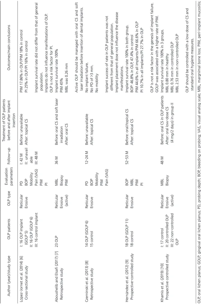
Only four studies with controls [6,8-10] and one retro- spective study [7] satisfying with the focused question were included in this review. The main features and conclusions of each study were summarized in Table 1 [6-10].

The use of topical/systemic corticosteroid was performed before and/or after implant placement in all included stud- ies. In the prospective studies, acute and erosive state of OLP was treated with corticosteroid before implant insertion

[9,10]. In one study, of 55 inserted implants in active OLP phase, 73% (42 implants) of implants failed after early load- ing [7]. After controlling of acute OLP with corticosteroid, new inserted 42 implants were survived and functioned well for 3 years. Implant survival rate was 100% in well- controlled OLP patients in all included studies. Success rate of implant among OLP patients did not seem to be differ- ent from that of general population [6,8]. Authors agreed that the well-controlled OLP is not a risk factor for peri-im- plantitis and implant failure and the implant also does not influence the manifestation of OLP. OLP with gingival in- volvement was associated with a higher rate of peri-implant mucositis [9].

DISCUSSION

1. Gingival OLP and Periodontal Conditions

OLP is a T-cell-mediated chronic inflammatory oral mu- cosal disease [11]. It affects most commonly the buccal mu- cosa, tongue, and gingiva [12]. The etiology of OLP is ex- actly unknown to date, but it is thought that intrinsic or ex- trinsic antigens trigger an inflammatory process, resulting in accumulation of T lymphocytes in the superficial lamina propria, liquefaction degeneration, and keratinocyte apop- tosis of in the basal layer [13].

Gingiva is one of the most frequently affected site in OLP patients. The prevalence of gingival involvement of OLP was 38.4%-48% [14,15]. Gingival OLP is characterized by atrophy, erythema, ulceration, whitish plaque-like le- sion and desquamation of attached and marginal gingiva.

We usually called this gingival lesion as desquamative gin- givitis. Patients with gingival OLP, expressed as DG, have trouble doing oral hygiene practices and accumulation of dental plaque [16]. Gingival OLP is usually misdiagnosed as plaque-induced periodontal diseases and combined with plaque-induced gingivitis or periodontal diseases.

There were several studies which compared the periodon- tal conditions between gingival OLP and healthy controls.

In a Ramón-Fluxiá’s study [17], the plaque and calculus in-

dices are significantly higher in the gingival OLP subjects

than non-gingival OLP subjects. But the periodontal disease

index was not significantly different between the groups

[17]. In other study, plaque index, bleeding on probing (BOP),

(3)

Table 1. The main featur es and conclusions of included studies Author (year)/study type OLP patients OLP type Evaluation parameters Follow-up Tr eatment of OLP patients befor e and after implant insertion Outcomes/main conclusions Ló pez-Jor net et al. (2014) [6] Cr oss-sectional study I: 16 OLP implant (GOLP 5) II: 16 OLP (GOLP 6) III: 16 contr ol implant

Reticular Er osive PD BOP Mobility Pain (V AS) PIM PI

I: 42 M II: unavail able III: 48 M

Befor e: unavailable After: topical CS PIM 17.86% in OLP/PIM 18% in contr ol PI 25% in OLP/PI 16% in contr ol Implant survival rate did not dif fer fr om that of general population. Implants do not influence manifestations of OLP . OLP is not a risk factor for PI. Aboushelib and Elsafi (2017) [7] Retr ospective study 23 OLP Reticular Er osive (active)

MBL Biopsy PIM

36 M Befor e: oral CS and soft laser irradiation After: oral CS

Implant survival rate 100% PIM 45% MBL was 0.26 mm Active OLP should be managed with oral CS and soft laser irradiation befor e insertion of dental implants. Czer ninski et al. (2013) [8] Retr ospective study 14 OLP (GOLP 6) 15 contr ol Reticular Er osive PD BOP Mobility PIM Pain (V AS)

12-24 M Befor e: unavailable After: topical CS No implant failur e. No PD of >3 mm. No mobility Implant success of rate in OLP patients was not dif fer ent fr om that of general population. Implant placement dose not influence the disease manifestations. Her n ández et al. (2012) [9] Pr ospective-contr olled study 18 OLP (GOLP 11) 18 contr ol Reticular Er osive BOP MBL PIM PI

52-53 M Befor e: oral/topical CS After: topical CS Implant survival rate 100% in both gr oup. BOP: 46.8% in OLP , 47.4% in contr ol PIM 44.6% in all implants/PIM 66.6% in OLP PI 10.7% in all implants/PI 27.7% in OLP OLP is not a risk factor in the genesis of implant failur e. GOLP was associated with a higher rate of PIM. Khamis et al. (2019) [10] Pr ospective contr olled study I: 17 contr ol II: 20 contr olled OLP III: 22 non-contr olled OLP

Reticular Er osive (active) MBL Biopsy 48 M Befor e: oral CS in OLP patients After: oral low-dose CS (4 mg/2 days) in gr oup II

Implant survival rate 100% in 3 gr oups. MBL 0.80 mm in healthy MBL 0.76 mm in contr olled OLP MBL 2.53 mm in non-contr olled OLP OLP should be contr olled with a low dose of CS and standar d oral hygiene measur es. OLP , oral lichen planus; GOLP , gingival oral lichen planus; PD, pr obing depth; BOP , bleeding on pr obing; VAS, visual analog scale; MBL, margnimal bone loss; PIM, peri-implant mucositis; PI, peri-implantits; M, month; CS, corticoster oid.

(4)

and clinical attachment level were worse in gingival OLP than healthy controls [16]. There were studies to compare the DG-positive sites and DG-negative sites in OLP or MMP patients. Russo et al. [18] revealed that probing depth (PD), clinical attachment loss, full mouth plaque score, and full mouth bleeding score were not significantly different be- tween the DG-positive sites and DG-negative sites. The oth- er study of them [19], total bacterial load showed no differ- ence between DG-positive and DG-negative sites. Gingival OLP per se may not be able to cause periodontal disease, but more gingival inflammation [16,17]. It seems that the critical factor to determine periodontal status in one patient is the host’s character such as ability of oral hygiene and disease severity and duration.

2. Gingival OLP and Dental Implant

Dental implants are widely used to replace missing teeth and offer a success rate of about 83% after 15 years [20].

Healthy peri-implant mucosal condition is critical for the epithelium to adhere and seal implantation site. It has been postulated that gingival OLP may directly alter the nature of the barrier, affecting the long-term success of the im- plants [21]. To date, oral mucocutaneous diseases such as gingival OLP, MMP, and PV are not absolute contraindica- tions for implant insertion.

Previous studies about dental implants and OLP are limit- ed to comparison between OLP groups and healthy controls [6-10].

There was a well-designed prospective-controlled study.

Hernández et al. [9] conducted a study to evaluate the im- plant survival rate in OLP patients. Fifty-six implants in 18 OLP patients (gingival OLP, 11 patients) were evaluated for a mean follow-up period of 53 months. OLP patients show- ing acute erosive/ulcerative phase were treated with topi- cal/systemic corticosteroid before implant insertion. All implants were placed in remission period of OLP. The OLP patients did not show any signs of delayed wound heal- ing after surgical procedure. The implant survival rate was 100.0% for the OLP patients. Peri-implant mucositis (BOP, PD ≥4 mm and no bone loss) was 66.6% and peri-implanti- tis (BOP or pus, bone loss ≥3 threads) [22] was 27.7% of the OLP patients. Both of them were not significantly differ- ent between OLP group and controls. It is difficult to draw

clear conclusion because of lack of previous study includ- ing homogeneous gingival OLP subjects. Though, in the Hernández’s study, the presence of gingival involvement was associated with a higher rate of peri-implant mucositis.

Gingival OLP would have possibility to accelerate the long- term progression to peri-implantitis. They concluded that the presence of OLP is not associated with a higher preva- lence of implant failure, peri-implant mucositis, peri-im- plantitis or immediate postsurgical complications, but pa- tient with gingival OLP should be carefully followed-up. As previously described, the relation between gingival OLP and dental implant is similar to that of gingival OLP and peri- odontal conditions.

Aboushelib and Elsafi [7] evaluated the implant and ac- tive OLP. In active OLP phase, high degree of inflamma- tory cell infiltration and cell-mediated immune reaction are present. Of 55 inserted implants in active OLP phase, 73%

of implants failed after early loading. It appeared to be a di- rect correlation between the accumulation of immune cells at the gingival tissue interface and normal wound healing around dental implants. The authors recommended that im- plant insertion should be done in remission period of OLP and oral corticosteroid therapy and soft tissue laser are ad- ministrated in advance.

Khamis et al. [10] also evaluated the long-term prognosis of dental implant in active OLP patients. They evaluated the marginal bone loss (MBL) of dental implants in active OLP patients for 4 years. There was no significant difference in MBL between healthy and controlled OLP patients who taking low-dose (4 mg/48 hours) of corticosteroids. But, uncontrolled OLP patients showed increased MBL which reached 2.53 mm after 4 years. The authors suggested that OLP patients receiving dental implants should be con- trolled on a low-dose of corticosteroids to prevent MBL and to maintain remission period. However, the use of long- term systemic corticosteroid should be perfomed with cau- tion due to side effects. Adequate management of gingival OLP lesions before and after implant insertion is the most important factor in reducing inflammation and associated bone loss.

In active erosive-atrophic gingival OLP lesion in denture-

bearing area, the use of denture triggers pain and erosion

due to mechanical tissue irritation. In this case, it is better

(5)

for the patient to receive an implant-supported overdenture to improve stability of denture and reduce soreness [5].

3. Keratinized Mucosa Around Implant

The presence of keratinized mucosa around teeth is im- portant factor to maintain periodontal health. More than 2 mm width of keratinized mucosal had been needed to maintain periodontal health [23]. But, from several studies, the current consensus is that provided with adequate oral hygiene, periodontal stability could be maintained even without adequate keratinized mucosa [24-26]. The presence of keratinized mucosa around implant is also significant to decrease peri-implant inflammation. Many studies tried to provide scientific evidences for this topic. Reduced keratin- ized mucosa width around dental implants is a risk factor for peri-implant mucositis [27]. The presence of at least 1-2 mm wide keratinized mucosa might be beneficial in de- creasing plaque accumulation, tissue inflammation, muco- sal recession, and attachment loss [25]. If the gingival OLP patients have not adequate keratinized mucosa at the site of implantation, free gingival graft is required.

4. Plaque Control and Gingival OLP

Most of OLP patients come to see a doctor for pain.

Pain affects the ability of patient’s oral hygiene practices.

Impaired oral hygiene results in plaque accumulation and increases prevalence of gingivitis. This will possibly lead to periodontal disease and clinical attachment loss in the long- term aspects.

Active plaque control affects the progression of OLP.

Studies [28-30] reported that the plaque control improved the painful symptoms and clinical features of gingival OLP.

Likewise, active plaque control and regular follow-up pro- gram should be important procedures in the management of gingival OLP with dental implant.

5. Proposal of Clinical Management

Based on the foregoing, following guidelines for dental implant treatment in OLP patients with gingival involve- ment are suggested.

1) Implant insertion should be done in remission period of OLP. If necessary, systemic corticosteroid therapy is ad- ministrated in advance.

2) Minimally traumatic surgical procedures are should be considered.

3) The presence of adequate (at least 1-2 mm) keratinized mucosa around implant is important to maintain peri-im- plant mucosal health.

4) In the early retention phase, topical/systemic cortico- steroid therapy should be required to prevent the recurrence of active OLP and MBL.

5) Active plaque control is essential in gingival OLP pa- tients to prevent peri-implant mucositis.

CONCLUSION

Clinician should be familiar with the clinical aspects of gingival OLP because of the high rate of gingival involve- ment in OLP. The implant survival and success rates in well-controlled OLP patients did not different from that of non-OLP healthy subjects. The presence of OLP is not asso- ciated with a higher prevalence of implant failure or imme- diate postsurgical complications, but the gingival OLP is as- sociated with higher rate of peri-implant mucositis. For the long-term healthy state of implant, active plaque control and frequent follow-up is required.

CONFLICT OF INTEREST

No potential conflict of interest relevant to this article was reported.

ORCID

YoungJoo Shim

https://orcid.org/0000-0001-7514-5974

REFERENCES

1. Lo Russo L, Fedele S, Guiglia R, et al. Diagnostic pathways and clinical significance of desquamative gingivitis. J Periodontol 2008;79:4-24.

2. Sugerman PB, Barber MT. Patient selection for endosseous dental implants: oral and systemic considerations. Int J Oral Maxillofac Implants 2002;17:191-201.

3. Fu L, Liu Y, Zhou J, Zhou Y. Implant-retained overdenture for a patient with severe lichen planus: a case report with 3 years’

follow-up and a systematic review. J Oral Maxillofac Surg

(6)

2019;77:59-69.

4. Reichart PA. Oral lichen planus and dental implants. Report of 3 cases. Int J Oral Maxillofac Surg 2006;35:237-240.

5. Esposito SJ, Camisa C, Morgan M. Implant retained overdentures for two patients with severe lichen planus: a clinical report. J Prosthet Dent 2003;89:6-10.

6. López-Jornet P, Camacho-Alonso F, Sánchez-Siles M. Dental im- plants in patients with oral lichen planus: a cross-sectional study.

Clin Implant Dent Relat Res 2014;16:107-115.

7. Aboushelib MN, Elsafi MH. Clinical management protocol for dental implants inserted in patients with active lichen planus. J Prosthodont 2017;26:29-33.

8. Czerninski R, Eliezer M, Wilensky A, Soskolne A. Oral lichen pla- nus and dental implants--a retrospective study. Clin Implant Dent Relat Res 2013;15:234-242.

9. Hernández G, Lopez-Pintor RM, Arriba L, Torres J, de Vicente JC.

Implant treatment in patients with oral lichen planus: a prospec- tive-controlled study. Clin Oral Implants Res 2012;23:726-732.

10. Khamis AK, Aboushelib MN, Helal MH. Clinical management protocol for dental implants inserted in patients with active lichen planus. Part II 4-year follow-up. J Prosthodont 2019;28:519-525.

11. Sugerman PB, Savage NW, Walsh LJ, et al. The pathogenesis of oral lichen planus. Crit Rev Oral Biol Med 2002;13:350-365.

12. Sciubba JJ. Autoimmune oral mucosal diseases: clinical, etio- logic, diagnostic, and treatment considerations. Dent Clin North Am 2011;55:89-103.

13. Scully C, Carrozzo M. Oral mucosal disease: Lichen planus. Br J Oral Maxillofac Surg 2008;46:15-21.

14. Camacho-Alonso F, López-Jornet P, Bermejo-Fenoll A. Gingival involvement of oral lichen planus. J Periodontol 2007;78:640- 644.

15. Mignogna MD, Lo Russo L, Fedele S. Gingival involvement of oral lichen planus in a series of 700 patients. J Clin Periodontol 2005;32:1029-1033.

16. Azizi A, Rezaee M. Comparison of periodontal status in gingival oral lichen planus patients and healthy subjects. Dermatol Res Pract 2012. doi: 10.1155/2012/561232. [Epub ahead of print]

17. Ramón-Fluixá C, Bagán-Sebastián J, Milián-Masanet M, Scully C.

Periodontal status in patients with oral lichen planus: a study of 90 cases. Oral Dis 1999;5:303-306.

18. Lo Russo L, Guiglia R, Pizzo G, et al. Effect of desquamative gin- givitis on periodontal status: a pilot study. Oral Dis 2010;16:102- 107.

19. Lo Russo L, Gallo C, Pellegrino G, et al. Periodontal clinical and microbiological data in desquamative gingivitis patients. Clin Oral Investig 2014;18:917-925.

20. Adler L, Buhlin K, Jansson L. Survival and complications: A 9- to 15-year retrospective follow-up of dental implant therapy. J Oral Rehabil 2019. doi: 10.1111/joor.12866. [Epub ahead of print]

21. Esposito M, Hirsch JM, Lekholm U, Thomsen P. Biological fac- tors contributing to failures of osseointegrated oral implants. (II).

Etiopathogenesis. Eur J Oral Sci 1998;106: 721-764.

22. Roos-Jansåker AM, Lindahl C, Renvert H, Renvert S. Nine- to fourteen-year follow-up of implant treatment. Part II: presence of peri-implant lesions. J Clin Periodontol 2006;33:290-295.

23. Lang NP, Löe H. The relationship between the width of keratin- ized gingiva and gingival health. J Periodontol 1972;43:623-627.

24. Miyasato M, Crigger M, Egelberg J. Gingival condition in areas of minimal and appreciable width of keratinized gingiva. J Clin Periodontol 1977;4:200-209.

25. Lin GH, Chan HL, Wang HL. The significance of keratinized mucosa on implant health: a systematic review. J Periodontol 2013;84:1755-1767.

26. Lindhe J, Echeverria J. Consensus report of session II. In: Lang NP, Karring T, eds. Proceedings on the 1st European workshop on periodontology. Berlin: Quintessence; 1994. pp. 210-214.

27. Grischke J, Karch A, Wenzlaff A, Foitzik MM, Stiesch M, Eber- hard J. Keratinized mucosa width is associated with severity of peri-implant mucositis. A cross-sectional study. Clin Oral Im- plants Res 2019;30:457-465.

28. Salgado DS, Jeremias F, Capela MV, Onofre MA, Massucato EM, Orrico SR. Plaque control improves the painful symptoms of oral lichen planus gingival lesions. A short-term study. J Oral Pathol Med 2013;42:728-732.

29. Guiglia R, Di Liberto C, Pizzo G, et al. A combined treatment regimen for desquamative gingivitis in patients with oral lichen planus. J Oral Pathol Med 2007;36:110-116.

30. López-Jornet P, Camacho-Alonso F. Application of a motivation-

behavioral skills protocol in gingival lichen planus: a short-term

study. J Periodontol 2010;81:1449-1454.

수치

Table 1. The main features and conclusions of included studies Author (year)/study typeOLP patientsOLP typeEvaluation  parametersFollow-upTreatment of OLP patients before and after implant  insertionOutcomes/main conclusions López-Jornet et al

참조

관련 문서