JCSJournal of Chest Surgery
Clinical Research Prognostic Analysis of Primary Pulmonary Malignant
Mesenchymal Tumors Treated Surgically
Muhammet Sayan, M.D., Aykut Kankoc, M.D., Dilvin Ozkan, M.D., Ali Celik, M.D., Ismail Cuneyt Kurul, M.D., Abdullah Irfan Tastepe, M.D.
Department of Thoracic Surgery, Faculty of Medicine, Gazi University, Ankara, Turkey
ARTICLE INFO
Received April 19, 2021 Revised May 31, 2021 Accepted June 15, 2021 Corresponding author Muhammet Sayan Tel 90-5071927600 Fax 90-3122025890
E-mail [email protected] ORCID
https://orcid.org/0000-0002-5402-9031
Background: Primary pulmonary malignant mesenchymal tumors are rare, constituting only 0.4% of all lung cancers. Since sarcomas are chemo/radio-resistant, surgical resection is the optimal treatment choice for patients with suitable medical conditions and tumor stage. In the present study, we analyzed the surgical outcomes and survival of primary pulmonary malignant mesenchymal tumors treated surgically.
Methods: We retrospectively examined the records of patients with primary pulmonary malignant mesenchymal tumors who underwent surgical resection at our department between January 2010 and December 2020. Patient data were analyzed according to age, sex, tumor grade and stage, resection completeness, surgical type, and tumor histopathol- ogy.Results: Twenty patients were included in the study. There were 13 men (65%) and 7 women (35%). The median survival rate was 36 months (range, 19–53 months), and the 5-year overall survival rate was 37%. Unfavorable prognostic factors for overall survival included parietal pleural invasion (p=0.02), high tumor grade (p=0.02), advanced tumor stage (p=0.02), and extensive parenchymal resection (pneumonectomy and bilobectomy, p=0.01). The median length of disease-free survival was 31 months (interquartile range, 21–41 months), and the 5-year disease-free survival rate was 32%. The most unfavorable prognostic factors for recurrence were parietal pleural invasion (p=0.02), high tumor grade (p=0.01), and tumors requiring lung resection with chest wall resection (p=0.02).
Conclusion: Primary malignant mesenchymal lung tumors are aggressive and have a high mortality rate. However, acceptable overall and disease-free survival rates can be ob- tained with surgical therapy.
Keywords: Malignant, Mesenchymal, Sarcoma, Pneumonectomy, Lung cancer
Copyright©2021, The Korean Society for Thoracic and Cardiovascular Surgery
This is an Open Access article distributed under the terms of the Creative Commons Attribution Non-Commercial License (http://creativecommons.org/licenses/
Introduction
Primary pulmonary malignant mesenchymal tumors (PPMMTs) are rare, accounting for 0.4% of all lung cancers according to the literature. Sarcomas detected in the lungs are often metastatic, and PPMMTs have generally been re- ported in case reports or small case series [1]. Terminologi- cal confusion may exist between PPMMTs and pulmonary sarcomatoid carcinomas, including carcinosarcoma, pleo- morphic carcinoma, pulmonary blastoma, giant cell carci- noma, and spindle cell carcinoma. Pulmonary sarcomatoid carcinomas are derived from epithelial tissue and include sarcoma components to varying degrees. They were in-
cluded in the group of epithelial tumors in the World Health Organization 2004 and 2015 classifications [2].
PPMMTs contain derived mesenchymal cells of the lung parenchyma and are aggressive [3]. Herein, we present an analysis of the surgical outcomes and survival rates of PPMMTs treated surgically.
Methods
The study was approved by the Gazi University Ethics Committee (IRB approval no., 2021.529). Written informed consent for publication was not obtained from the patients because this was a retrospective study based on medical
https://doi.org/10.5090/jcs.21.032 pISSN: 2765-1606 eISSN: 2765-1614 J Chest Surg. 2021;54(5):356-360
Muhammet Sayan, et al. Primary Pulmonary Malignant Mesenchymal Tumors JCS
records.
Patient selection
After the approval of the local ethics committee, the re- cords of patients with pulmonary mesenchymal tumors who underwent surgery at our department were reviewed retrospectively. Patients with metastatic sarcoma, benign inflammatory myofibroblastic tumors, pulmonary sarco- matoid tumors, and sarcomas arising in the chest wall or mediastinum, as well as those whose follow-up records were not available, were not included in the study. Patients who underwent surgical biopsy only for diagnostic purpos- es were not included in the study, while those with PPM- MTs who underwent surgical resection for treatment pur- poses were included. Patients were clinically staged in the preoperative period. Those with an appropriate clinical stage and medical condition for surgery and resectable tu- mors were considered suitable for surgery. Clinical staging was conducted based on a physical examination, thoracic and abdominal computed tomography, cranial magnetic resonance imaging and positron emission tomography–
computed tomography, endobronchial ultrasound-guided transbronchial needle aspiration, and mediastinoscopy if required. Patients with metastatic cancer, those who were medically inoperable, and those with unresectable masses were referred to chemoradiotherapy. Adjuvant treatment was not given to patients who underwent complete resec- tion (R0) and had low-grade tumors. Adjuvant chemora- diotherapy was planned for those with microscopic incom- plete resection or lymph node metastasis. Other patients received adjuvant chemotherapy. Patient data were ana- lyzed according to age, sex, type of surgery, tumor diame- ter and stage, resection completeness, and whether adju- vant therapy was applied.
Statistical analysis
All analyses were conducted using IBM SPSS ver. 20.0 (IBM Corp., Armonk, NY, USA). Descriptive data were ex- pressed as median (range), mean±standard deviation (SD), or number and percentage. The chi-square test was used for categorical variables, and the log-rank test was used for continuous variables. The distributions of numerical data were evaluated with histograms and the Kolmogorov- Smirnov test. Mean values with SDs were used for variables with a normal distribution and median values with ranges (minimum–maximum) were used for those with a skewed distribution. The overall survival (OS) time (months) was
calculated from the surgery date to the date of death for patients who died and the date of the study for those who survived.
Disease-free survival (DFS) was calculated from surgery to the date of recurrence or the study date. The OS and DFS were analyzed using the Kaplan-Meier method. The differences in survival rates between the groups were ana- lyzed using the log-rank test or Cox regression analysis.
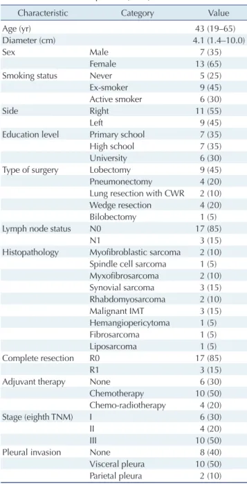
Table 1. Characteristics of patients (n=20)
Characteristic Category Value
Age (yr) 43 (19–65)
Diameter (cm) 4.1 (1.4–10.0)
Sex Male 7 (35)
Female 13 (65)
Smoking status Never 5 (25)
Ex-smoker 9 (45)
Active smoker 6 (30)
Side Right 11 (55)
Left 9 (45)
Education level Primary school 7 (35)
High school 7 (35)
University 6 (30)
Type of surgery Lobectomy 9 (45)
Pneumonectomy 4 (20)
Lung resection with CWR 2 (10)
Wedge resection 4 (20)
Bilobectomy 1 (5)
Lymph node status N0 17 (85)
N1 3 (15)
Histopathology Myofibroblastic sarcoma 2 (10) Spindle cell sarcoma 1 (5) Myxofibrosarcoma 2 (10) Synovial sarcoma 3 (15) Rhabdomyosarcoma 2 (10)
Malignant IMT 3 (15)
Hemangiopericytoma 1 (5)
Fibrosarcoma 1 (5)
Liposarcoma 1 (5)
Complete resection R0 17 (85)
R1 3 (15)
Adjuvant therapy None 6 (30)
Chemotherapy 10 (50)
Chemo-radiotherapy 4 (20)
Stage (eighth TNM) I 6 (30)
II 4 (20)
III 10 (50)
Pleural invasion None 8 (40)
Visceral pleura 10 (50)
Parietal pleura 2 (10)
Values are presented as median (range) or number (%).
CWR, chest wall resection; IMT, inflammatory myofibroblastic tumor;
TNM, tumor-node-metastasis.
https://doi.org/10.5090/jcs.21.032
JCS
The significance analysis and hazard ratios of the indepen- dent prognostic factors for OS and DFS were conducted using the multivariate Cox regression method. All analyses were conducted with a 95% confidence interval (CI).
Two-sided p-values were calculated, and p-values <0.05 were considered to indicate statistical significance.
Results
Twenty patients who met the inclusion criteria were in- cluded in the study. The general characteristics of the pa- tients are shown in Table 1. The median follow-up period was 36 months (range, 3–105 months). There were 13 men (65%) and 7 women (35%). The most common histopatho- logical types were malignant inflammatory myofibroblas- tic tumors and synovial sarcomas (15% each). The most common surgical procedure was lobectomy with mediasti- nal lymph node sampling or dissection in 9 patients (45%).
Complete resection (R0) was achieved in 17 patients (85%).
Postoperative complications specific to lung resections were also observed in our series, including prolonged air leakage in 4 patients (20%), atrial fibrillation in 2 patients (10%), pneumonia in 1 patient (5%), and prolonged thora- cotomy pain in 1 patient (5%).
The median OS was 36 months (interquartile range [IQR], 19–53 months), and the 5-year OS was 37% (Fig. 1).
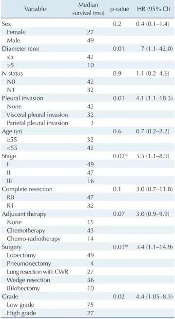
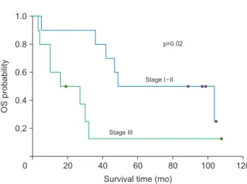
Age, tumor size, and lymph node invasion had no signifi- cant effects on OS (Table 2). However, parietal pleural in- vasion and stage III tumors based on the eighth tu- mor-node-metastasis (TNM) staging system were poor prognostic factors for OS (p=0.01 and p=0.02, respectively) (Table 2, Fig. 2). Additionally, the OS was significantly worse in patients who underwent major parenchymal re-
section (pneumonectomy and bilobectomy) (p=0.01) (Table 2). Histopathologically, the best median OS was detected in malignant inflammatory myofibroblastic tumors and the worst median OS in the leiomyosarcoma and hemangiope- riostoma groups. Nonetheless, the survival difference was not statistically significant (97 months and 10 months, re- spectively; p=0.08). A statistically significant difference was found between high-grade and low-grade tumors de- fined according to mitosis number, nuclear atypia, and dif-
1.0
0.8
0.6
0.4
0.2
120
OSprobability
Survival time (mo) 0
n=20
Median survival=36 mo 5-Year OS=37%
20 40 60 80 100
Fig. 1. Survival curve calculated using the Kaplan-Meier method.
OS, overall survival.
Table 2. Outcomes of Cox regression analyses for overall survival
Variable Median
survival (mo) p-value HR (95% CI)
Sex 0.2 0.4 (0.1–1.4)
Female 27
Male 49
Diameter (cm) 0.01 7 (1.1–42.0)
≤5 42
>5 10
N status 0.9 1.1 (0.2–4.6)
N0 42
N1 32
Pleural invasion 0.01 4.1 (1.1–18.3)
None 42
Visceral pleural invasion 32 Parietal pleural invasion 3
Age (yr) 0.6 0.7 (0.2–2.2)
≥55 32
<55 42
Stage 0.02a) 3.5 (1.1–8.9)
I 49
II 47
III 16
Complete resection 0.1 3.0 (0.7–11.8)
R0 47
R1 32
Adjuvant therapy 0.07 3.0 (0.9–9.9)
None 15
Chemotherapy 43
Chemo-radiotherapy 14
Surgery 0.01b) 3.4 (1.1–14.9)
Lobectomy 49
Pneumonectomy 4
Lung resection with CWR 27
Wedge resection 36
Bilobectomy 10
Grade 0.02 4.4 (1.05–8.3)
Low grade 75
High grade 27
HR, hazard ratio; CI, confidence interval; CWR, chest wall resection.
a)The patients whose with pathological tumor stage III had poorer prognoses than those with stage I and II. b)The overall survival of patients who underwent extensive parenchymal resection (pneumonectomy and bilobectomy) was significantly worse than their counterparts.
Muhammet Sayan, et al. Primary Pulmonary Malignant Mesenchymal Tumors JCS
ferentiation according to the criteria of the French Federation of Cancer Centers Sarcoma Group–Tumor Differentiation Score by histologic type (p=0.02) (Table 2, Fig. 3). We did not perform low-medium-high or grade 0–3 subgrouping due to the small number of patients. The criteria for high- grade tumors were a mitosis number of more than 9 in each area, the presence of necrosis, and the presence of dif- ferentiation [4].
The median DFS was 31 months (IQR, 21–41 months), and the 5-year DFS was 32%. The median DFS was signifi- cantly worse in patients with parietal pleural invasion (p=
0.02), those who underwent lobectomy with chest wall re- section (p=0.02), and those who had high-grade tumors (p=0.01). DFS was poorer in female patients (p=0.07) and those with a high tumor stage (p=0.1), advanced age (p=0.4), and fibrosarcoma as the histopathological tumor type (p=0.1) than in their respective counterparts; howev- er, those differences were not statistically significant.
Discussion
In this study, we aimed to present the surgical and sur- vival outcomes of PPMMTs, which are extremely rare. Due to the rarity of PPMMTs, very few studies have investigat- ed their surgical treatment and survival outcomes. In their study, Bacha et al. [1] reported 23 cases with a median age of 51 years and a 65% proportion of male patients. In a large database study by Spraker et al. [5], the median age was 63 years, and 56% of patients were men. However, an- other study reported that male patients were in the minori- ty and had a worse prognosis [6]. In our study, the median age was 43 years, and the percentage of male patients was 65%. Age and sex had no significant effects on OS.
As in other sarcomas, R0 resection has been associated with good survival in patients with PPMMTs. The rate of R0 resection was 89% in a study conducted by Petrov et al.
[7]. They reported that R0 resection was a significant pre- dictor of OS [7]. Similarly, some other studies found that R0 was a strong marker for OS [1,6,8,9]. In our series, R0 resection was achieved in 17 patients (85%). We found that the median OS was better in the R0 group, but the survival difference was not statistically significant. The reason for this result is the very small number of patients in the R1 resection group.
Generally, poor median and 5-year OS outcomes have been reported in related studies. Robinson et al. [9] report- ed that the median OS was 39 months, and the 5-year OS was 28.7%. They emphasized that the OS was better in the surgical group [9]. Yamada et al. [8] found that the 5-year OS was 50%, and significant prognostic factors were tumor diameter, stage, and grade and incomplete resection. The median and 5-year OS were 22 months and 27%, respec- tively, in a study conducted by Golota et al. [6], who found that tumor stage and hemoptysis were poor prognostic fac- tors. The results for the median and 5-year OS observed in this study are consistent with the literature.
Typically, regional lymph node metastasis in sarcomas is not expected. Instead, local recurrence or distant hematog- enous metastasis is more likely. Spraker et al. [5] reported that the rate of lymph node invasion was 16%, and that lymph node invasion constituted a significant prognostic factor for OS. In other studies, the lymph node invasion rates were between 8% and 22.5% [7-9]. Our study showed no metastasis in the N2 station, and the rate of N1 invasion was 15%. There was no significant difference in OS be-
1.0
0.8
0.6
0.4
0.2
120
OSprobability
Survival time (mo) 0
p=0.02
Stage I II
Stage III
20 40 60 80 100
Fig. 2. Comparison of survival rates between patients with early and advanced-stage tumors. OS, overall survival.
1.0
0.8
0.6
0.4
0.2
120
OSprobability
Survival time (mo) 0
p=0.02
Low grade
High grade
20 40 60 80 100
Fig. 3. Graphic representation of the difference in survival rates between patients with low-and-high-grade tumors. OS, overall survival.
https://doi.org/10.5090/jcs.21.032
JCS
tween the N1 and N0 groups. The negative effect of ad- vanced tumor stage on OS could be explained by tumor diameter rather than lymph node invasion. In addition, our study showed that parietal pleural invasion, an indirect indicator of local invasion, was a negative prognostic indi- cator for OS and DFS.
Although articles related to non-small cell lung cancer treated surgically have reported pneumonectomy to be a poor prognostic factor, we could not find any studies showing this effect in PPMMTs in the literature [10,11]. In our study, the median OS was worse in patients who un- derwent pneumonectomy. This result may be explained by the larger diameter of the tumors in the pneumonectomy group. However, the better DFS rate of pneumonectomy group contradicts that argument. The literature shows that the DFS of PPMMT patients varies from 15 to 65 months [3,6-8]. The DFS of our study is consistent with that re- ported in the literature, and we found that parietal pleural and chest wall invasion had a negative effect on DFS.
There are several limitations to our study. Principally, this was a retrospective, single-center study that included a small number of cases. In our study, the factors affecting OS and DFS were found as expected. Although no new prognostic factors were found, we think that it is vital to present the surgical and survival results of this rare tumor group.
In conclusion, malignant mesenchymal tumors are ag- gressive and chemo/radio-resistant. The OS and DFS after surgical resection are acceptable in patients with primary pulmonary sarcoma who have suitable medical conditions and tumor stage.
Conflict of interest
No potential conflict of interest relevant to this article was reported.
ORCID
Muhammet Sayan: https://orcid.org/0000-0002-5402-9031 Aykut Kankoc: https://orcid.org/0000-0001-5048-6115 Dilvin Ozkan: https://orcid.org/0000-0002-7149-5982
Ali Celik: https://orcid.org/0000-0001-5385-6492 Ismail Cuneyt Kurul: https://orcid.org/0000-0002-9480-010X Abdullah Irfan Tastepe: https://orcid.org/0000-0002-2032-7444
References
1. Bacha EA, Wright CD, Grillo HC, et al. Surgical treatment of prima- ry pulmonary sarcomas. Eur J Cardiothorac Surg 1999;15:456-60.
2. Sayan M, Bas A, Valiyev E, et al. Prognostic factors for sarcomatoid carcinomas of lung: a single-centre experience. Lung India 2020;37:
506-10.
3. Duran-Moreno J, Kokkali S, Ramfidis V, et al. Primary sarcoma of the lung: prognostic value of clinicopathological characteristics of 26 cases. Anticancer Res 2020;40:1697-703.
4. Guillou L, Coindre JM, Bonichon F, et al. Comparative study of the National Cancer Institute and French Federation of Cancer Centers Sarcoma Group grading systems in a population of 410 adult patients with soft tissue sarcoma. J Clin Oncol 1997;15:350-62.
5. Spraker MB, Bair E, Bair R, Connell PP, Mahmood U, Koshy M. An analysis of patient characteristics and clinical outcomes in primary pulmonary sarcoma. J Thorac Oncol 2013;8:147-51.
6. Golota J, Osowiecka K, Orlowski T. Primary pulmonary sarcoma:
long-term treatment outcomes and prognostic factors. Kardiochir Torakochirurgia Pol 2018;15:162-9.
7. Petrov DB, Vlassov VI, Kalaydjiev GT, et al. Primary pulmonary sarcomas and carcinosarcomas: postoperative results and compara- tive survival analysis. Eur J Cardiothorac Surg 2003;23:461-6.
8. Yamada Y, Kaplan T, Soltermann A, et al. Surgical outcomes and risk analysis of primary pulmonary sarcoma. Thorac Cardiovasc Surg 2021;69:101-8.
9. Robinson LA, Babacan NA, Tanvetyanon T, Henderson-Jackson E, Bui MM, Druta M. Results of treating primary pulmonary sarcomas and pulmonary carcinosarcomas. J Thorac Cardiovasc Surg 2021;
162:274-84.
10. Park JS, Yang HC, Kim HK, et al. Sleeve lobectomy as an alternative procedure to pneumonectomy for non-small cell lung cancer. J Tho- rac Oncol 2010;5:517-20.
11. Deslauriers J, Gregoire J, Jacques LF, Piraux M, Guojin L, Lacasse Y.
Sleeve lobectomy versus pneumonectomy for lung cancer: a compar- ative analysis of survival and sites or recurrences. Ann Thorac Surg 2004;77:1152-6.