• 검색 결과가 없습니다.

Analysis of Sternal Fixation Results According to Plate Type in Sternal Fracture

N/A
N/A
Protected

Academic year: 2021

Share "Analysis of Sternal Fixation Results According to Plate Type in Sternal Fracture"

Copied!
5
0
0

로드 중.... (전체 텍스트 보기)

전체 글

(1)

ISSN: 2233-601X (Print) ISSN: 2093-6516 (Online)

Received: September 1, 2015, Revised: November 2, 2015, Accepted: November 3, 2015, Published online: October 5, 2016

Corresponding author: Hyun Min Cho, Department of Trauma Surgery, Pusan National University Hospital Trauma Center, 179 Gudeok-ro, Seo-gu, Busan 49241, Korea

(Tel) 82-51-240-7369 (Fax) 82-51-240-7719 (E-mail) [email protected]

© The Korean Society for Thoracic and Cardiovascular Surgery. 2016. All right reserved.

This is an open access article distributed under the terms of the Creative Commons Attribution Non-Commercial License (http://creativecommons.org/

licenses/by-nc/4.0) which permits unrestricted non-commercial use, distribution, and reproduction in any medium, provided the original work is properly cited.

Analysis of Sternal Fixation Results According to Plate Type in Sternal Fracture

Chun Sung Byun, M.D. 1 , Il Hwan Park, M.D., Ph.D. 1 , Wan Jin Hwang, M.D. 2 , Yeiwon Lee, M.D. 2 , Hyun Min Cho, M.D. 3

1

Department of Thoracic and Cardiovascular Surgery, Wonju Severance Christian Hospital, Yonsei University Wonju College of Medicine,

2

Department of Thoracic and Cardiovascular Surgery, Konyang University Hospital, Konyang University

College of Medicine,

3

Department of Trauma Surgery, Pusan National University Hospital Trauma Center

Background: Sternal fractures are relatively rare, and caused mainly by blunt anterior chest wall trauma. In most cases, sternal fractures are treated conservatively. However, if the patient exhibits problematic symptoms such as intractable chest wall pain or bony crepitus due to sternal instability, surgical correction is indicated.

But no consensus exists regarding the most appropriate surgical method. We analyzed the results of surgical fixation in cases of sternal fracture in order to identify which surgical method led to the best outcomes.

Methods: We retrospectively reviewed the medical records of patients with sternal fractures from December 2008 to December 2011, and found 19 patients who underwent open reduction and internal fixation of the sternum with a longitudinal plate (L-group) or a T-shaped plate (T-group). We investigated patients’ charac- teristics, clinical details regarding each case of chest trauma, the presence of other associated injuries, the type of open reduction and fixation, whether a combined operation was performed, and postoperative complications. Results: Of the 19 patients, 10 patients (52.6%) were male, and their average age was 56.8 years (range, 32 to 82 years). Seven patients (36.8%) had isolated sternal fractures, while 12 (63.2%) had other associated injuries. Seven patients (36.8%) were in the L-group and 12 patients (63.2%) were in the T-group. Three patients in the L-group (42.9%) showed a loosening of the fixation. In all patients in the T-group, the fracture exhibited stable alignment. Conclusion: Open reduction and internal fixation with a T-shaped plate in sternal fractures is a safer and more efficient treatment method than treatment with a longitudinal plate, especially in patients with a severely displaced sternum or anterior flail chest, than a lon- gitudinal plate.

Key words: 1. Sternum

2. Thoracic injuries 3. Fracture fixation 4. Bone plates

Introduction

Sternal fractures are relatively rare, accounting for

<0.5% of total bone fractures and 8% to 10% of cases of blunt thoracic trauma [1-4]. Blunt anterior

chest wall contusions, in which the chest collides

with the steering wheel in traffic accidents, are the

main cause of sternal fractures. Morbidity and mor-

tality from sternal fractures are both directly related

to the degree of co-injury, and have no clear relation

http://dx.doi.org/10.5090/kjtcs.2016.49.5.361

(2)

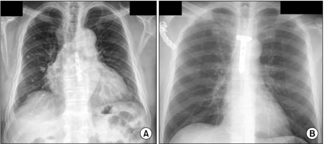
Fig. 1. Open reduction and internal fixation of sternum with (A) longi- tudinal plate with stainless steel wiring and (B) T-shaped plate.

to the sternal fracture itself [5,6]. Sternal fractures are treated conservatively in most cases. However, no reports have assessed the incidence of long-term complications after conservative treatment of sternal fractures. According to reports published over the last 50 years, if the internal organs sustain no dam- age in a severe sternal fracture, the mortality rate is low. No consensus exists regarding the most appro- priate methods of surgical correction if a patient ex- hibits problematic symptoms such as intractable chest wall pain or bony crepitus due to sternal instability or a chest wall deformity [7,8]. Therefore, we ana- lyzed the results of surgical fixation in patients with severe sternal fractures to determine the most ap- propriate surgical methods for sternal fractures.

Methods

We retrospectively reviewed the medical records of 117 patients with sternal fractures who were treated at Konyang University Hospital from December 2008 to December 2011. Of these patients, 19 pa- tients underwent open reduction and internal fixation of the sternum. Patient characteristics such as age and gender, as well as information regarding the du- ration of admission, chest X-ray findings, chest com- puted tomography (CT) findings, and operative re- cords, were collected. Clinical information including the mechanism of injury, time of injury, type of ster- nal fixation, co-injury site, surgical information for the co-injury site, and postoperative complications was also included. For patients with a sternal frac- ture, the surgical indications were complaints of se- vere pain and functional limitations with severe dis-

location of the sternum in a chest X-ray or chest CT, or a flail motion that caused difficulty in weaning them from a mechanical ventilator.

Surgery was performed with a 10-cm mid-sternal incision between the sternal notch and the xyphoid process under general anesthesia. After dissection of the fractured sternum, the fractured site was cor- rected manually and fixed with a titanium plate (Hankil Tech Medical Co. Ltd, Hwaseong, Korea) with cortical screws (Hankil Tech Medical Co. Ltd). 7 patients were treated with a longitudinal plate that was occa- sionally reinforced by a stainless steelwire (the L-gr- oup) (Fig. 1A). 12 patients, a T-shaped plate without wiring was used (the T-group) (Fig. 1B). Following surgery, we checked for hemothorax or pneumotho- rax on an immediate postoperative chest X-ray. When patients became ambulatory, we confirmed sternal their fixation status via a lateral sternum X-ray.

Incomplete fixation was defined as screw loosening in a radiologic examination after sternal fixation dur- ing the follow-up period.

Statistical analysis was performed using PASW SPSS statistics ver. 17.0.2 (SPSS Inc., Chicago, IL, USA). The Fisher exact test was used to evaluate dif- ferences between the groups, with p-values <0.05 considered statistically significant.

Results

Of the 19 patients who underwent surgical fixation

of the sternum using a titanium plate and screws, 10

patients were male (52.6%), and their average age

was 56.8 years (range, 32 to 82 years). The mean

follow-up time was 9.2 months (range, 2 to 22

(3)

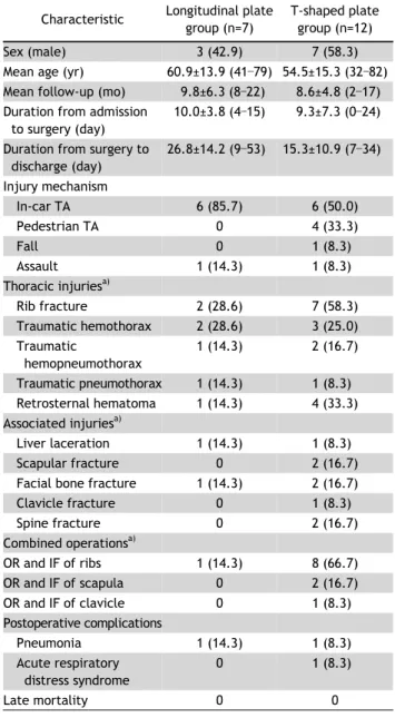
Table 1. General characteristics of patients (n=19) Characteristic Longitudinal plate

group (n=7)

T-shaped plate group (n=12)

Sex (male) 3 (42.9) 7 (58.3)

Mean age (yr) 60.9±13.9 (41 –79) 54.5±15.3 (32–82) Mean follow-up (mo) 9.8±6.3 (8 –22) 8.6±4.8 (2 –17) Duration from admission

to surgery (day)

10.0±3.8 (4 –15) 9.3±7.3 (0 –24)

Duration from surgery to discharge (day)

26.8±14.2 (9 –53) 15.3±10.9 (7–34)

Injury mechanism

In-car TA 6 (85.7) 6 (50.0)

Pedestrian TA 0 4 (33.3)

Fall 0 1 (8.3)

Assault 1 (14.3) 1 (8.3)

Thoracic injuries

a)

Rib fracture 2 (28.6) 7 (58.3)

Traumatic hemothorax 2 (28.6) 3 (25.0) Traumatic

hemopneumothorax

1 (14.3) 2 (16.7)

Traumatic pneumothorax 1 (14.3) 1 (8.3) Retrosternal hematoma 1 (14.3) 4 (33.3) Associated injuries

a)

Liver laceration 1 (14.3) 1 (8.3)

Scapular fracture 0 2 (16.7)

Facial bone fracture 1 (14.3) 2 (16.7)

Clavicle fracture 0 1 (8.3)

Spine fracture 0 2 (16.7)

Combined operations

a)

OR and IF of ribs 1 (14.3) 8 (66.7)

OR and IF of scapula 0 2 (16.7)

OR and IF of clavicle 0 1 (8.3)

Postoperative complications

Pneumonia 1 (14.3) 1 (8.3)

Acute respiratory distress syndrome

0 1 (8.3)

Late mortality 0 0

Values are presented as numbers (%) or mean±standard deviation (range).

TA, traffic accident; OR, open reduction; IF, internal fixation.

a)

Multiple responses.

Table 2. Comparison of surgical parameters between the longi- tudinal plate and T-shaped plate groups (n=19)

Characteristic

Longitudinal plate group

(n=7)

T-shaped plate group

(n=12)

p-value

Type of sternal fracture 0.045

Isolated 5 (71.4) 2 (16.7)

Combined injuries 2 (28.6) 10 (83.3) Fracture contour of sternal

fixation site

0.364

Linear 7 (100.0) 8 (66.7)

Oblique 0 2 (16.7)

Segmental 0 2 (16.7)

Location of sternal fixation 0.850

Manubrium 0 1 (8.3)

Sternal angle 0 2 (16.7)

Upper body 3 (42.9) 2 (16.7)

Mid-body 3 (42.9) 5 (41.7)

Lower body 1 (14.3) 2 (16.7)

Postoperative complications 0.523

Yes 1 (14.3) 2 (16.7)

Result of surgical fixation 0.036

Complete 4 (57.1) 12 (100.0)

Incomplete 3 (42.9) 0

Values are presented as numbers (%). Group comparisons were analyzed by the Fisher exact test.

months), and no cases of mortality were associated with sternal fixation. Table 1 shows the general char- acteristics of the L-group (7 patients, 36.8%) and the T-group (12 patients, 63.2%). The average period from admission to surgery was 10.0 days for the L-group (range, 4 to 15 days) and 9.3 days for the T-group (range, 0 to 24 days). The average period from surgery to discharge was 26.8 days for the L-group (range, 9 to 53 days) and 15.3 days for the

T-group (range, 7 to 34 days). A total of 7 isolated

sternum fractures took place in 19 patients. The as-

sociated intrathoracic injuries included were large

numbers of rib fractures, hemothoraces, hemopneu-

mothoraces, pneumothoraces, and retrosternal hema-

tomas. The associated extrathoracic injuries were cla-

vicle fractures, facial bone fractures, liver lacerations,

scapular fractures, and spine fractures. Open reduc-

tion and internal fixation of the ribs was concomi-

tantly performed in 1 patient (14.3%) in the L-group

and in 9 patients (66.7%) in the T-group. Operations

to treat scapular fractures were performed in 2 pa-

tients (16.7%) in the T-group, and an operation to

treat a clavicle fracture was performed in 1 patient

(8.3%), also in the T-group. Postoperatively, pneumo-

nia occurred in 1 patient in both groups, and acute

respiratory distress syndrome (ARDS) occurred in 1

patient in the T-group. No postoperative mortalities

occurred in either group. The patient who developed

ARDS exhibited anterior flail chest, received mechan-

ical ventilator care, and then progressed to ARDS; a

tracheostomy was therefore performed 8 days after

(4)

the sternal fixation.

When we compared the two groups defined ac- cording to plate type, differences were found in the frequency of isolated sternal fractures; 5 patients in the L-group (71.4%) did not have associated injuries, in comparison to 2 patients (16.7%) in the T-group.

No significant differences were found in the fracture contour of the sternal fixation sites, the locations of sternal fixation, and the incidence of postoperative complications. However, there was a difference in the frequency of complete fixation between the two groups;

3 patients in the L-group (42.9%) underwent com- plete fixation, while all the T-group patients did so (Table 2). During the follow-up period, 2 patients had conservative treatment for incomplete fixation. One patient had the plate removed 8 months after sur- gery, and the sternum was fused completely after removal. In this patient, the anterior chest wall pain was much reduced and he experienced few limi- tations in terms of posture or daily activity.

Discussion

Surgical fixation of the sternum is performed in a very limited number of patients, because only few patients present with surgical indications of sternal fracture, such as severe dislocation of the fractured site or anterior flail motion. In our study, severe sternal fracture patients had numerous associated in- trathoracic injuries, such as pneumothorax, hemo- thorax, and multiple rib fractures. Also, all patients’

cardiac enzyme levels were tested and, if elevated levels of any enzymes were found jointly with an electrocardiography abnormality, we performed echo- cardiography. All echocardiograms showed insignif- icant cardiac trauma in this study. Furthermore, all retrosternal hematomas were spontaneously absorbed after surgical fixation of the sternum. This implies that pulmonary complications such as pleural effu- sion, atelectasis, pneumonia, and ARDS occurred more frequently than cardiovascular injury in sternal frac- ture patients.

When early surgical fixation fails in patients with severe dislocations, a sternal fracture can progress to pseudoarthrosis, impaired ventilation, and transfigu- ration of the anterior chest wall. Early detection and treatment of sternal fractures is important for good quality of life after anterior chest wall trauma. For

example, pseudoarthrosis after a sternal fracture can aggravate severe anterior chest wall pain leading to limitations in daily activity; additionally, delayed sur- gical correction of pseudarthrosis is very difficult for physician.

A non-invasive technique of sternal reduction is hyperextension of the thoracic spine. However, surgi- cal experience in the treatment of severe sternal fractures has accumulated, and a variety of surgical techniques have been developed for sternal fractures.

Prior to 2008, we treated most sternal fracture pa- tients conservatively, even in cases of severe dis- placed fracture. However, since 2008, most displaced fractures have been treated with open reduction and internal fixation with titanium plate and screws.

Many internal fixation methods exist for the treat- ment of sternal fractures, including a Steinmann pin for segmental fractures, stainless steel wiring for dis- placed fractures, and the use of a titanium plate and screws [9-14]. Especially in patients with severe dis- location of the sternal fracture site, a titanium plate and screws are more stable than figure-8 wiring.

Titanium plates and screws for sternal fixation are both used for traumatic sternal fractures as well as chronic nonunion of the sternum after median ster- notomy. Sternal fixation with a titanium plate and screws can more securely facilitate rapid bone union, and minimizes the dissection of the mediastinal soft tissue without causing damage to collateral blood vessels and the internal organs around the sternum.

Furthermore, open reduction and internal fixation of a displaced sternal fracture can reduce anterior chest wall pain, improve pulmonary function, and prevent pseudoarthrosis and chest wall deformity.

In our study, the T-group patients exhibited more associated injuries and more oblique and segmental sternal fracture lines than the patients in the L-group;

however, postoperative complications did not differ

significantly between the two groups. This is because

postoperative complications depend on the severity

of the associated lung injury rather than the material

and methods used in surgical fixation. In terms of

fixation stability, the T-shaped plate was better than

the longitudinal plate for internal fixation of the frac-

tured sternum due to its low frequency of in-

complete fixation. The use of a different plate type

with similar surgical methods resulted in more in-

complete fixations in the L-group, suggesting that

(5)

T-shaped plates are better able to resist the ten- sion-compression force in the sternum than longi- tudinal plates. However, the use of two longitudinal plates leads to more secure sternum fixation than the use of a single longitudinal plate [ 15]. Transverse tension resistance between each longitudinal plate is absent; in contrast, the T-shaped plate is more re- sistant against the tension-compression force from the sternum while maintaining the bone union of the fracture site [16]. The convex shape on the anterior side of the sternum is exposed to a tension force during deep inspiration, and the concave shape on the posterior side of the sternum increases the com- pression pressure. However, a T-shaped plate located on the anterior side of the sternum can overcome the bending force better than a longitudinal plate, and it reduces the compression pressure of the pos- terior side of the sternum to maintain contact with the fractured bone. In the early period of the sternal fixation procedures analyzed in this study, we only employed longitudinal plates; however, the T-shaped plate became preferred in the later period of this study. This change was attributable to the initial learning curve in open reduction and internal fix- ation on the part of the attending surgeons.

The present study was limited by its non-random- ized and small sample size. In addition, although no significant differences were found in postoperative complication rates, the underlying general character- istics, trauma vectors, and associated injuries differed between the two groups, reducing the statistical power of the study. However, this study showed the results of sternum fracture fixation according to plate type, which can help guide sternum fracture management strategies in the future.

In conclusion, open reduction and internal fixation with a T-shaped plate in sternal fractures is a safer and more efficient method than fixation with a longi- tudinal plate, especially in patients with a severely displaced sternum or anterior flail chest. This con- clusion needs to be verified by additional prospective studies with larger populations, in order to provide clinically relevant findings regarding the short-term results of bone union and the long-term functional and cost advantages of fixation with a T-shaped plate.

Conflict of interest

No potential conflict of interest relevant to this ar- ticle was reported.

Acknowledgments

This study was supported by a Grant of the Samsung Vein Clinic Network (Daejeon, Anyang, Cheongju, Cheonan; Fund No. KTCS04-057).

References

1. Potaris K, Gakidis J, Mihos P, Voutsinas V, Deligeorgis A, Petsinis V. Management of sternal fractures: 239 cases.

Asian Cardiovasc Thorac Ann 2002;10:145-9.

2. Mayba II. Non-union of fractures of the sternum. J Bone Joint Surg Am 1985;67:1091-3.

3. Helal B. Fracture of the manubrium sterni. J Bone Joint Surg Br 1964;46:602-7.

4. Brookes JG, Dunn RJ, Rogers IR. Sternal fractures: a retro- spective analysis of 272 cases. J Trauma 1993;35:46-54.

5. Gouldman JW, Miller RS. Sternal fracture: a benign entity?

Am Surg 1997;63:17-9.

6. Trinca GW, Dooley BJ. The effects of mandatory seat belt wearing on the mortality and pattern of injury of car oc- cupants involved in motor vehicle crashes in Victoria. Med J Aust 1975;1:675-8.

7. Sadaba JR, Oswal D, Munsch CM. Management of isolated sternal fractures: determining the risk of blunt cardiac injury. Ann R Coll Surg Engl 2000;82:162-6.

8. Harley DP, Mena I. Cardiac and vascular sequelae of ster- nal fractures. J Trauma 1986;26:553-5.

9. Van Sterkenburg SM, Brutel de la Riviere A, Vermeulen FE.

Sternal fixation with resorbable suture material. Eur J Cardiothorac Surg 1990;4:345.

10. Kitchens J, Richardson JD. Open fixation of sternal frac- ture. Surg Gynecol Obstet 1993;177:423-4.

11. Molina JE. Evaluation and operative technique to repair isolated sternal fractures. J Thorac Cardiovasc Surg 2005;

130:445-8.

12. Hills MW, Delprado AM, Deane SA. Sternal fractures: asso- ciated injuries and management. J Trauma 1993;35:55-60.

13. Bonney S, Lenczner E, Harvey EJ. Sternal fractures: ante- rior plating rationale. J Trauma 2004;57:1344-6.

14. Athanassiadi K, Gerazounis M, Moustardas M, Metaxas E.

Sternal fractures: retrospective analysis of 100 cases.

World J Surg 2002;26:1243-6.

15. Schulz-Drost S, Oppel P, Grupp S, et al. Surgical fixation of sternal fractures: preoperative planning and a safe surgi- cal technique using locked titanium plates and depth lim- ited drilling. J Vis Exp 2015;(95):e52124.

16. Al-Qudah A. Operative treatment of sternal fractures.

Asian Cardiovasc Thorac Ann 2006;14:399-401.

수치

Fig. 1. Open reduction and internal  fixation of sternum with (A)  longi-tudinal plate with stainless steel  wiring and (B) T-shaped plate.
Table 2. Comparison of surgical parameters between the longi- longi-tudinal plate and T-shaped plate groups (n=19)

참조

관련 문서

Purpose: Calcaneal fracture is a rare fracture, which accounts for about 2% of all fractures, but is one of the most common fractures in the ankle bone.. There is

Conclusion: Aromatherapy with neroli essential oil had a difference in brain wave and brain utilization ability according to the MBTI personality type

The analysis results of differences in tenacity according to the career of winning a prize indicate some differences in desire for victory, fighting spirit,

High complication rate in locking plate fixation of lower periprosthetic distal femur fractures in patients with total knee arthroplasties.. Management and

 A crack in a thin sheet  a steeper R curve than a crack in a thick plate because of a low degree of stress triaxiality (3축) at the crack tip in the thin sheet, while

3,4 Therefore, to date, studies have been conducted to examine the absorbable fixation plate manufactured using polymer and a fixation screw that can

Conclusion: In Neer type II distal clavicle fracture treatment, both groups showed satisfactory result in clinical and radiological outcomes, but the hook plate

The feed is commonly a solution in a solvent like ethanol or t-butanol, and the nonsolvent is water..