웨어러블 기기를 이용한 발작의 비-뇌파적 감시
강원대학교병원 소아청소년과
노병호
Submitted: 1 December, 2017 Revised: 27 December, 2017 Accepted: 27 December, 2017
Correspondence to Byoungho H Noh, MD
Department of Pediatrics, Kangwon National University Hospital, Baengnyeong-ro 156, Chuncheon-Si, Gang- won-Do, 24289, Korea
Tel: +82-33-258-9020, Fax: +82-33-258-2418 E-mail: [email protected]
Non-EEG Seizure Detection Using Wearable Device
Electroencephalography (EEG) is the gold standard for seizure detection. However, EEG systems are usually expensive and require well-trained technicians for data ac- quisition. Detection of seizures is important in patients with refractory epilepsy, es- pecially when the seizure is prolonged and unwitnessed. Sudden unexpected death in epilepsy (SUDEP) is an important problem in patients with refractory epile psy be cause the risk of SUDEP decreases the quality of life. There are several alterna tives to EEG systems that are used to detect seizure-related pathophysiological changes.
Measurements of heart rate and heart rate variability can be used to detect seizure- related cardiologic phenomena. Respiratory signals obtained based on respiration rate and respiratory chest and abdominal movements may also be used for seizure detection. Ictal movements can be detected using video analysis or wearable devices, such as accelerometers and gyroscopes. In addition, electrodermal activity (EDA) measurements are currently under investigation as a way to alert others to an ictal period. Although these technologies have limitations, they are affordable and easily wearable. They are thus appropriate for use in patients during daily activity. These devices may be potentially useful to detect the occurrence of unwitnessed seizures, and to increase the quality of life.
Key Words: Seizure, Diagnosis, Quality of life, SUDEP, Electroencephalography
Byoungho H Noh, MD
Department of Pediatrics, Kangwon National Uni
versity Hospital, Chuncheon, Korea
Copyright © 2017 by The Korean Child Neurology Society
http://www.cns.or.kr
Introduction
Patients with epilepsy have unpredictable seizures. Higher numbers of seizures in these patients are associated with lower quality of life. In addition, more fre
quent seizures are associated with an increased risk of sudden unexpected death in epilepsy (SUDEP). Preventing seizures is undoubtedly the most important mis
sion in treating epilepsy. Nevertheless, because almost 30% of patients with epile
psy have medically refractory epilepsy, the prediction or even early detection of seizures require urgent attention, as they may decrease seizure burden and sei
zurerelated consequences, such as SUDEP1).
Electroencephalography (EEG) is the gold standard used to identify seizures because seizures are electrical cerebral phenomena. However, the use of EEG devices during daily life outside of a hospital has several limitations. First, these devices are large, as EEG recordings are usually performed using headsets or electrodes, an attached jackbox, and a fullsize computer system. These devices are thus not easy to carry. Second, detection of seizures requires interpretation
by welltrained experts. Finally, these systems are very expen
sive. Because of these limitations, there is a need for less expen
sive versions of seizure alarm systems that are easier to use.
Due to the rapid progress in engineering and technology, novel trials to detect and analyze biological signals have been perfor
med in different medical fields2). There are several current medical studies to investigate the use of commercial products, such as smart watches or fitness trackers. In the field of epilepsy research, there are trials designed to investigate seizure tracking using wearable devices3). Seizure alarms using wearable devices would be easy to use at home, especially for patients with intractable epilepsy. SUDEP management would benefit from using wearable technologies in addition to the traditional gold standard of EEG.
EEG-based seizure detection/prediction
There are several commercial handheld or wearable EEG re
cording devices3,4). Because the detection of ictal electrographic signals is the best way to detect seizures, current potential sei
zure detection devices for daily use rely on these signals. Several reports suggest that it is possible to measure brain electrical ac
tivity using wearable device during daily activity57). Although various kinds of psychiatric disease are investigated using these devices, no reliable peerreviewed publications discussing epile
psy treatment are as yet available.
Non-EEG based seizure detection
Many different biological signals are produced that are reflec
tive of the body’s condition. Some of these signals can consis
tently be used to identify pathophysiological conditions. For ex
ample, electrocardiography (EKG) is one of the most important diagnostic tools for the detection of myocardial infarction. In the field of epilepsy, several biological signals are potential indicators of periictal conditions. For these signals, sensitivity is slightly more important than specificity, as missing a major seizure is less desirable than producing a false alarm when the goal is to pre
vent SUDEP.
Different signals require the use of different sensors and de
tection devices. The different biological signals may be catego
rized as changes in metabolism, heart function, respiratory func
tion, physical movement, and autonomic nervous system. Thanks to considerable improvement in technology, sensor sizes have been reduced, and data collection systems such as smart phones and smart watches are now widely available and affordable8,9). There are commercially available EEG and nonEEG based wea
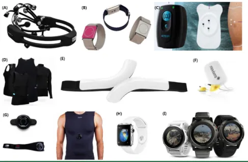
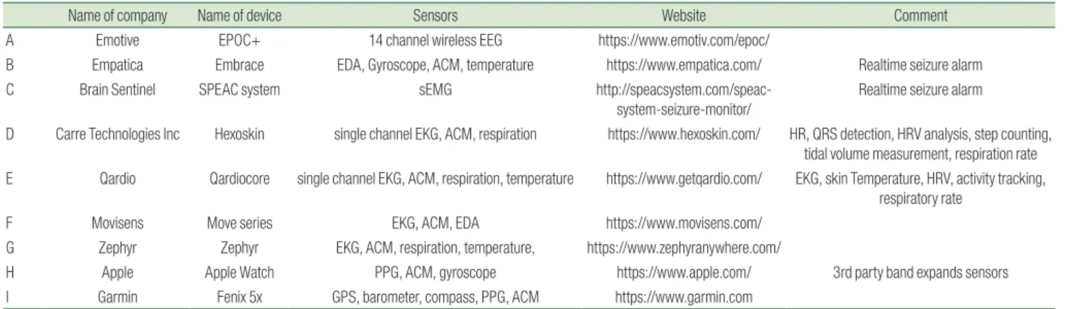
r able devices (Table 1, Fig. 1)
1. Cardiac signals
Heart rhythms can be detected using technologies such as EKG and photoplethysmography (PPG). The types of data from the sensors are heart rate (HR) and heart rate variability. EKG can be used to more precisely detect electrical cardiac activity when compared to PPG. PPG uses lightemitting diodes and optical
Fig. 1. Commercially available wearable devices, (A) Emotive EPOC+, (B) Empatica Embrace, (C) Brain
sentinel SPEAC system, (D) Carre technologies Inc. Hexoskin, (E) Qardio Qardiocore, (F) Movisens Movie
series, (G) Zephy, (H) Apple watch, (I) Garmin Fenix 5 series.
sensors to detect hemodynamic changes, as in a saturation mo
nitor use. This technology is commonly used in commercial de
vices, such as most fitness trackers and smart watches that mea
sure heart rate. These devices include the Samsung Gear series, Apple Watch, and Garmin products. In contrast, EKG recorders are used as patches or in bandaid form in hospitals. Commercial EKG patches are available to monitor patients who have suffered recent myocardial infarctions and are at risk for a recurrence of the attack at home10). PPG devices are less accurate than EKG de
vices, but are also less sensitive to motion artifacts.
Periictal HR changes are more noticeable during generalized tonicclonic seizures, temporal lobe seizures, and frontal lobe seizures because these types of seizures are accompanied by ta
chycardia, bradycardia, and even asystole11,12). QRS abnormalities, such as QT prolongation may also accompany the above sei
zures. One study has reported a linear correlation between elec
trocorticography and EKG during seizure13). The automated algo
rithms used have a seizure detection rate of 9098% and positive predictive values greater than 50%14,15). In addition to the EKG, combination of other types of signals such as, oxygen saturation, accelerometers (ACMs), and Electrodermal activity (EDA) can increase sensitivity and specificity16).
Devices monitoring heart signals are ideal for use in wireless systems using smart phone applications and enable the easy col lection and processing of data in realtime. Continuous long
term monitoring is important because collecting individualized data patterns during normal activities, such as sleep, resting, and exercise may be important in recognizing sudden periictal HR changes.
2. Respiration
Respiratory abnormalities are thought to be major causes of SUDEP17). More than onethird of patients with seizure have ictal
central apnea and ictal tachycardia. Changes in respiration rate and oxygen saturation can be used to detect respiratory abnor
malities. These abnormalities are usually temporary and rever
sible, but are at times prolonged and inevitably lethal.
1) Respiratory belts and nasal air flow detection
Abdominal or chest belts sense movements of the widening chest wall during respiration. Nasal thermistors detect tempera
ture changes produced by respiratory airflow. These devices are commonly used in polysomnography to detect sleep apnea. Ictal apnea can be easily captured using the same devices.
2) Oxygen saturation
Medicalgrade oxygen saturation monitors are very expensive.
However, there are many other lessexpensive oxygen saturation monitors. Some oxygen saturation sensors can be accessed wi
re lessly using a smart phone. This enables the device to alarm the caregiver if the patient is in a hypoxic condition. For example, portable oximeters with Bluetooth connectivity approved by the Food and Drug Administration of the United States are sold for around 200 to 400 USD in the market.
3. Motion detection using video and motion sensors
Changes in movement are the most noticeable changes during seizures. The vast majority of movements during seizures are rhythmic and repetitive movements comprising kicking, reaching, jerking, or a combination of the above. Nevertheless, many types of seizures, including absence seizures and atonic seizures, have negative symptoms. Clearly, motion detection techniques are designed to detect positive symptoms. In this sense, recognition of ictal movements as distinct from nonepileptic movements would be required for a gold standard technique.
Table 1. Commercially Available Wearable Devices
Name of company Name of device Sensors Website Comment
A Emotive EPOC+ 14 channel wireless EEG https://www.emotiv.com/epoc/
B Empatica Embrace EDA, Gyroscope, ACM, temperature https://www.empatica.com/ Realtime seizure alarm
C Brain Sentinel SPEAC system sEMG http://speacsystem.com/speac-
system-seizure-monitor/
Realtime seizure alarm
D Carre Technologies Inc Hexoskin single channel EKG, ACM, respiration https://www.hexoskin.com/ HR, QRS detection, HRV analysis, step counting, tidal volume measurement, respiration rate E Qardio Qardiocore single channel EKG, ACM, respiration, temperature https://www.getqardio.com/ EKG, skin Temperature, HRV, activity tracking,
respiratory rate
F Movisens Move series EKG, ACM, EDA https://www.movisens.com/
G Zephyr Zephyr EKG, ACM, respiration, temperature, https://www.zephyranywhere.com/
H Apple Apple Watch PPG, ACM, gyroscope https://www.apple.com/ 3rd party band expands sensors
I Garmin Fenix 5x GPS, barometer, compass, PPG, ACM https://www.garmin.com
EEG, electroencephalography; EDA, electrodermal activity; EMG, electromyography; PPG, photoplethysmography; ACM, accelerometer; HR, heart rate; HRV, heart rate variability; GPS, global positioning system.
1) Video
Video analysis can be easily adopted in clinical settings be
cause it does not require contact. In addition, large amounts of data can be collected based on videoEEG measurements. Auto
mated motion tracking of short video clips from ictal videoEEG showed more than 90% of sensitivity and specificity for focal motor seizure18,19). However, during daily activity, the patient may be invisible, as when he or she is under a blanket. Adding infrared cameras or even thermographic cameras to video monitoring devices may thus improve seizure detection, although the addi
tion of such devices increases cost without improving detection resolution. There is not realtime video analysis for seizure de
tection currently available.
2) Motion sensors
Almost all new smart phones have ACMs, gyroscopes, and ma
gnetometers, which are very sensitive and can be used to analyze motion patterns. Most fitness trackers, activity trackers, and smart watches also have the abovementioned sensors and can send motion data to smart phones. ACMs can be used to detect sei
zures2024), although the sensitivities of these devices are highly dependent on seizure type. Specifically, they have been reported to have a sensitivity close to 100% and a 50% positive predictive value for GTC seizures24). In contrast, they have been reported to have a sensitivity of 1634% for other types of seizure23).
4. Electrodermal activity (EDA)
Autonomic nervous function is dramatically altered in the peri
ictal period. Autonomic changes affect skin perfusion and per
spiration, which are parameters that can be measured based on changes in electrical signals. As a result, EDA or galvanic skin response is considered as an indicator of physiological arousal followed by the activation of sympathetic nervous system. Sweat on the skin leads to changes in electrical resistance or EDA during
seizures16,25). In fact, a considerable increase in the amplitude of electrical resistance of greater than 30fold has been reported during GTC seizures25). Several studies have reported the coexi
stence of seizure and EDA markers8,9,25). There is one commercial product that mainly focuses on seizure detection based on EDA parameters25,26).
Discussion
1. Seizure detection for SUDEP prevention
The remarkable physiological changes during seizure include sympathetic activation, which leads to increases in heart rate, blood pressure, respiratory rate, etc. These phenomena are fol
lowed by parasympathetic activation, which can decrease heart rate and blood pressure27). The known risk factors for SUDEP are generalized tonicclonic (GTC) seizures, male sex, age at seizure onset, epilepsy duration, and seizure frequency28). In addition, coexistence of intellectual disability and central nervous system lesions can increase the risk of SUDEP, especially in children with epilepsy. SUDEP may occur during sleep or during daytime. Early detection of seizure may enable early intervention to stop seizure.
Accurate estimation of seizure burden including unwitnessed seizures can attribute to establish better treatment plan.
2. Limitations
However, there is still a long way for these technologies to go.
First, there is no large qualified studies in epileptology yet. Al
though many studies suggest the potential benefits, they were conducted only for the carefully selected small number of pop
ulation. Second, the commercially available devices are limited in epilepsy medicine. Because the best result of these devices is basically less than that of EEG, the studies are mainly focusing to alarm seizure with blind to EEG. So, the commercial devices
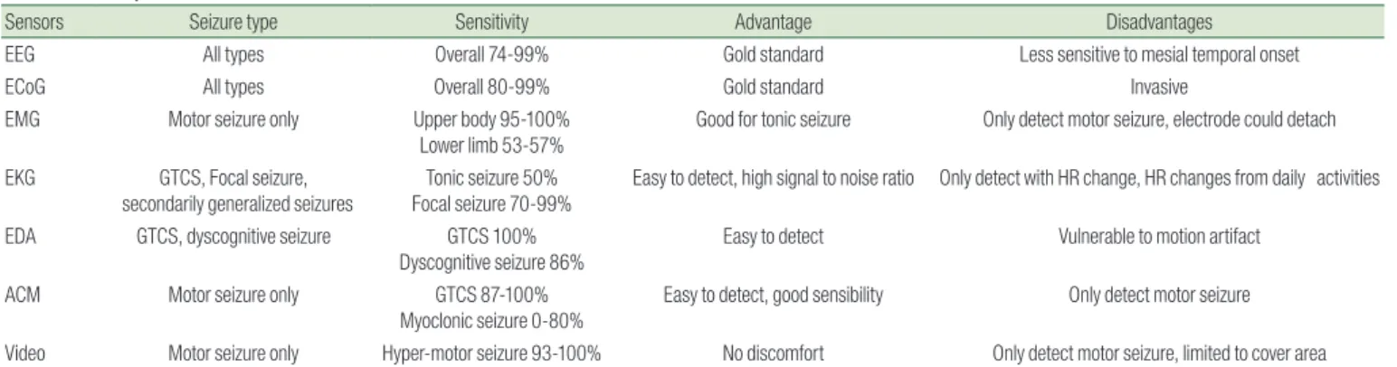
Table 2. Comparison of the Sensors and Seizure Detection
Sensors Seizure type Sensitivity Advantage Disadvantages
EEG All types Overall 74-99% Gold standard Less sensitive to mesial temporal onset
ECoG All types Overall 80-99% Gold standard Invasive
EMG Motor seizure only Upper body 95-100%
Lower limb 53-57%
Good for tonic seizure Only detect motor seizure, electrode could detach EKG GTCS, Focal seizure,
secondarily generalized seizures Tonic seizure 50%
Focal seizure 70-99% Easy to detect, high signal to noise ratio Only detect with HR change, HR changes from daily activities
EDA GTCS, dyscognitive seizure GTCS 100%
Dyscognitive seizure 86% Easy to detect Vulnerable to motion artifact
ACM Motor seizure only GTCS 87-100%
Myoclonic seizure 0-80%
Easy to detect, good sensibility Only detect motor seizure
Video Motor seizure only Hyper-motor seizure 93-100% No discomfort Only detect motor seizure, limited to cover area Electroencephalography (EEG), Electrocorticography (ECoG), electromyography (EMG), Electrocardiography (EKG), electrodermal activity (EDA), accelerometer (ACM), heart rate (HR), generalized tonic clonic seizures (GTCS)
focus on larger market, such as individuals with cardiac disease, or even professional sports trainers. Third, seizure types decide the usability. The devices can detect easily GTCS, however they are almost useless for the other types of seizure, such as focal behavioral changes (Table 2).
Conclusion
The detection of seizures is a major issue in the management of epilepsy. However, detecting every seizure is not only techni
cally difficult, but also timeconsuming for caregivers. Better te
chniques for the recognition of seizures that are easy to perform on a daily basis are required to reduce the risk of SUDEP. Portable devices are good alternatives to EEG monitoring. Although these devices are far less accurate than EEG, they enable the conti
nuous collection of data over 24 hours. Nevertheless, using a single approach is not an appropriate solution for seizure moni
toring. There are many commercial products with various kinds of sensors which is not currently focusing seizure detection.
Several commercial products combine more than two different types of signals to recognize ictal patterns26). These signals in
clude HR, EDA, and data from ACMs. Combining data from such devices in the future would enable detection of seizures using wearable medical devices. These devices may be ultimately used for epilepsy treatment.
요약
뇌파는 발작을 발견하는 최선의 방법이다. 하지만 뇌파기는 대체로 고가이며 뇌파데이터를 기록하는 데 잘 훈련된 인력이 필요하다. 발작 을 감시하는 것은 난치성 뇌전증 환자에게 중요하며, 목격자가 없거나 발작이 길어지는 경우 특히 그렇다. 뇌전증에서 돌발성 사망(SUDEP) 은 난치성 뇌전증 환자에게 중요한 문제이며, 돌발성 사망의 위험은 삶의 질을 떨어뜨리기 때문이다. 뇌파 이외에 발작을 감지하기 위한 병태생리학적인 변화를 감지하는 여러 대체 기술들이 연구되어 왔다.
심박과 심박 변이도는 발작과 관련한 심혈관계 현상을 이용한다. 호흡 수와 호흡운동이 또한 이용될 수 있다. 발작기의 움직임은 영상분석 을 통해서 혹은 가속도센서, 회전계(자이로스코프) 등을 통해서 감지 할 수 있다. 또한 피부전도도가 발작기를 알려주는데 연구되고 있다.
이러한 기술들은 각각 한계가 있다. 하지만 이 기술들은 가격이 저렴 하며 환자들이 매일 착용하고 사용하는 것이 간편하다. 이러한 장점 을 통해 목격자 없는 발작을 발견하는데 유용할 수 있으며 삶의 질을 높이는 데에 도움을 줄 수 있을 것이다.
References
1) Devinsky O. Sudden, unexpected death in epilepsy. N Engl J Med 2011;365:1801-11.
2) Reeder B, David A. Health at hand: a systematic review of smart watch uses for health and wellness. J Biomed Inform 2016;63:269-76.
3) Maskeliunas R, Damasevicius R, Martisius I, Vasiljevas M. Con- sumer-grade EEG devices: are they usable for control tasks? PeerJ 2016;4:e1746.
4) Krigolson OE, Williams CC, Norton A, Hassall CD, Colino FL.
Choosing MUSE: validation of a low-cost, portable EEG System for ERP research. Front Neurosci 2017;11:109.
5) Li G, Chung W-Y. Estimation of eye closure degree using EEG sensors and its application in driver drowsiness detection. Sen- sors 2014;14:17491-515.
6) Regan SO, Faul S, Marnane W, editors. Automatic detection of EEG artefacts arising from head movements using gyroscopes.
2010 3rd International Symposium on Applied Sciences in Bio- medical and Communication Technologies (ISABEL 2010); 2010 7-10 Nov. 2010.
7) Badcock NA, Mousikou P, Mahajan Y, de Lissa P, Thie J, McAr- thur G. Validation of the Emotiv EPOC® EEG gaming system for measuring research quality auditory ERPs. PeerJ 2013;1:e38.
8) Sarkis RA, Thome-Souza S, Poh M-Z, Llewellyn N, Klehm J, Mad- sen JR, et al. Autonomic changes following generalized tonic clonic seizures: an analysis of adult and pediatric patients with epilepsy. Epilepsy Res 2015;115:113-8.
9) Picard RW, Migliorini M, Caborni C, Onorati F, Regalia G, Fried- man D, et al. Wrist sensor reveals sympathetic hyperactivity and hypoventilation before probable SUDEP. Neurology 2017;89:
633-5.
10) Barrett PM, Komatireddy R, Haaser S, Topol S, Sheard J, Encinas J, et al. Comparison of 24-hour Holter monitoring with 14-day novel adhesive patch electrocardiographic monitoring. Am J Med 2014;127:95. e11-7.
11) Jansen K, Varon C, Van Huffel S, Lagae L. Peri-ictal ECG changes in childhood epilepsy: implications for detection systems. Epile- psy Behav 2013;29:72-6.
12) Nilsen KB, Haram M, Tangedal S, Sand T, Brodtkorb E. Is elevated pre-ictal heart rate associated with secondary generalization in partial epilepsy? Seizure 2010;19:291-5.
13) Osorio I, Manly BFJ. Is seizure detection based on EKG clinically relevant? Clin Neurophysiol 2014;125:1946-51.
14) van Elmpt WJC, Nijsen TME, Griep PAM, Arends JBAM. A model of heart rate changes to detect seizures in severe epilepsy. Seizure 2006;15:366-75.
15) Osorio I. Automated seizure detection using EKG. Int J Neural Sys 2014;24:1450001.
16) Cogan D, Nourani M, Harvey J, Nagaraddi V, editors. Epileptic seizure detection using wristworn biosensors. Conf Proc IEEE
23) Patterson AL, Mudigoudar B, Fulton S, McGregor A, Poppel KV, Wheless MC, et al. SmartWatch by smartmonitor: assessment of seizure detection efficacy for various seizure types in children, a large prospective single-center study. Pediatr Neurol 2015;53:
309-11.
24) Van de Vel A, Cuppens K, Bonroy B, Milosevic M, Van Huffel S, Vanrumste B, et al. Long-term home monitoring of hypermotor seizures by patient-worn accelerometers. Epilepsy Behav 2013;
26:118-25.
25) Poh MZ, Loddenkemper T, Reinsberger C, Swenson NC, Goyal S, Madsen JR, et al. Autonomic changes with seizures correlate with postictal EEG suppression. Neurology 2012;78:1868-76.
26) Onorati F, Regalia G, Caborni C, Migliorini M, Bender D, Poh M- Z, et al. Multicenter clinical assessment of improved wearable multimodal convulsive seizure detectors. Epilepsia 2017;58:1870- 9.
27) Jones LA, Thomas RH. Sudden death in epilepsy: insights from the last 25 years. Seizure 2017;44:232-6.
28) Devinsky O, Hesdorffer DC, Thurman DJ, Lhatoo S, Richerson G.
Sudden unexpected death in epilepsy: epidemiology, mecha- nisms, and prevention. Lancet Neurol 2016;15:1075-88.
Eng Med Biol Soc 2015; 2015:5086-9.
17) Singh K, Katz ES, Zarowski M, Loddenkemper T, Llewellyn N, Manganaro S, et al. Cardiopulmonary complications during pe- diatric seizures: a prelude to understanding SUDEP. Epilepsia 2013;54:1083-91.
18) Rémi J, Silva Cunha JP, Vollmar C, Bilgin Topçuoğlu Ö, Meier A, Ulowetz S, et al. Quantitative movement analysis differentiates focal seizures characterized by automatisms. Epilepsy & Behavior 2011;20:642-7.
19) Karayiannis NB, Xiong Y, Tao G, Frost JD, Wise MS, Hrachovy RA, et al. Automated Detection of videotaped neonatal seizures of epileptic origin. Epilepsia 2006;47:966-80.
20) Nijsen TME, Arends JBAM, Griep PAM, Cluitmans PJM. The po- tential value of three-dimensional accelerometry for detection of motor seizures in severe epilepsy. Epilepsy Behav 2005;7:74-84.
21) Beniczky S, Polster T, Kjaer TW, Hjalgrim H. Detection of genera- lized tonic–clonic seizures by a wireless wrist accelerometer: A prospective, multicenter study. Epilepsia 2013;54:e58-e61.
22) Lockman J, Fisher RS, Olson DM. Detection of seizure-like move- ments using a wrist accelerometer. Epilepsy & Behavior 2011;20:
638-41.