INTRODUCTION
Congenital leukemia, defined as a leukemia that occurs within 4 to 6 weeks of birth, is rare. Excluding Down syn- drome-related transient neonatal myeloproliferation, its inci- dence is less than 1% of all childhood leukemia (1, 2). The diagnostic criteria include the presence of immature leukemic blasts in the blood and in extrahematopoietic tissues and the absence of congenital infections (syphilis, toxoplasmosis, her- pes simplex, cytomegalovirus, rubella, and bacterial infec- tions), hypoxia, and hemolytic disease, which may produce a similar clinical and hematological picture. In addition, there may be absence of chromosomal disorders that may be asso- ciated with unstable hematopoiesis, such as trisomy 21 (3).
Although its biology and natural history are still under investigation, it is clear that the leukemic process originat- ed in utero even in infants diagnosed within the first few months of life (2). In infants with congenital leukemia under the age of 1 month, the 6-month survival rate is only one third despite aggressive chemotherapy (4). It has a higher mortal- ity rate than any other congenital cancer, but recently some reports showed spontaneous remissions (3, 5). These findings implicate therapeutic dilemmas on deciding which patients to treat or to wait.
Here we present a case of congenital myeloid leukemia (M5) initially characterized by the presence of leukemia cutis and hyperleukocytosis with chromosomal abnormality (double
translocation of t[8;16][p11;p13.2] and t[17;19][?;q13.3]). With the fear of toxic effect of chemotherapeutic agents, par- ents did not agree to chemotherapy at first. Three weeks later we started chemotherapy but the prognosis was poor. We report our experience and review previous cases in view of treatment dilemmas.
CASE REPORT
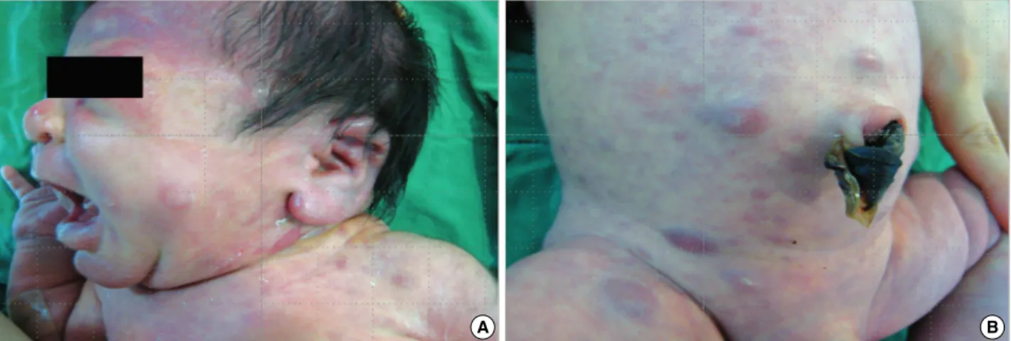
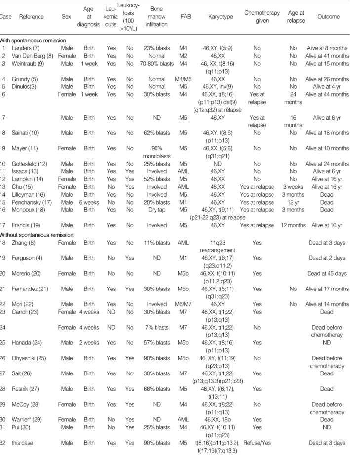
A term male infant weighing 3,360 gram was born to a 32-yr-old woman after uncomplicated pregnancy. There was no history of exposure to radiation, any known teratogens, smoking, drinking, or drug use from his mother. A cesarean section was done for previous section. Gross examination of the infant noted at birth to have an unusual purpuric rash on his whole body looking like ‘‘blueberry-muffin’’ rash (Fig. 1).
Physical examination showed no hepatosplenomegaly and there were no dysmorphic features. Skin examination revealed widespread, scattered, yellowish-colored firm nodules with multiple purpuric macules and papules predominantly on the face and proximal extremities with a few present on the trunk. A punch-biopsy of a nodule showed proliferation of atypical hematologic cells in the dermis. Blood count at birth revealed white blood cell count of 173×109/L with 58.8%
neutrophils, 5.3% lymphocytes, 26.1% monocytes, and 0.5%
eosinophils; hemoglobin of 13.9 g/dL; platelets of 133×109/L.
945
Tae-Jung Sung1, Dae-Hyoung Lee1, Soon-Ki Kim2, and Yong-Hoon Jun2
Department of Pediatrics1, College of Medicine, Hallym University Medical Center, Seoul; Department of Pediatrics2, College of Medicine, Inha University Medical Center, Inchoen, Korea
Address for Correspondence Tae-Jung Sung, M.D.
Department of Pediatrics, College of Medicine, Hallym University Medical Center, 2 Dwitsan-gil, Yungdungpo-gu, Seoul 150-950, Korea Tel : +82.2-829-5142, Fax : +82.2-829-4469 E-mail : [email protected]
Congenital Acute Myeloid Leukemia with t(8;16) and t(17;19) Double Translocation: Case Presentation and Literature Review
Congenital leukemia is uncommon and excluding transient myeloproliferation asso- ciated with Down syndrome, makes up approximately 1% of childhood leukemias.
A newborn boy was born with multiple subcutaneous nodules and large purpuric papules. Skin biopsy revealed proliferation of atypical hematologic cells in the der- mis. Bone marrow morphology was consistent with acute myeloid leukemia (M5) and cytogenetic studies revealed t(8;16) and t(17;19) double translocation. Although prognosis of congenital leukemia is known to be dismal, recent reports showed spon- taneous remissions. With the fear of chemotherapy-related toxicity, to treat or not to treat may be a dilemma both to parents and pediatricians. We report our experi- ence and review the literature.
Key Words : Leukemia; Translocation, Genetic; Leukemic Infiltration; Drug Therapy
Received : 24 November 2008 Accepted : 17 February 2009
ⓒ 2010 The Korean Academy of Medical Sciences.
This is an Open Access article distributed under the terms of the Creative Commons Attribution Non-Commercial License (http://creativecommons.org/licenses/by-nc/3.0) which permits unrestricted non-commercial use, distribution, and reproduction in any medium, provided the original work is properly cited.
TORCH titers (against toxoplasmosis, cytomegalovirus, her- pes simplex virus, rubella, and syphilis) were all negative and blood cultures were also negative. Peripheral blood morphol- ogy showed marked leukocytosis with 32% of immature cells and monocytosis. Immunophenotyping showed that blasts were positive for CD14, CD33 and HLA-DR(+). Flow cytom- etry on the bone marrow showed the blasts to be acute mye- loid leukemia (M5) in the French-American-British classifi- cation based on the morphologic features and special stains.
Bone marrow aspiration and biopsy also showed 90% of leu- kemic blasts. The results of special stain were peroxidase (+), PAS (-), ANAE with NaF inhibition (+). Cytogenetic anal- ysis revealed 46, XY karyotype with multiple chromosomal defect, t(8;16)(p11;p13.2), t(17;19)(?;q13.3). Serial white blood cell count showed a progressive rise up to 256×109/L with thrombocytopenia of 88×109/L. Leukemia cutis also progressed and it became more pronounced on the face and somewhat hardened. Parents only wanted conservative treat- ment at first. Three weeks after birth, parents agreed to che- motherapy. Initial chemotherapy was started with daunoru- bicin, etoposide, and cytarabine, but, he died at 27 days of age.
DISCUSSION
Leukemia is the most common malignancy presented dur- ing the childhood, however, congenital leukemia is a very rare disease, representing less than 1% of all childhood leukemia.
It should be differentiated from transient leukemoid reaction and other small round cell tumors (3).
Several risk factors are known to be associated with the development of congenital leukemia. Maternal alcohol con- sumption, tobacco smoking, maternal exposures to radiation, high birth weight, high levels of insulin-like growth factors, maternal consumption of topoisomerase II inhibitors, such as fruits and vegetables, coffee, tea, cocoa, wine, and soybeans are those factors (2, 6). However, as in our case, some had been
reported to have no known risk factors. Frequently, reported cases of congenital leukemia with constitutional chromoso- mal abnormalities have led to suspicion whether congenital leukemia is a result of chromosomal fragility (2).
Congenital leukemia may reflect the early onset of intrauter- ine leukemia, that is, leukemic process originated in utero even in infants diagnosed within the first few months of life.
Since embryonic hematopoiesis begins in undifferentiated mesenchyme starting the third week after fertilization, leu- kemia cutis may be a primary event and the first manifesta- tion of congenital leukemia (6).
Prognosis of congenital leukemia is dismal and has a pro- gressive downhill course, even with chemotherapy. However, there are some reported cases of spontaneous remission (Table 1) (3, 7-19). The reason that spontaneous remissions may occur in newborn infants is unclear (1). The period of spon- taneous remission varied widely from months to years, and several of the children with relapses remained in prolonged remission after chemotherapy (3, 15). There appear to be no common clinical features between those who progressed and those that had spontaneous remissions. Neither bone marrow involvement at diagnosis nor hyperleukocytosis appears to increase the risk of relapse (3, 4, 15-17, 19). Chromosome abnormalities affecting chromosome 11 band q23 are involv- ed in the majority of infant leukemia cases. The major trans- locations involving the 11q23 locus are t(4;11) and t(11;19) (13, 16). Translocation involving 11q23 band or reciprocal translocations involving chromosomes 8 and 16 are known to be mapped with oncogene(s) and are known to be associ- ated with a poor prognosis (6, 10, 30). Hence, even cytoge- netic abnormality of 11q23 rearrangement of skin without involvement of bone morrow was also treated aggressively (6).
Moreover, AML with the t(8;16) is associated with a young age at diagnosis, myelomonocytic (FAB M4) or monocytic (FAB M5) morphology, erythrophagocytosis, disseminated intravascular coagulation and a poor outcome (9).
The gene at 11q23, known as MLL (ALL-1, HRX, HTRX,
Fig. 1. Gross view of the skin. (A, B) There are widespread, scattered, yellowish-colored firm nodules with multiple purpuric macules and papules predominantly on the face and the trunk.
A B
*One case of Warrier had incomplete data and omitted it.
ND, no data.
Case Reference
Bone marrow infiltration Sex
Age at diagnosis
Leu- kemia
cutis
FAB Karyotype Chemotherapy given
Age at
relapse Outcome Leukocy-
tosis (100
>109/L) With spontaneous remission
1 Landers (7) Male Birth Yes No 23% blasts M4 46,XY, t(5;9) No No Alive at 8 months
2 Van Den Berg (8) Female Birth Yes No Normal M2 46,XX No No Alive at 41 months
3 Weintraub (9) Male 1 week Yes No 70-80% blasts M4 46, XX, t(8;16) No No Alive at 15 months (q11;p13)
4 Grundy (5) Male Birth Yes No Normal M4/M5 46,XX No No Alive at 26 months
5 Dinulos(3) Male Birth Yes No Normal M5 46,XY, inv(9) No No Alive at 4 yr
6 Female 1 week Yes No 30% blasts M4 46,XX, t(8;16) Yes at 24 Alive at 44 months
(p11;p13) del(9) relapse months (q12;q32) at relapse
7 Male Birth Yes No ND M5 46,XY Yes at 16 Alive at 6 yr
relapse months
8 Sainati (10) Male Birth Yes No 62% blasts M5 46,XY, t(8;6) No No Alive at 18 months
(p11;p13)
9 Mayer (11) Female Birth Yes No 90% M5 46,XX, t(5;6) No No Alive at 10 months
monoblasts (q31;q21)
10 Gottesfeld (12) Male Birth Yes No 25% blasts M5 ND No No Alive at 24 months
11 Issacs (13) Male Birth Yes Yes Involved AML 46,XY No No Alive at 6 yr
12 Lampkin (14) Female Birth Yes Yes 52% blasts M5 46,XX No No Alive at 16 yr
13 Chu (15) Female Birth No Yes Involved AML 46,XX Yes at relapse 3 weeks Alive at 16 yr 14 Lilleyman (16) Male Birth Yes No Involved M5 46,XY Yes at relapse 3 months Dead 15 Penchansky (17) Male 6 weeks No No 20% blasts M1 46,XY Yes at relapse 12 yr Dead 16 Monpoux (18) Male Birth Yes No Dry tap M5 46,XY, t(9;11) Yes at relapse 3 months Dead
(p21-22;q23) at relapse
17 Francis (19) Male Birth Yes No Involved M5 46,XY Yes at relapse 12 months Alive at 10 yr Without spontaneous remission
18 Zhang (6) Female Birth Yes No 11% blasts AML 11q23 Yes Dead at 3 days
rearrangement
19 Ferguson (4) Male Birth No Yes ND M1 46,XY, t(6;17) Yes Dead at 2 days
(q23;q11.2)
20 Morerio (20) Female Birth No No ND M5b 46,XX, t(10;11) Yes Dead at 45 days
(p11.2;q23)
21 Fernandez (21) Male Birth Yes Yes 30% blasts M5b 46,XY, t(5;11) Yes No Alive at 17 months (q31;q23)
22 Mori (22) Male Birth Yes No Involved M6/M7 46,XY Yes No Alive at 14 months
23 Carroll (23) Female 4 weeks ND No 30% blasts M7 46,XX, t(1;22) Yes Dead
(p13;q13)
24 Female 4 weeks ND No 7% blasts M7 46,XX, t(1;22) No Dead before
(p13;q13) chemotheray
25 Hanada (24) Male 2 weeks Yes No 57% blasts M5b 46,XY, t(8;16) Yes ND
(p11;p13)
26 Ohyashiki (25) Male Birth Yes Yes 90% blasts M5b 46, XY, t(11;19) No Dead before
(q23;p13) chemotherapy
27 Sait (26) Male Birth Yes No 30% blasts M7 46,XY, t(1;22) Yes Dead
(p13;q13.3)(p21;p23)
28 Resnik (27) Male Birth Yes Yes 68% blasts M5 46,XY, t(6;17), Yes Dead
t(13;11)
29 McCoy (28) Female Birth Yes Yes ND M4 46,XX, t(8;22) No Dead before
(p11;q13) chemotherapy
30 Warrier* (29) Female Birth No Yes ND AML 46,XX, 18p Yes Dead
31 Pui (30) Male Birth No Yes 25% blasts M4 46,XY, t(10;11) Yes ND
(p11;q23)
32 this case Male Birth Yes Yes 90% blasts M5 t(8;16)(p11;p13.2), Refuse/Yes Dead at 3 days t(17;19)(?;q13.3)
Table 1. Reported cases of congenital nonlymphocytic leukemia from 1980 to 2007
or Hu-ets-1), is required for the production of normal num- bers of hematopoietic precursors and for their proper differ- entiation (2, 13). Chromosomal translocations can theoreti- cally occur between any gene loci. 11q23-MLL gene rearrange- ments after DNA damage occur most frequently with AML.
Translocations involving MLL may fuse with other genes and hence MLL usually retains at least two DNA binding domains (2, 13). Since there are a few reports of double translocations as our case, exact meaning of double gene translocation is yet to be discussed.
Because of the rarity of this disease, there is no standard pro- tocol of chemotherapy. Treating congenital leukemia means exposure of the toxic chemotherapeutic agents to the neonate.
Although there had been reported cases of prolonged periods of remission, rapidly downhill course were also noted in some cases and some of them died after relapse during the course of chemotherapy (16-18). While some cases with t(8; 16) remit- ted spontaneously without treatment (3, 9, 10), paradoxical- ly, those who had chromosomal abnormalities died after ini- tiation of chemotherapy as our case (4, 6, 20, 23, 26-29). It seems like that karyotypic findings in blasts in the neonatal period may not be predictive of whether or not a spontaneous remission will occur (2).
For our case, hyperleukocytosis, progressive leukemia cutis, bone marrow involvement, double translocation involving (8;16) made us to decide to treat at first. However, with the fear of unknown toxic effect of chemotherapeutic agents, par- ents refused to treat. Three weeks later after parents’ agree- ment, we treated with daunorubicin, etoposide, and cytara- bine but he died of pulmonary hemorrhage three days after induction chemotherapy. He may died of leukemia itself but, we are not sure whether he died due to chemotherapy or he could have survived with only conservative management as other spontaneous remitted cases. That is because it is the first case of congenital AML with double translocation of (8;16) and (17;19) to be reported in the English literature to the best of our knowledge.
Although congenital leukemia remains a rare disorder, an international collection of data or register system is indispens- able for establishing an optimal treatment protocols.
REFERENCES
1. Lampkin BC. The newborn infant with leukemia. J Pediatr 1997;
131: 176-7.
2. Sande JE, Arceci RJ, Lampkin BC. Congenital and neonatal leuke- mia. Semin Perinatol 1999; 23: 274-85.
3. Dinulos JG, Hawkins DS, Clark BS, Francis JS. Spontaneous remis- sion of congenital leukemia. J Pediatr 1997; 131: 300-3.
4. Ferguson EC, Talley P, Vora A. Translocation (6;17)(q23;q11.2): a novel cytogenetic abnormality in congenital acute myeloid leukemia?
Cancer Genet Cytogenet 2005; 163: 71-3.
5. Grundy RG, Martinez A, Kempski H, Malone M, Atherton D. Spon-
taneous remission of congenital leukemia: a case for conservative treatment. J Pediatr Hematol Oncol 2000; 22: 252-5.
6. Zhang IH, Zane LT, Braun BS, Maize J Jr, Zoger S, Loh ML. Con- genital leukemia cutis with subsequent development of leukemia. J Am Acad Dermatol 2006; 54 (2 Suppl): S22-7.
7. Landers MC, Malempati S, Tilford D, Gatter K, White C, Schroed- er TL. Spontaneous regression of aleukemia congenital leukemia cutis. Pediatr Dermatol 2006; 22: 26-30.
8. van den Berg H, Hopman AH, Kraakman KC, de Jong D. Sponta- neous remission in congenital leukemia is not related to trisomy 21:
case presentation and literature review. Pediatr Hematol Oncol 2004; 21: 135-44.
9. Weintraub M, Kaplinsky C. Amariglio N, Rosner E, Brok-Simoni F, Rechavi G. Spontaneous regression of congenital leukaemia with an 8;16 translocation. Br J Haematol 2000; 111: 641-3.
10. Sainati L, Bolcato S, Cocito MG, Zanesco L, Basso C, Montaldi A, Piovesan AL. Transient acute monoblastic leukemia with recipro- cal (8;16)(p11;p13) translocation. Pediatr Hematol Oncol 1996;
13: 151-7.
11. Mayer JL, Seashore MR, Hajjar FM. Translocation (5;6) associated with spontaneously remitting congenital leukemia. Cancer Genet Cytogenet 1995; 81: 38-41.
12. Gottesfeld E, Silverman RA, Coccia PF, Jacobs G, Zaim MT. Tran- sient blueberry muffin appearance of newborn with congenital mono- blastic leukemia. J Am Acad Dermatol 1989; 21: 347-51.
13. Isaacs H Jr. Fetal and neonatal leukemia. J Pediatr Hematol Oncol 2003; 25: 348-61.
14. Lampkin BC, Peipon JJ, Price JK, Bove KE, Srivastava AK, Jones MM. Spontaneous remission of presumed congenital acute nonlym- phocytic leukemia (ANLL) in a karyotypically normal neonate. Am J Pediatr Hematol Oncol 1985; 7: 346-51.
15. Chu JY, O’Connor DM, Gale GB, Silberstein MJ. Congenital leu- kemia: two transient regressions without treatment in one patient.
Pediatrics 1983; 71: 277-9.
16. Lilleyman JS. Congenital monocytic leukaemia. Clin Lab Haematol 1980; 2: 243-5.
17. Penchansky L, Wollman MR, Gartner JC, Wenger SL. Spontaneous remission of infantile acute nonlymphocytic leukemia for 11 years in a child with a normal karyotype. Cancer 1993; 71: 1928-30.
18. Morerio C, Rosanda C, Rapella A, Micalizzi C, Panarello C. Is t(10;
11)(p11.2;q23) involving MLL and ABI-1 genes associated with con- genital acute monocytic leukemia? Cancer Genet Cytogenet 2002;
139: 57-9.
19. Fernandez MC, Weiss B, Atwater S, Shannon K, Matthay KK. Con- genital leukemia: successful treatment of a newborn with t(5;11)(q31;
q32). J Pediatr Hematol Oncol 1999; 21: 152-7.
20. Mori T, Kaneko H, Kumagai MA, Miyauchi J, Kaneko Y, Fujimoto J, Tsunematsu Y. Congenital leukemia with a mixed phenotype of megakaryoblasts and erythroblsts: a case report and characteriza- tion of the blasts. Br J Haematol 1997; 96: 740-2.
21. Monpoux F, Lacour JP, Hatchuel Y, Hofman P, Raynaud S, Sudaka I, Ortonne JP, Mariani R. Congenital leukemia cutis preceding mono- blastic leukemia by 3 months. Pediatr Dermatol 1996; 13: 472-6.
22. Carroll A, Civin C, Schneider N, Dahl G, Pappo A, Bowman P,
Emami A, Gross S, Alvarado C, Phillips C, Krischer J, Crist W, Head D, Gresik M, Ravindranath Y, Weinstein H. The t(1;22)(p13;q13) is nonrandom and restricted to infants with acute megakaryoblastic leukemia: A pediatric oncology group study. Blood 1991; 78: 748-52.
23. Hanada T, Ono I, Minosaki Y, Moriyama N, Nakahara S, Ohtsu A.
Translocation t(8;16)(p11;p13) in neonatal acute monocytic leu- kaemia. Eur J Peditr 1991; 150: 323-4.
24. Francis JS, Sybert VP, Benjamin DR. Congenital monocytic leuke- mia: report of a case with cutaneous involvement and review of the literature. Pediatr Dermatol 1989; 6: 306-11.
25. Ohyashiki K, Ohyashiki JH, Nomura K, Ryan DH, Kinniburgh AJ, Sandberg AA. Hu-ets-1 gene in congenital leukemia with t(11;19)(q23;
p13). Cancer Genet Cytogenet 1988; 30: 233-8.
26. Sait SN, Brecher ML, Green DM, Sandberg AA. Translocation t(1;
22) in congenital acute megakaryocytic leukemia. Cancer Genet Cytogenet 1988; 34; 277-80.
27. Resnik KS, Brod BB. Leukemia cutis in congenital leukemia. Arch Dermatol 1993; 129: 1301-6.
28. McCoy JP Jr, Travis SF, Blumstein L, Birdsall PB, Schroeder K, Overton WR, McMorrow LE, Campbell T, Wineburg M, Greenbaum BH. Congenital leukemia: report of two cases. Cytometry 1995; 22:
89-92.
29. Warrier RP, Ravindranath Y, Inoue S, Kaplan J, Emami A. Congen- ital leukemia: two transient regressions without treatment in one patient. Pediatrics 1983; 72: 916-7.
30. Pui CH, Raimondi SC, Murphy SB, Ribeiro RC, Kalwinsky DK, Dahl GV, Crist WM, Williams DL. An analysis of leukemic cell chro- mosomal features in infants. Blood 1987; 69: 1289-93.