1980년대 중반이래 여러가지 혈액종양 즉 급성 및 만성 백 혈병, 임파종, 다발성 골수암과, 재생불량성빈혈 및 유방암, 폐 암, 고환암 등의 완치적 요법으로 골수이식(bone marrow transplantation, BMT)이 시행되고 있다(1-3). 환자들의 생존 율 또한 증가되어 5년 후에도 60% 이상의 환자가 혈액질환이 재발되지 않은 상태로 살아간다(4, 5). 이들 환자에게서 계속 되는 이환율과 사망률을 높이는 두가지 원인 질환은 후기 감 염과 면역편대 숙주질환(graft versus host disease)이며 그 외에 이식받은 환자의 약 10% 정도에서 골괴사가 만성 합병 증으로 나타난다(5-7). 이중 약 60-70% 에서 고관절이 침 범되며 대개는 양측성으로 상당히 골괴사가 진행된 상태로 발 견된다(4, 5, 8). 골괴사를 좀 더 조기에 발견하는 경우 보존 적 치료 또는 핵심감압천공술등의 치료로 고관절을 상당기간 보존하는 수술 방법을 선택할 수 있으므로 골괴사를 조기에 진 단할 필요성이 있다(4, 8-12).
1987년 Atkinson 등(9)이 동종골수이식후 발생된 골괴사에 관하여 처음 기술한 이래 골괴사에 관한 몇가지 보고가 있기 는 하나 비혈연간의 골수 이식이나 말초혈액 자가조혈모세포 이식에 발생된 골괴사는 거의 보고되어 있지 않다(4, 8, 11, 13-15).
저자들은 골수이식 후 추적 관찰 중 대퇴골두 골괴사가 발 생된 환자의 임상 소견과 MR 영상 소견을 분석하였다.
대상과 방법
1996년부터 2000년 까지 5년 동안 조혈모세포 이식을 받 았던 1112명 중 고관절 MR 영상에서 대퇴골두에 골괴사가 발 견된 23명의 환자를 대상으로 임상소견과 MR 소견을 분석하 였다.
남자환자가 13명, 여자환자는 10명이었고 이들의 평균연령 은 31세(범위 20-47세)이었다 23명의 환자(재생불량성빈혈 5명, 백혈병 15명, 골수이형성증 2명, 임파종 1명)중 17명은 동종이식(allogeneic transplantation)이 시행되었고 4명은 비 혈연간이식(unrelated transplantation), 2명은 말초혈액 자가
골수이식후 발생된 대퇴골두의 골괴사
1박정미・전정수・박창숙・김용식2・권순용2・김유진3・김춘추3
목적: 골수이식을 받은 환자에서 생긴 대퇴골두 골괴사 소견을 분석하였다.
대상과 방법: 1996년부터 2000년까지 5년동안 조혈모세포이식을 받았던 1112명 중 대퇴골두
에 골괴사가 발견된 23명의 임상소견과 MR 영상소견을 분석하였다.
결과: 골괴사가 발생되었을 때의 환자의 나이는 20세에서부터 47세로 평균 31세이었고, 골수 이식후 골괴사가 발견되기까지의 기간은 평균 17개월이었다. 이식 후 전례에서 다양한 정도의 조직편대 숙주질환이 발생되었으며 급성 및 전신적인 조직편대 숙주질환을 보인 17명은 스테 로이드 펄스치료를 하였으며 국한된 부위에 조직편대 숙주질환을 보인 6명은 소량의 스테로이 드 투여를 지속하였다. 1명을 제외하고는 양측 대퇴골두에 골괴사가 있어서 총 45예의 대퇴골 두가 침범되었다. ARCO 분류상 II기의 골괴사 소견을 보인 예가 28예로 가장 많았고, I기는 15예, III기는 2예였으며 IV기는 1명도 없었다. 11명의 환자에서는 대퇴골두 외에 장골(ilium) 과 하부 요추 및 대퇴골간에서도 골괴사의 소견이 발견되었다. 우선적으로는 보존적 치료로서 체중부하를 줄이고 진통제를 투여하였으나 추적 관찰중 10예의 고관절은 핵심 감압천공술(core decompression), 5예는 혈관성 비골 이식술(vascularized fibular graft), 6예의 고관절은 인공 관절치환성형술을 시행하였다.
결론: 골수이식을 받은 환자에서 조직편대 숙주질환으로 인해 스테로이드 치료를 받은 경우 조 기에 골괴사를 진단하고 진행을 막기위해서 MR 검사가 필요하다.
1가톨릭대학교 의과대학 진단방사선과
2가톨릭대학교 의과대학 정형외과
3가톨릭대학교 의과대학 내과
이 논문은 2002년 12월 9일 접수하여 2003년 6월 11일에 채택되었음.
조혈모세포이식(autologous peripheral blood stem cell tran- plantation)이 시행되었다.
골수 이식후 골괴사가 발견되기까지의 기간, 임상적으로 고 관절 통증의 유무, 이식전 처치에 사용된 방법과 투여된 약물 의 종류, 조직편대숙주질환의 예방과 치료를 위한 약제를 검토 하였고, 골괴사후의 경과 및 수술적 치료유무를 알아보았다
사용한 자기공명영상 기기는 1.5T(Magnetom Vision Plus, Siemens, Erlangen, Germany) 초전도기종으로서 스핀에코기 법으로 고관절의 T1강조(TR/TE=750/12 msec), 고속 T2강 조(TR/TE=3000-4000/100-120 msec) 및 gadopentetate dimeglumine 0.2 ml/kg을 정맥주사한 뒤 지방억제 조영증강 T1강조(TR/TE=900/12 msec) 관상,축상 및 시상영상을 얻 었다. Matrix number는 228×256 또는 256×512, FOV는 330 mm, 절편두께는 4.0 mm 또는 5.0 mm로 하였고 절편사 이의 간격은 0.4 mm 또는 0.5 mm 이었다.
고관절 단순 사진과 MR상 골괴사를 ARCO(Association Research Circulation Osseous)의 분류 기준(16)에 따라 병 기를 결정하였고 골괴사부위가 대퇴골두에서 차지하는 범위에 따라 15% 미만을 A, 15-30% 범위의 대퇴골두를 침범하였을 때 B, 30% 이상 침범하였을 때를 C로 분류하였다.
결 과
골괴사가 발생되었을 때의 환자의 나이는 20세에서부터 47 세로 평균 31세이었다. 골수이식후 2개월에서부터 31개월사이 에 대퇴골두 괴사가 발견되어 이식후 평균 17개월에 골괴사가 발견되었다. 4명은 고관절의 통증이 없는 상태에서 골수상태 를 평가하기 위해서 MR 검사를 하다가 우연히 골괴사가 발견 되었고 다른 19명은 고관절 통증을 호소하였다. 10명은 양측 고관절 통증을 호소하였으나 9명은 일측성 통증을 호소하였다.
이식전 처치로서 18명은 전신방사선조사와 cyclophosphamide, busulfan, 또는 melphalan등을 병합하여 투여받았고 4명은 procarbazine, antithymocyte globulin, cyclophosphamide를 병 합하여 또는 BCNU, VP-16과 Ara-C를 병합하여 투여받았 다. 모든 환자에서 조직편대 숙주질환의 예방을 위해 이식 전 후에 cyclosporin이나 FK506과 methotrexate를 투여하였으나 이식 후 전례에서 다양한 정도의 조직편대 숙주질환이 발생되 었다. 급성 및 전신적인 조직편대 숙주질환을 보인 17명은 250-500 mg/day의 prednisolone을 5-7일 간 투여하고 서서 히 감량하는 스테로이드 펄스치료를 하였다. 국한된 부위에 조 직편대숙주질환을 보인 6명은 cyclosporin 과 소량의 스테로
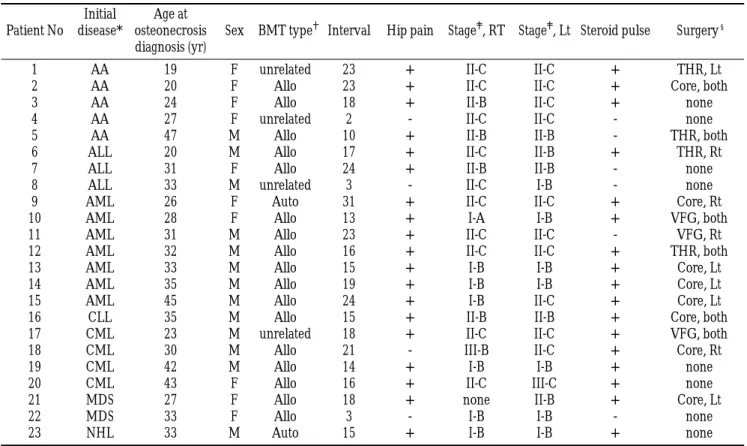
Table 1. Characteristics of 23 Patients Developing Osteonecrosis of the Femoral Head after Marrow Transplantation Initial Age at
Patient No disease* osteonecrosis Sex BMT type Interval Hip pain Stage , RT Stage , Lt Steroid pulse Surgery§ diagnosis (yr)
1 AA 19 F unrelated 23 + II-C II-C + THR, Lt
2 AA 20 F Allo 23 + II-C II-C + Core, both
3 AA 24 F Allo 18 + II-B II-C + none
4 AA 27 F unrelated 2 - II-C II-C - none
5 AA 47 M Allo 10 + II-B II-B - THR, both
6 ALL 20 M Allo 17 + II-C II-B + THR, Rt
7 ALL 31 F Allo 24 + II-B II-B - none
8 ALL 33 M unrelated 3 - II-C I-B - none
9 AML 26 F Auto 31 + II-C II-C + Core, Rt
10 AML 28 F Allo 13 + I-A I-B + VFG, both
11 AML 31 M Allo 23 + II-C II-C - VFG, Rt
12 AML 32 M Allo 16 + II-C II-C + THR, both
13 AML 33 M Allo 15 + I-B I-B + Core, Lt
14 AML 35 M Allo 19 + I-B I-B + Core, Lt
15 AML 45 M Allo 24 + I-B II-C + Core, Lt
16 CLL 35 M Allo 15 + II-B II-B + Core, both
17 CML 23 M unrelated 18 + II-C II-C + VFG, both
18 CML 30 M Allo 21 - III-B II-C + Core, Rt
19 CML 42 M Allo 14 + I-B I-B + none
20 CML 43 F Allo 16 + II-C III-C + none
이드 투여를 지속하였다.
23명의 대퇴골두 골괴사 환자의 임상 및 MR 소견은 Table 1에 요약되어 있다.
1명을 제외하고는 양측 대퇴골두에 골괴사가 있어서 총 45 개의 대퇴골두가 침범되었다. ARCO 분류상 II기의 골괴사 소 견을 보인 예가 28예로 가장 많았고, I기는 15예, III기는 2예 였으며 IV기는 1예도 없었다. 골괴사의 침범부위가 대퇴골두 의 30% 미만인 경우가 24예, 30% 이상을 침범한 경우는 21 예이었다. 11명의 환자에서는 대퇴골두 외에 장골(ilium)과 하 부 요추 및 대퇴골간에서도 골괴사의 소견이 발견되었다 (Fig.
1).
모든 환자에서 우선적으로는 보존적 치료로서 체중부하를 줄 이고 진통제를 투여하였으나 21예의 고관절은 수술적 치료를 하였다. 즉 10예의 고관절은 핵심감압천공술(core decom- pression), 5예는 혈관성 비골 이식술(vascularized fibular graft), 6예의 고관절은 인공 관절치환성형술을 시행하였다. 핵
심 감압천공술을 받은 3명과 비골 이식술을 받았던 2명은 추 적관찰 중 대퇴골두의 허탈이 진행되어 첫번째 수술 후 7개월 에서 25개월사이에 인공 고관절 치환성형술을 다시 시행하였 다.
고 찰
골괴사는 여러가지 다양한 질환에서 동반되는데 외상, 혈색 소질환(hemoglobinopathy), 내인성 또는 외인성 스테로이드 투여, 알코올중독, 췌장염, 감압증 등에서 병발할 수 있으며, 특 히 전신성 스테로이드 과투여의 중요한 합병증으로 골괴사가 일어난다(16-19). 1987년 Atkinson 등(9)이 동종 골수이식 후 발생된 골괴사에 관하여 처음 기술한 이래 골괴사에 관한 몇가지 보고가 있기는 하나(4, 8, 9, 11, 13-15) 주로 동종 골수 이식 후에 발생된 것으로서 비혈연간의 골수 이식이나 말 초혈액 자가조혈모세포이식에 발생된 골괴사는 거의 보고되어
A B
C
Fig.1. A thirty-two-year old male with acute myelocytic leukemia received allogeneic marrow transplant 16months ago and steroid pulse therapy was done immediately after the trasnplantation because of acute graft versus host disease. Coronal T1-weighted pre-contrast (A), post-contrast (B) and T2-weighted images (C) of the hip show large irregular defects in both femoral heads anterosuperiorly. In addition to osteonecrosis in both femoral heads, there are multiple medullary infarcts in both iliums (hol- low arrows), sacrum (white arrows), and femoral diaphyses (black ar- rows).
있지 않다. 동종 골수 이식후 골괴사의 빈도는 10.4%로 보고 되었고(8, 9), 특히 100명의 동종 골수 이식환자의 고관절 MR 에서 19%의 환자에서 대퇴골두에 골괴사가 발견되었다 (20).
다른 보고에 비해 본 연구에서 골괴사의 빈도가 낮은 것은 골 수이식을 받았던 모든 환자를 추적 검사하였거나 일정한 기간 동안 모든 골수이식환자를 대상으로 조사한 것이 아니라 골수 이식후 회복이 늦어 골수상태를 평가하기 위한 골수 MR검사 에서 골괴사가 우연히 발견되었거나 관절통을 호소한 환자를 중심으로 MR을 시행한 결과이기 때문에 골수 이식환자에서 실 제 골괴사의 빈도를 추정할 수 있는 것은 아니라고 생각된다.
골괴사는 골수이식후 비교적 늦게 발생되는 합병증으로서 진 단이 늦어짐에 따라 골괴사로 인한 이환율이 높아질 수 있어 골괴사를 조기에 진단해야 할 필요성이 높아지고 있다. 골 괴 사는 초기에는 임상증상이 있음에도 불구하고 단순 X-선사진 에서 발견되지 않는 경우가 많으므로 골괴사를 조기에 진단하 여 그 진행을 예방하기 위해서는 MR 검사가 필수적이며 또한 추적검사에서 그 진행을 평가하는 데에도 유용하다(21, 22).
저자들의 증례에서도 15예의 고관절에서 단순 X-선 사진상으 로는 정상이었으나 MR상 골괴사가 있는 병기 I이었으며 병기 II가 28예, 병기 III은 2예, 병기 IV는 한 명도 없어서 비교적 조기에 골괴사를 MR 영상으로 진단할 수 있었다.
골수 이식 후 골괴사를 일으키는 위험인자로서 나이가 많은 것, 이식 전처치로서 전신 방사선조사를 받은 것, 면역편대 숙 주질환의 예방적 처치로 cyclosporin이나 methotrexate를 투 여받은 경우, 급성 또는 만성 면역편대 숙주질환과 연관된 미 세혈관질환(microangiopathy)등이 지적되고 있는데 특히 면역 편대 숙주질환과 이의 예방과 치료를 위해 스테로이드를 과다 하게 사용하므로써 골괴사의 위험이 높아진다고 알려져 있다 (4, 8, 12, 20, 23, 24). 저자들의 증례에서는 모든 환자들이 면역편대 숙주질환의 치료를 위해 스테로이드를 투여받았는데 17명은 스테로이드 펄스요법을 시행하였고 다른 6명은 소량 의 스테로이드와 cyclosporin만을 투여받았다. 보고자마다 다 르지만 골수이식후 골괴사가 발생된 환자에서 스테로이드 누 적 총용량은 60-840 mg/kg이었으나(4), 스테로이드 치료용 량이 단지 14 mg/kg인 경우에도 골괴사가 발생하였다(9).
골괴사가 대퇴골두에서 많이 발생되나 체내 다른 부위에서 도 골괴사가 일어날 수 있는 점을 생각해 볼 때 골수 이식후 골괴사의 발생빈도를 정확히 평가하기 위해서는 전신적인 검 사가 필요하다. 그러나 다른 관절에 비해 체중 부하가 많은 고 관절에 골괴사가 발생된 경우 좀 더 골관절염으로의 진행이 빠 르므로 고관절이 우선적인 검사부위가 되어야 할 것이다.
본 연구에서 환자의 나이와 성별, 골수이식후 골괴사가 진단
로 사용된다(28). 외과의들은 진단이 초기에 이루어진 경우 감 압시술이 아주 효과적이라고 느끼고 있지만 그 치료효과에 관 한 보고는 다양하여서 병기 I이나 II의 조기 골괴사를 핵심감 압 천공을 시행한 경우 약 40%는 완전히 치료되거나 변화가 없으나 환자의 60%에서는 골괴사가 진행된다(29-32). 절골 술은 병기 II 또는 III의 환자에서 시행될 수 있으나(33), 골수 이식후에 발생된 골괴사에서의 치료효과에 관해서는 잘 알려 져 있지 않다. 대퇴골두의 침범부위가 넓은 병기 III이나 병기 IV 환자의 경우에는 인공 고관절 치환성형술이 우선적이 치료 법이다(34, 35). 그러나 골수이식후 발생된 대퇴골두 골괴사 의 경우 일반적인 다른 원인에 의한 골괴사보다 대퇴골두의 허 탈이 조기에 빨리 진행되므로 우선적인 외과적 치료 방법으로 인공관절 치환성형술을 권하는 보고도 있다(5).
골수이식후의 심각한 합병증인 조직편대 숙주질환의 치료를 위해 과다하게 스테로이드를 사용한 경우 대퇴골두의 골괴사 가 대부분 빨리 진행되어 그에 따른 이환율이 증가되는 까닭 에 MR등의 검사로 조기에 진단하여 적절한 치료를 해야한다.
참 고 문 헌
1. Thomas ED, Storb R, Clift RA, et al. Bone marrow transplantation (second of two parts). N Eng J Med 1975;292:895-902
2. Thomas ED. Bone marrow transplantation: past experiences and future prospects. Semin Oncol 1992;19(Suppl 7):3-6
3. Crouch MA, Ross JA. Current concepts in autologous bone mar- row transplantation. Semin Oncol Nurs 1994;10:12-19
4. Socie G, Selimi F, Sedel L, et al. Avascular necrosis of bone after al- logeneic bone marrow transplantation: clinical findings, incidence and risk factors. Br J Hematol 1994;86:624-628
5. Bizot P, Nizard R, Socie G, Gluckman E, Witvoet J, Sedel L.
Femoral head osteonecrosis after bone marrow transplantation.
Clin Orthop 1998;357:127-134
6. Atkinson K. Chronic graft-versus-host disease. Bone Marrow Transplant 1990;5:69-82
7. Deeg HJ. Delayed complications and long-term effects after bone marrow transplantation. Hematol Oncol Clin North Am 1990;4:641- 657
8. Enright H, Haake R, Weisdorf D. Avascular necrosis of bone: a common serious complication of allogeneic bone marrow trans- plantation. Am J Med 1990;89:733-738
9. Atkinson K, Cohen M, Biggs J. Avascular necrosis of the femoral head secondary to corticosteroid therapy for graft-versus-host dis- ease after marrow transplantation: effective therapy with hip arthroplasty. Bone Marrow Transplant 1987;2:421-426
10. Russell JA, Blahey WB, Stuart TA, Edwards G, Card RT. Avascular necrosis of bone in bone marrow transplant patients. Med Pediatr Oncol 1989;17:140-143
11. Mascarin M, Giavitto M, Zanazzo GA, et al. Avascular necrosis of
study of 27 consecutive THAs with a minimal two-year follow-up.
J Bone Joint Surg Br 1996;78B:878-883
15. Wiesmann A, Pereira P, Bohm P, Faul C, Kanz L, Einsele H.
Avascular necrosis of bone following allogeneic stem cell trans- plantation: MR screening and therapeutic options. Bone Marrow Transplant 1998;22:565-569
16. Resnick D, Sweet DE, Madewell JE: Osteonecrosis: Pathogenesis, diagnostic techniques, specific situations, and complications. In:
Resnick D ed. Diagnosis of Bone and Joint Disorders. Ed 4.
Philadelphia: WB Saunders, 2002:3599-3685
17. Ficat RP. Idiopathic bone necrosis of the femoral head. Early diag- nosis and treatment. J Bone Joint Surg 1985;67:3-9
18. Arlet J. Nontraumatic avascular necrosis of the femoral head. Past, present, and future. Clin Orthop 1992;277:12-21
19. Mankin HJ. Nontraumataic necrosis of bone (osteonecrosis). N Engl J Med 1992;326:1473-1479
20. Torii Y, Hasegawa Y, Kubo T, et al. Osteonecrosis of the femoral head after allogeneic bone marrow transplantation. Clin Orthop 2001;382:124-132
21. Mitchell DG, Rao VM, Dalinka MK, et al. Femoral head avascular necrosis: correlation of MR imaging, radiographic staging, radionu- clide imaging, and clinical findings. Radiology 1987;162:709-715 22. Glickstein MF, Burk DL Jr, Schiebler ML, et al. Avascular necrosis
versus other diseases of the hip: sensitivity of MR imaging.
Radiology 1988;169:213-215
23. Mori A, Hashino S, Kobayashi S, et al. Avascular necrosis in the femoral head secondary to bone marrow infarction in a patient with graft-versus-host disease after unrelated bone marrow trans- plantation. Ann Hematol 2001;80:238-242
24. Fink JC, Leisenring WM, Sullivan KM, Sherrard DJ, Weiss NS.
Avascular necrosis following bone marrow transplantation: a case- control study. Bone 1998;22:67-71
25. Zizic TM, Marcoux C, Hungerford DS, Dansereau JV, Stevens MB.
Corticostreoid therapy associated with ischemic necrosis of bone in systemic lupus erythematosus. Am J Med 1985;79;596-604 26. Feletti C, Di Felice A, Scolari MP, Bonomini V. Glucocorticoids
and avascular bone necrosis in renal transplantation. Adv Exp Med Biol 1984;171:361-368
27. Isono SS, Woolson ST, Schurman DJ. Total joint arthroplasty for steroid-induced osteonecrosis in cardiac transplant patients. Clin Orthop 1987;217:201-208
28. Saito S, Ohzono K, Ono K. Joint-preserving operations for idiopath- ic avascular necrosis of the femoral head. Results of core decom- pression, grafting and osteotomy. J Bone Joint Surg Br 1988;70:78- 84
29. Markel DC, Miskovsky C, Sculco TP, Pellicci PM, Salvati EA. Core decompression for osteonecrosis of the femoral head. Clin Orthop 1996;323:226-233
30. Hopson CN, Siverhus SW. Ischemic necrosis of the femoral head.
Treatment by core decompression. J Bone Joint Surg Am 1988;70:
1048-1051
31. Camp JF, Colwell CW Jr. Core decompression of the femoral head for osteonecrosis. J Bone Joint Surg Am 1986;68:1313-1319 32. Mont MA, Carbone JJ, Fairbank AC. Core decompression versus
nonoperative management for osteonecrosis of the hip. Clin Orthop 1996;24:169-178
33. Inao S, Ando M, Gotoh E, Matsuno T. Minimum 10-year results of Sugioka’s osteotomy for femoral head osteonecrosis. Clin Orthop 1999;368:141-148
34. Hartley WT, McAuley JP, Culpepper WJ, Engh CA Jr, Engh CA Sr.
Osteonecrosis of the femoral head treated with cementless total hip arthroplasty. J Bone Joint Surg Am 2000;82:1408-1413
35. Callaghan JJ, Albright JC, Goetz DD, Olejniczak JP, Johnston RC.
Charnley total hip arthroplasty with cement. Minimum twenty- five-year follow-up. J Bone Joint Surg Am 2000;82:487-497
J Korean Radiol Soc 2003;49:51-56
Address reprint requests to : Jeongmi Park, M.D., Department of Radiology, St. Mary’s Hospital, College of Medicine, The Catholic University of Korea, 62 Youido-dong, Yongdungpo-gu, Seoul 150-713, Korea.
Tel. 82-2-3779-2037 Fax. 82-2-783-5288 E-mail: jmpark@catholic.ac.kr
Osteonecrosis of the Femoral Head after Bone Marrow Transplantation
1Jeongmi Park, M.D., Jeongsu Jun, M.D., Changsuk Park, M.D., Yong-Sik Kim, M.D.2, Soon-Yong Kwon, M.D.2, Yoojin Kim, M.D.3, Chun-Choo Kim, M.D.3
1Department of Radiology, St. Mary’s Hospital, College of Medicine, The Catholic University of Korea
2Department of Orthopedic Surgery, St. Mary’s Hospital, College of Medicine, The Catholic University of Korea
3Department of Internal Medicine, St. Mary’s Hospital, College of Medicine, The Catholic University of Korea
Purpose:To retrospectively review finding of osteonecrosis of the femoral head after bone marrow transplan- tation.
Materials and Methods:We reviewed the clinical and MR findings of osteonecrosis of the femoral head in 23 of 1112 patients who underwent marrow transplantation during a five-year follow-up period lasting from 1996 to 2000.
Results:Mean age at the time of diagnosis was 31 (range, 20-47) years, and the mean time from transplant to diagnosis was 17 months. All patients developed variable graft-versus-host disease and seventeen were treated with high-dose prednisolone and/or cysclosporin for severe acute or extensive chronic graft versus host dis- ease. Osteonecrosis was diagnosed by magnetic resonance (MR) imaging, which allowed early detection of dis- ease assessment of its stage. At the time of diagnosis, 15 hips were at stage I, 28 at stage II, two at stage III, and none at stage IV, according to the international ARCO classification system. Osteonecrosis of femoral diaphy- ses, the lower lumbar spine, or pelvic bones in the MR field was also found to have occurred in 11 patients.
Initial treatment was conservative: 21 hips underwent surgery [core decompression (n=10), vascularized fibu- lar bone graft (n=5), and joint replacement (n=6)].
Conclusion: In patients receiving high-dose steroids for the treatment of graft-versus-host disease, MR screen- ing might help detect osteonecrosis at an early stage.
Index words :Bone marrow, transplantation Bones, necrosis
Hip, MR
Steroids, complications