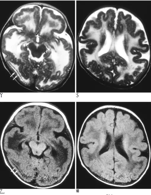
MR Imaging of Fukuyama Congenital Muscular Dystrophy: A Case Report
전체 글
수치

관련 문서
유체는 점성유체로 평상시에는 평범한 점성유체 이지만, 여 기에 전기, 자기장을 걸어주게 되면 유체 내부의 입자들이 규칙적으로 배열 하기 때문에 고체처럼
The index is calculated with the latest 5-year auction data of 400 selected Classic, Modern, and Contemporary Chinese painting artists from major auction houses..
소프트웨어 업그레이드는 http://www.comfile.co.kr/download.html 방문하셔서 PIC TOOL 관련 소프트웨어 및 사용설명서 다운로드에서 다운 받을 수
Preoperative evaluation of pelvic lateral lymph node of patients with lower rectal cancer: comparison study of MR imaging and CT in 53 patients?.
We investigated upper gastrointestinal tract involvement and characteristic endoscopic findings in scrub typhus, and we also determined the correlation between
The locations of aneurysms were middle cerebral artery in 15 patients, cerebral artery in 15 patients, cerebral artery in 15 patients, cerebral artery in
Utility of T1-and T2-weighted high-resolution vessel wall imaging for the diagnosis and follow up of isolated posterior inferior cerebellar artery dissection with
The proposed system includes an acupuncture training dummy linked to MR content, acupuncture controller linked to MR content, tracker, control interface
