Address reprint requests to Seong-Hwan Moon, M.D.
Department of Orthopaedic Surgery, Yonsei University College of Medicine
#134 Shinchon-dong, Soedaemun-gu, Seoul 120-752, Korea
Tel : 82-2-361-5649, Fax : 82-2-363-1139, E-mail : shmoon@yumc.yonsei.ac.kr
단순 전후 면 방사선 촬영과 Ferguson 촬영에 의한 요천추 간 후외측 유합의 평가
이환모・박문수・이상훈*・김기학・장준섭・문성환
연세대학교 의과대학 정형외과학교실, 연세대학교 의과대학 진단방사선학교실*
Evaluation of Posterolateral Fusion Mass at Lumbosacral Junction Using Standard AP and Ferguson Radiographs
Hwan-Mo Lee, M.D., Moon-Soo Park, M.D., Sang Hoon Lee, M.D.*, Kee-Hak Kim, M.D., Jun-Seop Jahng, M.D. and Seong-Hwan Moon, M.D.
Department of Orthopaedic Surgery, Yonsei University College of Medicine, Seoul, Korea Diagnostic Radiology, Yonsei University College of Medicine, Seoul, Korea*
– Abstract –
Purpose : To evaluate the reliance of standard AP radiograph and Ferguson radiograph in assessment of instrumented lum- bosacral fusion mass with interobserver and intraobserver reproducibilities.
Materials and Methods : Postoperative standard AP radiograph and Ferguson radiograph were used to evaluate the fusion mass at the lumbosacral region of 44 consecutive patients who underwent posterolateral L4-S1 or L5-S1 instrumented fusion with pedicle screws & autogenous iliac bone graft. Ferguson radiograph was performed with the x-ray beam oriented toward the cra- nial portion at 40˚ relative to the x-ray table. All observations were performed independently by three observers, blinded to the history, diagnosis, and patient identity. The fusion mass was graded as solid, pseudarthrosis or questionable. A second review was repeated at 2 weeks after index review. Interobserver and intraobserver reproducibilities were analyzed with Fleiss’m e t h o d . Results : Ferguson radiographs were more reliable than standard AP radiographs in detecting the fusion mass. Kappa values with the interobserver reproducibility were higher in Ferguson radiographs than in the standard AP radiographs. Kappa values with the intraobserver reproducibility of all three observers were higher in Ferguson radiographs than in the standard AP radi- ographs. The questionable fusion masses in the standard AP radiographs were revealed solid or pseudarthrosis in Ferguson radiographs in 67%.
Conclusion : Ferguson radiograph is a more reliable method than standard AP radiograph in evaluating instrumented postero- lateral fusion mass in lumbosacral region.
Key Words : Lumbosacral region, Posterolateral fusion, Radiologic evaluation, Ferguson radiograph
Journal of Korean Spine Surg.
Vol. 8, No. 3, pp 235~241, 2001
서 론
요추의 후방 유합술 또는 후외측 유합술은 여러 가지 척추 질환의 치료방법으로서 오래전부터 사용되어 왔
으며1 , 1 4 ), 단순 방사선 촬영이 요천추 간 후외측 유합술
후 골 유합을 평가하는 방법으로 널리 사용되고 있다.
일반적으로 요추부 후외측 유합술의 유합율은 단순 방 사선 촬영시 6 5 %에서 9 2 %까지로 알려져 있으나7 , 1 2 , 1 7 , 1 9 )
골 유합의 방사선 소견과 수술적 소견이 일치 하지 않아 단순 방사선 촬영으로 골 유합을 예측할 수 있는 범위는 6 4 %에서 6 9 %로 보고되고 있다2 , 3 ). 이러한 단점을 보완 하고 , 요천추 간 골 유합을 정확히 평가하기 위하여 E b r a h e i m과 X u9 )은 퍼거슨(Ferguson) 전후 면 방사선 촬 영을 보고하였으나, 지금까지 단순 전후 면 방사선 촬영 과 비교하여 퍼거슨 촬영의 신뢰도를 분석한 보고는 없 었다. 본 연구에서는 내고정 기기를 이용한 요천추 간 골 유합을 평가 시 단순 전후 면 방사선 촬영과 퍼거슨 촬영의 신뢰도를 알기 위하여 관찰자간 및 관찰자내 재 현성을 비교분석하였다.
연구 재료 및 방법
1995년 11월부터 2000년 1월까지 자가 이식골과 내고 정 기기를 이용하여 제 4 요추부터 제 1 천추까지 또는 제 5 요추에서 제 1 천추까지 후외측 유합술을 시행 받 은 44명의 환자를 추시하여 촬영한 단순 전후 면 방사선 사진과 40도 상방향으로 촬영하는 퍼거슨 사진을 이용 한 연구이다.
수술 당시의 환자의 연령은 14세부터 66세로 평균 연 령은 46.0세였다. 남자가 12명, 여자가 32명이었다. 수술 전 진단은 24예에서 협부형 척추 전방 전위증, 9예에서 척추관 협착증, 5예에서 퇴행성 척추 전방 전위증, 3예 에서 추간판 내장증, 2예에서 결핵성 척추염, 1예에서 추간판 탈출증이었다.
모든 수술은 관찰자가 아닌 동일한 의사에 의하여 진 행하였다. 모든 환자에서 골막하 절개로 횡돌기의 끝까 지 노출한 후 자가 이식골을 이용하여 양측성 후외측 유 합술을 시행하였다. 총 44명의 환자 중 15명에서는 제 4 요추부터 제 1 천추까지, 29명에서는 제 5 요추에서 제 1 천추까지 후외측 유합술을 시행하였다 . 31 명에서는 Moss Miami system(DePuy Motech Inc, Warsaw, IN)을 사 용하여 고정하였고, 11명에서는 Synergy system(Inter- pore Cross International Inc, Irvine, CA)으로, 2명에서는
기타 수술기구로 고정하였다.
추시 1년에 동일한 방사선과에서 피사체 거리를 표준 화하여 단순 전후 면 방사선 사진을 기립위에서 촬영하 였고, 앙아위에서 40도 상방향으로 퍼거슨 사진을 촬영 하였다.
관찰자들은 방사선과 전문의 한 명, 정형외과 전문의 한 명과 정형외과 4년차 전공의였으며, 각각의 관찰자 들은 독립적으로 요천추 간 골 유합 정도를 골 유합, 가 관절 또는 판정불가로 분류하였고, 제 5 요추의 횡돌기 와 천추 날개를 연결하는 이식골 내에 골소주가 양측에 서 관찰되거나 또는 단측에서는 골소주가 관찰되고 나 머지 단측은 불확실하면 골 유합으로 분류하였다5,20).이 식골 내 골소주가 양측 모두에서 관찰되지 않거나, 또는 단측에서는 골소주가 관찰되지 않고 나머지 단측은 관 찰되면 가관절로 분류하였다. 골소주가 양측 모두에서 불확실하면 판정불가로 분류하였다.
관찰자들은 환자의 과거력, 진단명 및 성명을 모르면 서 방사선 사진을 판독하였다. 방사선 사진들은 관찰자 가 판독하는 동안에만 보여졌고 관찰자 간의 상호 작용 은 없었으며, 일차 관찰 후 2주에 이차 관찰을 시행하였 다. 동일한 관찰자들이 동일한 방사선 사진을 이용하여 일차 관찰 순서와 다른 순서로 관찰하였다.
통 계
관찰자간 및 관찰자내 일치도를 계산하고 단순 전후 면 방사선 촬영과 퍼거슨 촬영의 관찰자간 및 관찰자내 재 현성을 K a p p a값으로 비교하였다. 관찰자간 일치도는 관 찰자 모두가 같은 관찰값으로 판정한 환자수를 분자로, 전체 환자수를 분모로 하여 정의하였고 관찰자내 일치도 는 한 관찰자가 두 차례 관찰할 때 같은 관찰값으로 판정 한 환자수를 분자로, 전체 환자수를 분모로 하여 정의하 였다. 관찰자간 재현성은 골 유합을 평가할 때 관찰자들 간의 동의로 정의하였으며 관찰자내 재현성은 한 관찰자 가 여러 번 관찰할 때 같은 관찰값으로 평가하는 것으로 정의하였다. 관찰자간 및 관찰자내 재현성을 Fleiss 방법 으로 계산된 K a p p a값을 이용하여 분석하였다1 0 ). Landis와 K o c h1 5 )가 제시한 대로 K a p p a값이 0이면 불량 신뢰도 (Poor), 0.00~0.20이면 근소한 신뢰도(Slight), 0.21~0.40이 면 공정한 신뢰도(Fair), 0.41~0.60이면 중등 신뢰도( M o d- erate), 0.61~0.80이면 확실한 신뢰도(Substantial), 0.80~1이 면완전한 신뢰도(Almost Perfect)로 분류하였다.
결 과
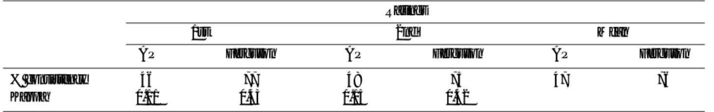
단순 전후 면 방사선 촬영에 의한 골 유합 판정에서 가 관절율은 1 3 . 3 % (방사선과 전문의 5/44, 정형외과 전문 의 6/44, 정형외과 전공의 7/44)였고 퍼거슨 촬영에 의한 가관절율은 6.1%(방사선과 전문의 1/44, 정형외과 전문 의 5/44, 정형외과 전공의 2/44)였다(Table 1).
본 연구에서 관찰자간 일치도는 단순 전후면 방사선
촬영시 평균 4 7 %에서 퍼거슨 촬영시 평균 7 6 %로 향상 하였다(Table 2). 관찰자간 재현성으로서 k a p p a값이 일차 관찰 시 단순 전후면 방사선 촬영의 0 . 1 1에서 퍼거슨 촬 영의 0 . 4 3으로, 이차 관찰시 0 . 1 5에서 0 . 4 2로 향상되어 중 등 신뢰도로 평가하였다(Table 2). 관찰자내 일치도는 관 찰자 모두에서 단순 전후면 방사선 촬영보다 퍼거슨 촬 영시 향상하였다. 관찰자내 재현성으로서 k a p p a값은 퍼 거슨 사진으로 평가할 때 향상되었다(Table 3). 단순 전 후면 방사선 촬영으로 평가시 판정 불가로 분류된 것 중 6 7 %에서 퍼거슨 촬영으로 평가시 골 유합이나 가관절
Table 1. Grading of fusion rating with two different radiographies.
AP Ferguson
Observers Fused Pseud Questionable Fused Pseud Questionable
1 83.0 11.4 05.6 91.1 02.9 6.0
2 61.9 12.5 25.6 85.7 10.5 3.8
3 80.7 15.9 03.4 93.6 04.8 1.6
Mean 75.2 13.3 11.5 90.1 06.1 3.8
1 : Radiologist 2 : Orthopaedic surgeon 3 : Orthopaedic 4th-year resident
AP : Standard anteroposterior radiography Ferguson : Ferguson radiography Pseud : Pseudarthrosis
Table 2. Range of interobserver consistencies in rating of cases on two different radiographies.
Ratings
1st 2nd Mean
AP Ferguson AP Ferguson AP Ferguson
% consistency 46 77 48 75 47 76
Kappa 0.11 0.43 0.15 0.42
AP : Standard anteroposterior radiography Ferguson : Ferguson radiography
Table 3. Range of intraobserver consistencies in rating of cases on two different radiographies : two-week interval.
Observers
1 2 3
AP Ferguson AP Ferguson AP Ferguson
% consistency 74 91 67 77 51 77
Kappa 0.61 0.83 0.35 0.63 0.20 0.57
1 : Radiologist 2 : Orthopaedic surgeon 3 : Orthopaedic 4th-year resident
AP : Standard anteroposterior radiography Ferguson : Ferguson radiography
로 분류되었으나, 33%에서는 동일하게 판정 불가로 분 류되었다. 퍼거슨 촬영으로 평가시 판정 불가로 분류된 것 중에서 단순 전후면 방사선 촬영으로 평가시 골 유합 이나 가관절로 분류된 것은 없었다.
증 례
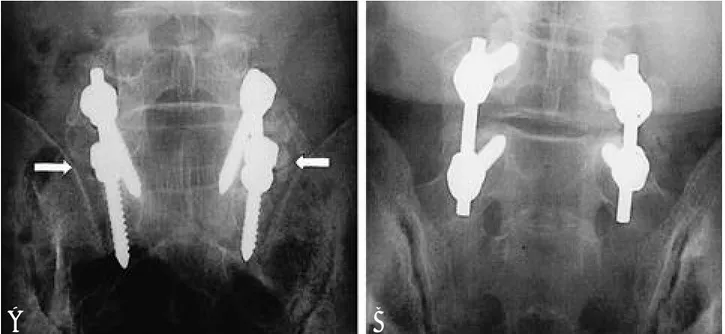
4 0세 여자 환자로 척추관 협착증 진단 하에 보존적 치 료를 하였으나, 호전되지 않아서 감압술 후 내고정 기기 및 자가 이식골을 이용하여 제 4 요추에서 천추까지 양 측성 후외측 유합술을 시행하였다. 단순 전후 면 방사선 사진으로 일차, 이차 관찰시 가관절로 분류되었으나( F i g . 1A) 퍼거슨 사진으로 일차, 이차 관찰시 모든 관찰자가 골유합으로 분류하였다(Fig. 1B).
고 찰
본 연구의 목적은 내고정 기기를 사용한 요천추 간 골 유합을 단순 전후 면 방사선 촬영과 퍼거슨 촬영으로 평 가 후 각 촬영법의 관찰자간 및 관찰자내 재현성을 비교 하여 각 촬영법의 신뢰도를 알기 위한 것이다.
본 연구에서 관찰자간 및 관찰자내 일치도는 단순 전후 면 방사선 촬영보다 퍼거슨 촬영시 향상되었으며 k a p p a 값은 관찰자간 및 관찰자내 재현성 모두 퍼거슨 촬영시 높았다. 특히 단순 전후면 방사선 촬영으로 평가 시 판정 불가로 분류된 것 중 6 7 %에서 퍼거슨 촬영으로 평가시
골 유합 또는 가관절로 분류되어 퍼거슨 촬영으로써 명 확히 평가할 수 있었다.
골 유합을 퍼거슨 촬영으로 평가시 관찰자간 재현성 의 kappa값은 0.43, 0.42로 Landis와 Koch의 중등 신뢰도 였다. 50예의 제 4,5 요추와 제 1 천추 간 유합술 후 단순 전후면 및 측면 방사선 사진으로 여섯 명의 관찰자가 골 유합을 평가한 결과 관찰자간 재현성은 L a n d i s와 K o c h 의 공정한 신뢰도였다13).
관찰자내 재현성의 k a p p a값은 관찰자간 재현성의 kappa값보다 높았으며 이 현상은 상기 두 촬영법에서 모 두 발생하였다. 여섯 명의 관찰자가 골 유합의 평가 시 관찰자들의 수련과정이 서로 다르므로 관찰자내 재현 성의 kappa값이 관찰자간 재현성의 kappa값보다 높았다 고 보고되었다13).
관찰자내 재현성의 kappa값은 퍼거슨 촬영 시 정형외 과 전공의의 경우 0.57, 정형외과 전문의의 경우 0.63으 로, 방사선과 전문의의 경우 0.83으로서 관찰자가 숙련 될수록 높았으며 단순 방사선 촬영에서도 관찰자가 숙 련될수록 높았다. 숙련도가 다른 관찰자들이 골 유합을 평가 시 관찰자내 재현성의 Kappa값이 정형외과 전공의 의 경우 0.18, 정형외과 전문의의 경우 0.44로서 숙련된 관찰자에서 더 높다고 보고되었다1 3 ). 그러므로 판독 경 험의 숙련도는 골 유합을 포함하는 일반적인 방사선 검 사 판독에 중요한 영향을 미치는 인자로 생각된다.
퍼거슨 촬영은 요천추 간의 전만각을 고려하여 상방 향으로 촬영하는 전후 면 방사선 촬영으로서 제 5 요추 횡돌기와 천추 날개의 간격을 넓히고9)제 5 요추와 제 1 천추의 척추체를 관상면으로 보이게 한다(Fig. 2B)16).따
Fig. 1. A 40-year-old woman with spinal stenosis from L4 to S1 not responding to conservative treatment underwent decompression and bilateral instrumented posterolateral fusion from L4 to sacrum 12 months ago. This anteroposterior view(A) illustrates bilateral pseudarthrosis recorded by all observers. Ferguson view(B) illustrates bilateral posterolateral fusion mass recorded by all observers.
A B
라서, 단순 전후 면 방사선 촬영에서는 요추 횡돌기와 천추 날개가 겹치는 것에 의해 골 유합 종괴가 명확히 관찰되지 않아 골 유합의 판단이 어려우며9)겹치지 않더 라도 횡돌기와 천추 날개의 피질골을 골 유합 종괴의 분 열 즉 가관절로 오인할 수 있는 단점이 있다(Fig. 2A). 또 한 단순 전후 면 방사선 촬영에서는 내고정된 척추경 나 사못의 두부가 횡돌기와 천추 날개의 사이에 위치하여 유합 종괴를 가리나(Fig. 2A)3)퍼거슨 촬영은 제 5 요추 체의 정 전후면 상을 얻기 위해 방사선 조사 각도를 조 절한 것이므로 척추경 나사못의 두부가 횡돌기에 위치 하여 횡돌기와 천추 날개 사이의 유합 종괴를 잘 보이게 한다(Fig. 2B). 또한 굴곡 신연 부하 촬영으로 유합 종괴 를 포함한 척추 분절의 운동을 증명하는 것이 단순 방사 선 검사로 가관절을 진단하는 표준방법이나2 1 ), 척추 분 절의 굴신 운동은 내고정 기기에 의해 제한되므로 굴곡 신연 부하 촬영은 내고정 기기가 삽입된 골 유합 종괴의 기능적 가관절의 진단에 적합하지 않다2,4,6).이외에 퍼거 슨 촬영은 경제적으로 추가 비용 없이 저렴하다9).
단순 방사선 검사는 후외측 유합술 후 골 유합을 평가 하는 방법으로 널리 사용되고 있으나2 ), 이 검사가 후외측 유합술 후 골 유합을 평가하는 가장 정확한 방법으로 인 정되지는 않으며2 1 )요추 골 유합의 방사선 소견과 수술적 소견도 일치 하지 않아 단순 방사선 검사로 골 유합을 예 측할 수 있는 범위는 6 4 %에서 6 9 %로 보고되고 있다2 , 3 ).
보고자에 따라 골 유합율이 다르게 보고되는 것도 방사 선 검사에 의한 골 유합을 평가하는 방법이 신뢰도가 낮 기 때문이다8 , 1 1 , 1 8 ). 이상적인 검사법은 모든 관찰자에 있어 서 관찰자내 및 관찰자간에서 재현할 수 있어야 한다.
퍼거슨 촬영이 단순 전후면 방사선 촬영보다 관찰자 간 및 관찰자내 재현성이 우수하였으며 특히 단순 전후 면 방사선 촬영으로 판정이 불가능한 것 중 67%에서 퍼 거슨 촬영으로 평가시 골 유합 또는 가관절로 명확히 분 류되었다. 그러므로, 퍼거슨 촬영에 의한 요천추 간 후 외측 골 유합 판정이 실제 임상에서 정확하면서 쉽게 사 용될 수 있다.
결 론
퍼거슨 전후 면 방사선 촬영이 단순 전후 면 방사선 촬 영보다 내고정 기기를 사용한 요천추간 후외측 골 유합 의 평가에 더 유용하였다.
REFERENCES
01) Albee FH : Transplantation of a portion of the tibia into the spine for Pott’s disease: A preliminary report. JAMA,
Fig. 2-A. The cortical bones of the transverse processes and sacral ala can be misdiagnosed as the cracks(pseudarthroses)(white arrows). The heads of pedicle screws tend to obscure the fusion mass between the L5 transverse processes and sacral ala.
Fig. 2-B. The use of the Ferguson view eliminates the superimposition of the L5 transverse process on the posterior part of the supe- rior ala of the sacrum, widens the L5-S1 intertransverse space, and enhances direct visualization. The heads of pedicle screws are located at the transverse processes and thus the fusion mass between the L5 transverse processes and sacral ala are well visualized.
A B
42(11):885-886, 1911.
02) Blumenthal SL and Gill K : Can lumbar spine radiographs accurately determine fusion in postoperative patients?: cor - relation of routine radiographs with a second surgical look at lumbar fusions. Spine, 18(9):1186-1189, 1993.
03) Brodsky AE, Evan SK and Momtaz AK : Correlation of radiographic assessment of lumbar spine fusions with sur - gical exploration. Spine, 16:S261-265, 1991.
04) Chafetz N, Cann CE, Morris JM, Steinbach LS and Goldbert HI : Pseudarthrosis following lumbar fusion;
Detection by direct coronal CT scanning. Radiology, 162:
803-805, 1987.
05) Christensen FB, Laursen M, Gelineck J, Eiskjær SP, Thomsen K and Bunger CE : Interobserver and intraob - server agreement of radiograph interpretation with and without pedicle screw implants; the need for a detailed classification system in posterolateral spinal fusion. Spine, 26(5):538-544, 2001.
06) Dawson EG, Clader TJ and Bassett LW : A comparison of different methods used to diagnose pseudarthrosis fol - lowing posterior spinal fusion for scoliosis. J Bone Joint Surg, 67-A:1153-1159, 1985.
07) Deguchi M, Rapoff AJ and Zdeblick T : P o s t e r o l a t e r a l fusion for isthmic spondylolithesis in adults: analysis of fu- sion rate and clinical results. J Spinal Disord, 11(6):459- 464, 1998.
08) DePalma AF and Rothman RH : The nature of pseudar- throsis. Clin Orthop, 59:113-118, 1968.
09) Ebraheim NA and Xu R : Assessment of lumbosacral fusion mass by angled radiography: technical notes. Spine, 23(7):842-843, 1998.
10) Fleiss JL : Statistical methods for rates and proportions.
2nd ed. New York, John Wiley & Sons, Inc.:229-232, 1981.
11) Frymoyer JW, Hanley EN, Howe J, Kuhlmann D and Matteri RE : A comparison of radiographic findings in fusion and nonfusion patients ten or more years following lumbar disc surgery. Spine, 4:435-440, 1979.
12) Greenfield RT, Capen DA, Thomas JC, Nelson R, Na- gelberg S, Rimoldi RL and Haye W : Pedicle screw fixa - tion for arthrodesis of the lumbosacral spine in the elderly:
an outcome study. Spine, 23(13):1470-1475, 1998.
13) Hamill CL and Simmons ED : Interobserver variability in grading lumbar fusions. J Spinal Disord, 10(5):387-390, 1 9 9 7 .
14) Hibbs RA : An operation for progressive spinal deformi - ties: A preliminary report of three cases from the service of the orthopaedic hospital. N Y Med J, 93(21):1013-1016, 1 9 1 1 .
15) Landis RJ and Koch GG : The measurement of observer agreement for categorical data. Biometrics, 33:159-174, 1977.
16) Lenke LG, Bridwell KH, Bullis D, Betz RR, Baldus C and Schoenecker PL : Results of in situ fusion for isthmic spondylolithesis. J Spinal Disord, 5(4):433-442, 1992.
17) Nachemson A, Zdeblick TA and O’Brien JP : C o n t r o - versy, Lumbar disc disease with discogenic pain; what sur - gical treatment is most effective? Spine, 21(15):1835-1838, 1 9 9 6 .
18) Rothman RH and Booth R : Failures of spinal fusion.
Orthop Clin North Am, 6:299-303, 1971.
19) Schwab FJ, Nazarian DG, Mahmud F and Michelsen C B : Effects of spinal instrumentation on fusion of the lumbosacral spine. Spine, 20(18):2023-2028, 1995.
20) Stauffer RN and Coventry MB : Posterolateral lumbar spine fusion. J Bone Joint Surg, 54-A:1195-1204, 1972.
21) Steinmann JC and Herkowitz HN : Pseudarthrosis of spine. Clin Orthop, 284:80-90, 1992.
연구목적 :내고정 기기를 이용한 요천추 간 후외측 유합을 평가시 단순 전후 면 방사선 촬영과 퍼거슨 촬영의 신뢰 도를 알기 위하여 관찰자간 및 관찰자내 재현성을 비교분석하였다.
대상 및 방법 :자가 이식골과 내고정 기기를 이용하여 제 4 요추부터 제 1 천추까지 또는 제 5 요추에서 제 1 천추까 지 후외측 골 유합술을 시행 받은 44명의 환자를 추시하고 단순 전후 면 방사선 사진과 40도 상방향으로 촬영한 퍼거 슨 사진을 이용한 연구이다. 3명의 관찰자들은 독립적으로 환자의 정보 없이 방사선 사진을 관찰하여 골 유합의 정 도를 골 유합, 가관절 또는 판정 불가로 분류하였다. 이차 관찰은 2주 후 시행하였다. 관찰자간 및 관찰자내 재현성을 Fleiss 방법을 이용하여 분석하였다.
결과 :단순 전후면 방사선 촬영보다 퍼거슨 촬영에서 관찰자간 및 관찰자내 재현성이 우수하였다. 일차 및 이차 관 찰 모두에서 관찰자간 재현성의 kappa값은 단순 전후 면 방사선 촬영보다 퍼거슨 촬영에서 높았다. 또한 동일한 관찰 자에 의한 일차 및 이차 관찰 간의 관찰자내 재현성의 kappa값은 관찰자 모두에서 단순 전후 면 방사선 촬영보다 퍼 거슨 촬영에서 높았다. 단순 전후면 방사선 촬영으로 평가시 판정 불가로 분류된 것 중 67%에서 퍼거슨 촬영으로 평 가시 골 유합 또는 가관절로 분류되었다.
결론 :퍼거슨 촬영이 단순 전후면 방사선 촬영보다 내고정 기기를 이용한 후외측 요천추 간 골 유합의 평가에 더 유 용한 것으로 사료된다.
색인단어 : 요천추부, 후외측 유합술, 방사선 촬영, 퍼거슨 전후 면 촬영 국 문 초 록
※ 통신저자 : 문 성 환
서울특별시 서대문구 신촌동 134 연세대학교 의과대학 정형외과학교실
Tel : 82-2-361-5649, Fax : 82-2-363-1139, E-mail : shmoon@yumc.yonsei.ac.kr