Background and Purpose The detection of aquaporin 4-IgG (AQP4-IgG) is now a critical diagnostic criterion for neuromyelitis optica spectrum disorder (NMOSD). To evaluate the se- rostatus of NMOSD patients based on the 2015 new diagnostic criteria using a new in-house cell-based assay (CBA).
Methods We generated a stable cell line using internal ribosome entry site-containing bicis- tronic vectors, which allow the simultaneous expression of two proteins (AQP4 and green flu- orescent protein) separately from the same RNA transcript. We performed in-house CBA using serum from 386 patients: 178 NMOSD patients diagnosed according to the new diagnostic criteria without AQP4-IgG, 63 high risk NMOSD patients presenting 1 of the 6 core clinical characteristics of NMOSD but not fulfilling dissemination in space, and 145 patients with other neurological diseases, including 66 with multiple sclerosis. The serostatus of 111 definite and high risk NMOSD patients were also tested using a commercial CBA kit with identical serum to evaluate the correlation between the 2 methods. All assays were performed by two independent and blinded investigators.
Results Our in-house assay yielded a specificity of 100% and sensitivities of 80% (142 of 178) and 76% (48 of 63) when detecting definite- and high risk NMOSD patients, respectively. The comparison with the commercial CBA kit revealed a correlation for 102 of the 111 patients:
no correlation was present in 7 patients who were seronegative using the commercial method but seropositive using the in-house method, and in 2 patients who were seropositive using the commercial method but seronegative using the in-house method
Conclusions These results demonstrate that our in-house CBA is a highly specific and sensi- tive method for detecting AQP4-IgG in NMOSD patients.
Key Words neuromyelitis optica spectrum disorder, aquaporin 4, aquaporin 4-IgG, cell-based immunofluorescence assay.
Large-Scale in-House Cell-Based Assay for Evaluating the Serostatus in Patients with Neuromyelitis Optica Spectrum Disorder Based on New Diagnostic Criteria
INTRODUCTION
Neuromyelitis optica (NMO) is a relapsing autoimmune disease of the central nervous sys- tem that predominantly affects the optic nerves and spinal cord. The discovery of an auto- antibody targeting water channel aquaporin 4 (AQP4), termed AQP4-IgG,1,2 has led to ma- jor advances in the understanding of NMO as a distinct disease entity with a fundamentally different etiology from that of multiple sclerosis (MS).3 Several studies have yielded evi- dence of an involvement of AQP4 in the pathogenesis of NMO.4-6 Based on determination of the highly specific AQP4-IgG, NMO has been recognized as a spectrum disease with a more-diverse clinical presentation that is not limited to optic neuritis or myelitis, and the new nomenclature of neuromyelitis optica spectrum disorder (NMOSD) is now widely Yeseul Kima,b*
Gayoung Kima,b* Byung Soo Konga,b Ji-Eun Leeb,d Yu-Mi Ohb,d Jae-Won Hyuna Su-Hyun Kima AeRan Jounga Byoung Joon Kimc Kyungho Choid Ho Jin Kima,b
a Department of Neurology, Research Institute and Hospital of National Cancer Center, Goyang, Korea
b Division of Translational and Clinical Research II, Research institute, National Cancer Center, Goyang, Korea
c Department of Neurology, Sungkyunkwan University School of Medicine, Seoul, Korea
d Department of Biochemistry and Molecular Biology, and Department of Biomedical Sciences, Seoul National University College of Medicine, Seoul, Korea
pISSN 1738-6586 / eISSN 2005-5013 / J Clin Neurol 2017;13(2):175-180 / https://doi.org/10.3988/jcn.2017.13.2.175
Received October 27, 2016 Revised December 22, 2016 Accepted December 23, 2016 Correspondence Ho Jin Kim, MD, PhD Department of Neurology, Research Institute and Hospital of National Cancer Center, 323 Ilsan-ro, Ilsandong-gu, Goyang 10408, Korea Tel +82-31-920-2438 Fax +82-31-905-5524 E-mail hojinkim@ncc.re.kr
*These authors are contributed equally to this work.
cc This is an Open Access article distributed under the terms of the Creative Commons Attribution Non-Com- mercial License (http://creativecommons.org/licenses/by-nc/4.0) which permits unrestricted non-commercial use, distribution, and reproduction in any medium, provided the original work is properly cited.
JCN
Open Access ORIGINAL ARTICLELarge-Scale in-House CBA for NMOSD
JCN
used. The detection of AQP4-IgG allows the early diagnosis of NMOSD even in patients who experience a single attack with the core clinical characteristics of NMOSD,7 and thus assaying AQP4-IgG is now considered an integral step in the diagnosis of NMOSD.
Considering devastating impact of NMOSD relapses, early accurate diagnosis of NMOSD is crucial, and thus there is an increased demand for an assay that has high sensitivity and specificity in detecting AQP4-IgG in patients. Several assays with varying sensitivities and specificities have been developed over the years, such as the following types of cell- based assays (CBAs) involving live or fixed cells: cell-based indirect immunofluorescence assay and cell-based flow cy- tometry, enzyme-linked immunosorbent assay, and tissue- based indirect immunofluorescence and fluorescence im- munoprecipitation assays. Among these assays, those based on AQP4-transfected cell lines have yielded the highest sen- sitivity and specificity.8
We built an in-house CBA using the M23-AQP4-trans- fected human embryonic kidney 293 (HEK293) cell line, generated by an internal ribosome entry site (IRES) vector.
We aimed to evaluate the sensitivity and specificity of this assay for detecting AQP4-IgG in NMOSD patients based on the 2015 new diagnostic criteria and in patients with other neurological diseases including MS. We also aimed to con- firm the reliability of the procedure by comparing results with those obtained using a commercial anti-AQP4 indirect im- munofluorescence assay (referred to as the commercial CBA henceforth).
METHODS
Patients
Serum samples from 386 patients were tested in duplicate, comprising 178 samples from patients who fulfilled the 2015 diagnostic criteria for NMOSD without the AQP4 serostatus (referred to as definite NMOSD here), 63 samples from pa- tients with 1 of the 6 core clinical characteristics of NMOSD but not fulfilling dissemination in space (referred to as a high risk NMOSD here), and 145 samples from patients with other neurological diseases, including 68 samples from MS patients who the fulfilled 2010 McDonald’s criteria.9 The de-
mographic and clinical characteristics of the NMOSD and MS patients are summarized in Table 1. The patients with other neurological diseases are grouped and summarized in Table 2. All patients provided written informed consent, and this study was approved by our Institutional Review Board.
Generation of M23-AQP4-HEK293 cells
M23-AQP4 cDNA was PCR-amplified from pET15b-M23- AQP4 plasmid and cloned into the NheI and SacI sites of the pIRES2-EGFP vector (Clontech, Mountain View, CA, USA). The cloned M23-AQP4 plasmid or the empty vector plasmid was transfected into HEK293 cells (ATCC, Manas- sas, VA, USA) using Lipofectamine 2000 reagent (Invitro- gen, Waltham, MA, USA). After 48 hours, the cells were split into the medium containing 2 mg/mL G418 (Invitrogen).
The G418-resistant cells were isolated after 2 weeks and then further cloned by limiting dilution. The individual sta- ble transfectant clones were screened for green fluorescent protein (GFP) expression using flow cytometry. M23-AQP4 protein expression in GFP-positive cells was confirmed by Western blotting with anti-AQP4 antibody (Abcam, Cam- bridge, UK). The following primer sequences were used for the PCR: forward, 5’-GGT ACC GCT AGC GCC ACC ATG GTG GCT TTC AAA GGG-3’; reverse, 5’-TCT AGA GAG CTC TCA TAC TGA AGA CAA TAC-3’. All of the sequences were confirmed by automatic sequencing.
Cell-based indirect immunofluorescence assay Stably transfected HEK293 cells expressing GFP-M23- AQP4 fusion protein or HEK293 cells stably transfected with empty vector were seeded into eight-well chamber slides (SPL Life Science, Pocheon, Korea), and incubated in 5%
Table 1. Demographic characteristics of the NMOSD and MS patients
Demographic Definite NMOSD (n=178) High risk NMOSD (n=63) MS (n=68)
Men:women (n:n) 20:158 13:50 20:48
Onset age (years, mean±SD) 31.8±11.5 40.4±11.0 27.7±9.2
Disease duration (years, mean±SD) 22.0±5.6 6.7±5.7 5.9±4.7
EDSS score (median) 3.0 2.5 1.5
EDSS: Kurtzke’s Expanded Disability Status Scale, MS: multiple sclerosis, NMOSD: neuromyelitis optica spectrum disorder, SD: standard deviation.
Table 2. Patients with other neurological diseases (ONDs) (n=145) Disease n
Multiple sclerosis 68
Idiopathic isolated myelitis 29
Optic neuritis 5
Isolated brain demyelination 19
Unknown central nervous system inflammation (DIS+) 5 Non inflammatory neurological disease 19 DIS: dissemination in space.
Kim Y et al.
JCN
CO2 at 37°C overnight. Culture medium was removed by gentle suction and the cells were washed three times with phosphate-buffered saline (PBS). Cells were blocked with blocking buffer (PBS containing 5% bovine serum albumin) at room temperature (RT) for 1 hour. Sera were diluted 1:20 using blocking buffer. Diluted serum (100 μL) was added to each well and incubated at RT for 2 hours. The cells were washed three times with PBS as described above, and then fixed with 2% paraformaldehyde at RT for 45 min. After washing with PBS, the cells were incubated with goat anti- human IgG conjugated with Alexa-594 (Jackson Immu- noresearch, West Grove, PA, USA; diluted 1:2,000 with PBS) for 1 hour at RT in the dark. The cells were then washed three times before being mounted with VECTASHIELD® antifade reagent with DAPI (Vector Laboratories, Burlingame, CA, USA) and covered with coverslips. Each experiment was performed in duplicate, and the green and red fluorescence on the cell membranes was examined by two investigators (K.G.Y. and K.Y.S.) who were blinded to the clinical and lab- oratory information of the studied patients, under a fluores- cence microscope (Zeiss, Oberkochen, Germany). The pres- ence of only green fluorescence was considered to indicate negativity for AQP4 antibodies and the presence of red flu- orescence that was not colocalized with green fluorescence was considered to indicate nonspecific binding, and hence negative for AQP4 antibodies. The presence of red fluores- cence on cell membranes and green fluorescence in the cyto- sol was considered to indicate positivity for AQP4-IgG.
Statistical analysis
The following calculations were performed as part of the sta- tistical analysis in this study:
1) Sensitivity was calculated as [true positives/(true posi- tives+ false negatives)].
2) Specificity was calculated at [true negatives/(true nega- tives+ false positives)].
3) Accuracy was calculated as [(true positives+true nega- tives)/total samples]×100.
4) The 95% confidence interval (CI) for sensitivity was calculated as √[(1-sensitivity)×sensitivity/(number of tests)]×
1.96.
5) The 95% CI for specificity was calculated as √[(1-spec- ificity)× specificity/(number of tests)]×1.96.
RESULTS
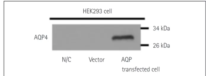
HEK293 cells transfected with M23-AQP4 were used in the in-house CBA, with successful transfection confirmed us- ing Western blotting (Fig. 1). The widely used CBA method was adopted, and fluorescence levels were measured using
fluorescence microscopy. Green fluorescence in the cytosol was observed in AQP4-transfected cells, and when patient serum that contained AQP4-IgG was added, red fluores- cence was observed through the cell membranes (Fig. 2). If the patient serum did not contain AQP4-IgG, no red fluo- rescence was observed (Fig. 2).
Sera from 387 patients were assayed. The 178 definite- NMOSD patients comprised 142 true positives and 36 false negatives, yielding a sensitivity of 80% (95% CI=73.1–
85.4%). The 63 high risk NMOSD patients comprised 48 true positives and 15 false negatives, yielding sensitivity of 76% (95% CI=63.8–86.0%). None of the 145 patients with other neurological diseases exhibited positivity, yielding specificity of 100% (95% CI=97.5–100%). The accuracy of the in-house CBA for detecting AQP4-IgG in definite NMOSD was 80% and in high risk NMOSD was 76%. This suggests that the in-house CBA is an accurate assay with high sensi- tivity and specificity (Table 3).
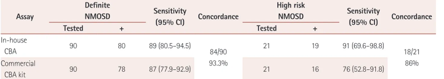
Sera from 90 definite-NMOSD and 21 high risk NMOSD patients were tested using both the in-house CBA and com- mercial CBA kit, and the results were compared (Table 4).
The in-house CBA yielded sensitivity of 89% (95% CI=
80.5–94.5%) and the commercial CBA yielded sensitivity of 87% (95% CI=77.9–92.9%) when sera from definite-NMOSD patients were assayed. The sensitivity for detecting AQP4-IgG in high risk NMOSD patients was higher for the in-house CBA was a more sensitive (sensitivity 91%, 95% CI=69.6–
98.8%) method of detecting AQP4-IgG in high risk NMOSD patients than for the commercial CBA (sensitivity 76%, 95% CI=52.8–91.8%). The specificity of each assay could not be evaluated since only NMOSD patients were tested.
The interassay concordance between the in-house CBA and the commercial CBA was determined for each patient group. The results of 84 samples (93%) from 90 definite- NMOSD patients correlated; in the other 6 samples, did not
Fig. 1. AQP4 overexpression in HEK293 cells using the AQP4-IRES2- EGFP vector. Western blot data confirming the successful transfec- tion of AQP4. Lanes containing either untransfected HEK293 cells or HEK293 cells transfected with the IRES2-EGFP vector only do not show any bands. The lane containing lysate of HEK293 cells trans- fected with the M23-AQP4 gene shows a band at ~30 kDa. AQP4:
aquaporin 4, EGFP: enhanced green fluorescent protein, HEK293: hu- man embryonic kidney 293, IRES: internal ribosome entry site, N/C:
untransfected cells, Vector: transfected IRES2-eGFP vector only.
HEK293 cell
34 kDa 26 kDa AQP4
N/C Vector AQP
transfected cell
Large-Scale in-House CBA for NMOSD
JCN
correlate as 4 were seropositive using the in-house CBA but seronegative using the commercial CBA, while 2 were sero- negative using the in-house CBA but seropositive using the commercial CBA. The interassay concordance was deter- mined to be 86% (18/21) when sera from high risk NMOSD patients were evaluated 3 samples did not correlate: 3 were seropositive using the in-house CBA but seronegative using
the commercial CBA.
DISCUSSION
The importance of AQP4-IgG is now widely known, and different assays have been developed to detect this autoan- tibody in patient sera. However, these assays reportedly have
Fig. 2. In-house CBA to detect AQP4-IgG in patient sera. Sera from healthy subjects (A-D), MS patients (E-H), and NMOSD patients (I-L) were added to AQP4-GFP-transfected HEK293 cells. HEK293 cells transfected with AQP4-GFP show green fluorescence in the cytosol under fluorescence mi- croscopy (B, F, and J). When the patient serum contained AQP4-IgG, red fluorescence was detected on the HEK293 cell membrane (K) due to bind- ing of AQ4-IgG to AQP4, which is expressed on the membrane of HEK293 cells. AQP4-IgG: aquaporin 4-IgG, CBA: cell based assay, GFP: green flu- orescent protein, HEK293: human embryonic kidney 293, MS: multiple sclerosis, NMOSD: neuromyelitis optica spectrum disorder.
GFP DAPI
Healthy
MS
NMOSD
Alexa594 Merge
Table 3. Sensitivity, specificity, and accuracy of the in-house CBA Assay
Definite
NMOSD Sensitivity (95% CI)
Accuracy (95% CI)
High risk
NMOSD Sensitivity (95% CI)
Accuracy (95% CI)
OND Specificity (95% CI)
Tested + Tested + Tested +
In-house
CBA 178 142 80
(73.1–85.4) 80
(73.1–85.4) 63 48 76
(63.8–86.0) 76
(63.8–86.0) 145 0 100
(97.5-100) CBA: cell based assay, CI: confidence interval, NMOSD: neuromyelitis optica spectrum disorder, OND: other neurological diseases.
Table 4. Interassay concordance between the in-house CBA and the commercial CBA kit Assay
Definite
NMOSD Sensitivity
(95% CI) Concordance
High risk
NMOSD Sensitivity
(95% CI) Concordance
Tested + Tested +
In-house
CBA 90 80 89 (80.5–94.5)
84/90 93.3%
21 19 91 (69.6–98.8)
18/21 Commercial 86%
CBA kit 90 78 87 (77.9–92.9) 21 16 76 (52.8–91.8)
CI: confidence interval, CBA: cell based assay, NMOSD: neuromyelitis optica spectrum disorder.
Kim Y et al.
JCN
varying sensitivities (33–90%).10 In the present fully blinded study, our in-house CBA detected AQP4-IgG in 80% of defi- nite-NMOSD patients and 76% of high risk NMOSD pa- tients, with 100% specificity. A systemic review on literatures reporting on different assays for detecting AQP4-IgG found that the CBA had the highest sensitivity (mean sensitivi- ty=76.7%, range=55.6–96.7%),27 while the mean accuracy was also higher for the CBA (76.5%) than for other methods (range=48.5–62.3%).
The commercial CBA kit is based on using cells that have already been fixed, which makes it a simple and easy meth- od. An international multicenter study found that the com- mercial CBA kit (EUROIMMUN, Luebeck, Germany) had a specificity of 100% and sensitivities of between 51.5% and 86.4%.10 A similar study found that the CBA using live cells expressing human M23-AQP4 was more sensitive than the CBA using fixed cells.8 We evaluated the interassay concor- dance between the in-house CBA and the commercial CBA kit using identical serum from 90 definite-NMOSD and 21 high risk NMOSD patients, and found a high interassay concordance. Moreover, the in-house CBA showed a higher sensitivity than the commercial CBA kit, and we suggest that this is due to our in-house CBA being based on live cells rather than fixed cells.
AQP4 is found on the astrocytic membrane in two iso- forms: M23 and M1. The M1 isoform is the full-length pro- tein while the M23 isoform is 22 amino acids shorter at the intracellular N-terminus.11 Some studies have found no dif- ferences between the M1 and M23 isoforms when detecting AQP4-IgG,8,12,13 while many other studies showed that the use of the M23 isoform provided a more sensitive substrate for AQP4-IgG assays than did the M1 isoform.14-21 A higher binding affinity of AQP4-IgG to the M23 isoform could be due to the formation of orthogonal arrays of particles (OAPs) at the cell surface by M23.22 In fact, when mutation was in- troduced to M23 protein and OAP formation was disrupted, the AQP4-IgG binding affinity was significantly reduced.23 Hence, in order to develop a more sensitive test, we used the M23 isoform of AQP4 when generating transfected HEK293 cells.
Live transiently transfected cells with fluorescently tagged AQP4 are often used due to the possibility that the expres- sion of the gene of interest could decrease over time as the cell replicates,24 and the maintenance of cell lines and trans- fection prior to its use could limit the reproducibility of the assay.13 Moreover, CBAs with transiently transfected cells are based on the colocalization of AQP4-IgG in patient serum to cells that express fluorescently tagged AQP4. Tagging AQP4 with GFP and the expression of GFP can be used to confirm successful transfection and monitor AQP4 gene
expression and protein localization.25 However, this method could restrict the binding between the antibody and anti- gen, since the addition of the 27-kDa GFP protein could change the structure of the AQP4 protein as well as the for- mation of OAP, leading to a reduced sensitivity. However, no difference in sensitivity between the M1 and M23 iso- forms was found when N-terminal GFP tag was added.26 Mader et al.14 used GFP tagged at the C-terminus of M23- AQP4 and found that the M23 isoform maintained superior sensitivity; however, fluorescent tags still could be cytotoxic and so influence the assay.
The current study applied a new approach of the IRES- containing bicistronic vector to generate a stable HEK293 cell line. In contrast to transient transfection, stable transfec- tion allows the gene of interest to be integrated into the host genome and the gene expression persists even as the cell replicates.24 Instead of a GFP tag, we used an IRES vector that is based on IRES controlling the downstream cistron ex- pression without affecting the expression of upstream cis- tron, thereby allowing the simultaneous expression of both the protein of interest (M23-AQP4) and a selectable marker (GFP) without interfering with the expression of one an- other.27 We were therefore able to determine the serostatus without any interference in the fluorescence of each protein.
Despite our in-house CBA yielding high sensitivity and specificity, our method could not overcome the innate limi- tation of cell based immunofluorescence assay and it can only produce semiquantitative results.
In conclusion, we have refined a previous method by uti- lizing an IRES vector to generate stably transfected M23- AQP4-HEK293 cell lines. The high sensitivity and specificity of this assay reconfirms that the CBA is an accurate and reli- able method for detecting both definite and high risk NMOSD patients.
Conflicts of Interest
Kim YS, Kim GY, Kong BS, Lee JE, Oh YM, Hyun JW, Kim SH, Joung AR, Kim BJ and Choi KH report no conflicts of interest. Dr. Kim HJ has lectured, consulted, and received honoraria from Bayer Schering Pharma, Biogen, Genzyme, HanAll BioPharma, MedImmune, Merck Serono, No- vartis, Teva-Handok, and UCB; received a grant from the Ministry of Science; and accepted research funding from ICT & Future Planning, Genzyme, Kael-GemVax, Merck Serono, Teva-Handok, and UCB. He serves on a steering committee for MedImmune and as a co-editor for the Multiple Sclerosis Journal-Experimental, Translational, and Clinical. He also serves as an associated editor for the Journal of Clinical Neurology.
Acknowledgements
This study was supported by the Bio & Medical Technology Develop- ment Program (M3A9B6069339) thourough the Ministry of Science, ICT & Future Planning, Republic of Korea.
Large-Scale in-House CBA for NMOSD
JCN
REFERENCES
1. Lennon VA, Wingerchuk DM, Kryzer TJ, Pittock SJ, Lucchinetti CF, Fujihara K, et al. A serum autoantibody marker of neuromyelitis op- tica: distinction from multiple sclerosis. Lancet 2004;364:2106-2112.
2. Lennon VA, Kryzer TJ, Pittock SJ, Verkman AS, Hinson SR. IgG marker of optic-spinal multiple sclerosis binds to the aquaporin-4 water channel. J Exp Med 2005;202:473-477.
3. Roemer SF, Parisi JE, Lennon VA, Benarroch EE, Lassmann H, Bruck W, et al. Pattern-specific loss of aquaporin-4 immunoreactivity dis- tinguishes neuromyelitis optica from multiple sclerosis. Brain 2007;
130:1194-1205.
4. Bradl M, Misu T, Takahashi T, Watanabe M, Mader S, Reindl M, et al.
Neuromyelitis optica: pathogenicity of patient immunoglobulin in vivo. Ann Neurol 2009;66:630-643.
5. Hinson SR, McKeon A, Lennon VA. Neurological autoimmunity tar- geting aquaporin-4. Neuroscience 2010;168:1009-1018.
6. Verkman AS, Phuan PW, Asavapanumas N, Tradtrantip L. Biology of AQP4 and anti-AQP4 antibody: therapeutic implications for NMO.
Brain Pathol 2013;23:684-695.
7. Wingerchuk DM, Banwell B, Bennett JL, Cabre P, Carroll W, Chitnis T, et al. International consensus diagnostic criteria for neuromyelitis optica spectrum disorders. Neurology 2015;85:177-189.
8. Waters PJ, McKeon A, Leite MI, Rajasekharan S, Lennon VA, Villalo- bos A, et al. Serologic diagnosis of NMO: a multicenter comparison of aquaporin-4-IgG assays. Neurology 2012;78:665-671; discussion 669.
9. Polman CH, Reingold SC, Banwell B, Clanet M, Cohen JA, Filippi M, et al. Diagnostic criteria for multiple sclerosis: 2010 revisions to the McDonald criteria. Ann Neurol 2011;69:292-302.
10. Waters P, Reindl M, Saiz A, Schanda K, Tuller F, Kral V, et al. Multi- centre comparison of a diagnostic assay: aquaporin-4 antibodies in neuromyelitis optica. J Neurol Neurosurg Psychiatry 2016;87:1005- 1015.
11. Hinson SR, Romero MF, Popescu BF, Lucchinetti CF, Fryer JP, Wol- burg H, et al. Molecular outcomes of neuromyelitis optica (NMO)-IgG binding to aquaporin-4 in astrocytes. Proc Natl Acad Sci U S A 2012;
109:1245-1250.
12. Kalluri SR, Illes Z, Srivastava R, Cree B, Menge T, Bennett JL, et al.
Quantification and functional characterization of antibodies to na- tive aquaporin 4 in neuromyelitis optica. Arch Neurol 2010;67:1201- 1208.
13. Jarius S, Wildemann B. Aquaporin-4 antibodies (NMO-IgG) as a se- rological marker of neuromyelitis optica: a critical review of the liter- ature. Brain Pathol 2013;23:661-683.
14. Mader S, Lutterotti A, Di Pauli F, Kuenz B, Schanda K, Aboul-Enein F, et al. Patterns of antibody binding to aquaporin-4 isoforms in neu-
romyelitis optica. PLoS One 2010;5:e10455.
15. Pisani F, Sparaneo A, Tortorella C, Ruggieri M, Trojano M, Mola MG, et al. Aquaporin-4 autoantibodies in neuromyelitis optica: AQP4 isoform-dependent sensitivity and specificity. PLoS One 2013;8:
e79185.
16. Kitley J, Woodhall M, Leite MI, Palace J, Vincent A, Waters P. Aqua- porin-4 antibody isoform binding specificities do not explain clinical variations in NMO. Neurol Neuroimmunol Neuroinflamm 2015;2:
e121.
17. Crane JM, Lam C, Rossi A, Gupta T, Bennett JL, Verkman AS. Binding affinity and specificity of neuromyelitis optica autoantibodies to aqua- porin-4 M1/M23 isoforms and orthogonal arrays. J Biol Chem 2011;
286:16516-16524.
18. Iorio R, Fryer JP, Hinson SR, Fallier-Becker P, Wolburg H, Pittock SJ, et al. Astrocytic autoantibody of neuromyelitis optica (NMO-IgG) binds to aquaporin-4 extracellular loops, monomers, tetramers and high order arrays. J Autoimmun 2013;40:21-27.
19. Sato DK, Nakashima I, Takahashi T, Misu T, Waters P, Kuroda H, et al.
Aquaporin-4 antibody-positive cases beyond current diagnostic cri- teria for NMO spectrum disorders. Neurology 2013;80:2210-2216.
20. Marignier R, Bernard-Valnet R, Giraudon P, Collongues N, Papeix C, Zéphir H, et al. Aquaporin-4 antibody-negative neuromyelitis optica:
distinct assay sensitivity-dependent entity. Neurology 2013;80:2194- 2200.
21. Rossi A, Ratelade J, Papadopoulos MC, Bennett JL, Verkman AS.
Neuromyelitis optica IgG does not alter aquaporin-4 water permeabil- ity, plasma membrane M1/M23 isoform content, or supramolecular assembly. Glia 2012;60:2027-2039.
22. Nicchia GP, Mastrototaro M, Rossi A, Pisani F, Tortorella C, Ruggieri M, et al. Aquaporin-4 orthogonal arrays of particles are the target for neuromyelitis optica autoantibodies. Glia 2009;57:1363-1373.
23. Tuller F, Holzer H, Schanda K, Aboulenein-Djamshidian F, Höftberger R, Khalil M, et al. Characterization of the binding pattern of human aquaporin-4 autoantibodies in patients with neuromyelitis optica spectrum disorders. J Neuroinflammation 2016;13:176.
24. Kim TK, Eberwine JH. Mammalian cell transfection: the present and the future. Anal Bioanal Chem 2010;397:3173-3178.
25. Chalfie M, Tu Y, Euskirchen G, Ward WW, Prasher DC. Green fluo- rescent protein as a marker for gene expression. Science 1994;263:
802-805.
26. Waters PJ, Pittock SJ, Bennett JL, Jarius S, Weinshenker BG, Winger- chuk DM. Evaluation of aquaporin-4 antibody assays. Clin Exp Neu- roimmunol 2014;5:290-303.
27. Gurtu V, Yan G, Zhang G. IRES bicistronic expression vectors for ef- ficient creation of stable mammalian cell lines. Biochem Biophys Res Commun 1996;229:295-298.