© 2017 Korean Breast Cancer Society. All rights reserved. http://ejbc.kr | pISSN 1738-6756
INTRODUCTION
Breast cancer is the most commonly diagnosed neoplasm and the leading cause of cancer mortality among women [1].
It is a heterogeneous disease; predicting its clinical behavior and preparing therapeutic plans are challenging despite vari- ous histopathological classifications at diagnosis [2]. Clinico- pathological factors such as American Joint Committee on Cancer (AJCC) stage, histological grade, estrogen receptor (ER) and progesterone receptor (PR) expression, and human epidermal growth factor receptor 2 (HER2) gene amplifica- tion are currently considered in the prognosis and manage- ment of breast cancer [3]. Based on emerging molecular and immunohistochemical methods, breast cancer is classified into four major subtypes: luminal A, luminal B, HER2+, and triple-negative. Luminal B is subdivided according to HER2
status (luminal B HER2− and luminal B HER2+) and Ki-67 labeling index [4].
Signaling by the Wnt family of secreted glycolipoproteins is an ancient and evolutionarily conserved pathway linked to cell proliferation, cell polarity, and fate determination [5].
Wnt7a, a member of the Wnt family, is normally expressed in several organs, including the lung, testis, lymph node, and brain [6]. As an oncogenic autocrine glycoprotein, Wnt7a promotes tumor invasion and distant metastasis with cancer- associated fibroblasts [7]. Interestingly, published data have shown that Wnt7a shows different expression patterns in dif- ferent types of malignancies [6]. Wnt7a exhibits tumorigenic potential and functions as an oncogene in endometrial and ovarian cancer, but ironically seems to suppress tumors in the cervix, lung, leukemia, kidney, and pleura [8-14]. Additional- ly, high Wnt7a expression is associated with a good prognosis in mesothelioma [14] but a poor prognosis in ovarian cancer [9]. Therefore, Wnt7a apparently has antithetical biological behaviors in several cancers.
In several breast cancer studies, only one demonstrated a correlation between Wnt7a expression and worse disease-free survival (DFS) in breast cancer, especially in the basal subtype [7]. Although Wnt7a expression was linked to low DFS rate,
Wnt7a Deficiency Could Predict Worse Disease-Free and Overall Survival in Estrogen Receptor-Positive Breast Cancer
Kijong Yi*, Kyueng-Whan Min*, Young Chan Wi, Yeseul Kim, Su-Jin Shin, Min Sung Chung1, Kiseok Jang, Seung Sam Paik
Departments of Pathology and 1Surgery, Hanyang University College of Medicine, Seoul, Korea ORIGINAL ARTICLE
Purpose: Wnt7a is a glycoprotein involved in embryonic develop- ment and the progression of different types of malignant tumors.
This study aimed to detect the level of Wnt7a expression in breast cancer and explore its role in the disease progression and prognosis. Methods: A total of 258 patients diagnosed with invasive ductal carcinoma of the breast were included in this study. Using tissue microarray and immunohistochemical stain- ing, we evaluated the association between Wnt7a expression and clinicopathological parameters, and the prognostic value of Wnt7a. Results: Wnt7a expression was significantly correlated with estrogen receptor (ER) expression (odds ratio, 3.95; 95%
confidence interval [CI], 1.99–7.80; p<0.001). On univariate and multivariate analyses, loss of Wnt7a expression was associated
with poor disease-free survival (DFS) (multivariate hazard ratio [HR], 9.12; 95% CI, 1.80–46.09; p=0.008), but not with poor overall survival (OS). In the ER-positive group (n=114), loss of Wnt7a expression was an independent prognostic factor for shorter DFS (multivariate HR, 13.54; 95% CI, 1.11–165.73; p=
0.042) and OS (multivariate HR, 4.76; 95% CI, 1.29–17.61;
p=0.019) on univariate and multivariate analyses. However, in the ER-negative group, there was no significant difference in DFS and OS according to Wnt7a expression. Conclusion: The loss of Wnt7a expression might be a meaningful factor in assessing DFS and OS, especially in ER-positive breast cancer.
Key Words: Breast neoplasms, Estrogen receptors, Prognosis, Wnt proteins
Correspondence to: Seung Sam Paik
Department of Pathology, Hanyang University College of Medicine, 222-1 Wangsimni-ro, Seongdong-gu, Seoul 04763, Korea
Tel: +82-2-2290-8252, Fax: +82-2-2296-7502 E-mail: [email protected]
*These authors contributed equally to this work.
Received: June 7, 2017 Accepted: October 16, 2017
Breast
Cancer
whether this association is accurate enough to predict the clinical outcome remains disputable when considering the multifactorial nature of breast cancer development.
Here, we investigated the prognostic validity of Wnt7a ex- pression and analyzed any statistical correlations with clinico- pathological parameters, DFS, and overall survival (OS) in patients with invasive ductal carcinoma (IDC) of the breast.
Moreover, we evaluated how Wnt7a and survival rate are in- terrelated according to ER, PR, and HER2 status.
METHODS
Patients and specimens
We collected 299 cases of breast cancer at Hanyang Univer- sity Hospital between January 2000 and December 2009.
Clinicopathological information was obtained from the pa- tients’ medical records and pathological reports. Among the patients, 41 were excluded due to unavailability of paraffin blocks or inadequate clinical history. The remaining 258 pa- tients diagnosed with IDC were selected as subjects. The me- dian age was 49 years (range, 27–79 years). Among them, 181 patients underwent modified radical mastectomy, 65 under- went breast-conserving operation, 11 underwent skin-sparing mastectomy, and one underwent a simple mastectomy.
The distribution of tumor (T) and node (N) stages was as follows: T1, 120 (46.5%); T2, 122 (47.3%); T3, 8 (3.1%); T4, 8 (3.1%); and N0, 140 (54.3%); N1, 54 (20.9%); N2, 33 (12.8%);
N3, 31 (12.0%). Overall, 90 patients received a combination of chemotherapy and tamoxifen, 53 received tamoxifen only, and 103 received chemotherapy only. Ten patients showed re- currence and 49 patients died during the follow-up. The 1-, 3-, and 5-year mortality rates were 11.6%, 8.2%, and 12.1%, re-
spectively. The recurrence rate at the 1-, 3-, and 5-year follow- ups were 0.8%, 2.5%, and 3%, respectively.
This study, along with the waiver of informed consent, was approved by the Institutional Review Board at Hanyang Uni- versity Hospital (HYUH 2016-05-009).
Tissue microarray construction and immunohistochemistry We punched a 2-mm core in the most cellular area of each paraffin-embedded tumor tissue block, and manually trans- ferred the specimen to a recipient block. Tissue microarray (TMA) sections (4-μm thick) were stained using anti-Wnt7a an- tibody (1:50 dilution; ab183653; Abcam, Cambridge, UK) after deparaffinization, heat-induced antigen retrieval with Bond epi- tope retrieval solution (Leica BioSystems, Newcastle, UK), and endogenous peroxidase blocking. We used a Bond-Max auto- mated immunostainer (Leica BioSystems). We incubated the sections in the primary antibody for 30 minutes at room tem- perature and developed the sections using the Bond Polymer Refine Detection kit (Leica BioSystems) and 3,3´-diaminobenzi- dine tetrahydrochloride as a chromogen. Immunohistochemical staining for ER, PR, and HER2 was performed using the follow- ing antibodies (Novocastra Laboratories, Newcastle, UK):
monoclonal mouse anti-ER (1:50), monoclonal mouse anti-PR (1:100), and monoclonal mouse anti-c-erbB-2 (1:800).
Interpretation of immunohistochemical staining
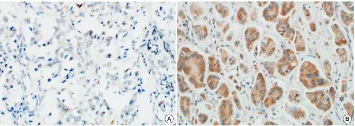
For this study, positive Wnt7a immunostaining was defined as cytoplasmic staining regardless of nuclear staining and graded according to both the intensity and percentage of posi- tively stained tumor cells [8,15]. Wnt7a staining intensity was scored on a scale of 0 to 3 (0, negative; 1, weak; 2, moderate; 3, strong). The percentage of Wnt7a-positive cells was also clas-
Figure 1. Representative photographs of Wnt7a expression in the cytoplasm of neoplastic cells. (A) Negative, (B) positive (immunohistochemical stain for Wnt7a, original magnification ×200).
A B
sified into four categories: 1 (0%–25%), 2 (26%–50%), 3 (51%–75%), and 4 (76%–100%). The level of Wnt7a staining was evaluated as an immunoreactive score (IRS), which was calculated by multiplying the staining intensity scores and the percentages of positive cells [16]. Based on the receiver oper- ating characteristic (ROC) curve, there was moderate dis- criminatory power for correlating recurrence rate with Wnt7a expression (area under the ROC, 0.636; sensitivity, 82.7%;
specificity, 50%). Wnt7a expression was determined as either
negative (IRS <1) or positive (IRS ≥1) (Figure 1). The distri- bution of the molecular subtypes using immunohistochemi- cal markers was as follows: luminal A (ER+ and/or PR+;
HER2−; Ki-67 <14%), 100 (38.8%); luminal B HER2− (ER+
and/or PR+; HER2−; Ki-67 ≥14%), 45 (17.4%); luminal B HER2+ (ER+ and/or PR+; HER2+), 21 (8.1%); HER2+ (ER−
and PR−; HER2+), 39 (15.1%); and triple-negative (ER− and PR−; HER2−), 53 (20.5%) [4].
Statistical analysis
Categorical variables were compared using the chi-square test. For ordinal variables, the Mantel-Haenszel method (also called linear-by-linear association) was used to determine trends. Continuous variables were compared using Student t-test. DFS and OS curves were generated using the Kaplan- Meier method and were compared by the log-rank test.
Meanwhile, multivariate analysis was performed to identify independent prognostic markers for DFS and OS using a Cox multistep regression model. A p<0.05 was considered statisti- cally significant. All statistical computations were performed with R version 3.2.2 (http://www.R-project.org/).
RESULTS
Relationship between Wnt7a expression and clinicopathological parameters and molecular subtypes
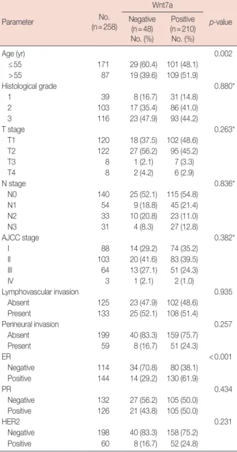
Wnt7a expression was associated with ER positivity (odds ratio [OR], 3.95; 95% confidence interval [CI], 1.99–7.80; p<
0.001), which was frequently observed in patients older than 55 years (OR, 3.6; 95% CI, 1.54–8.42; p=0.002). No signifi- cant difference existed with other parameters such as tumor size, lymphovascular invasion, perineural invasion, AJCC stage, histological grade, and HER2 expression (Table 1). Re- garding molecular subtypes [4], Wnt7a expression was more frequently observed in luminal A, luminal B, and HER2 groups than in the triple-negative group (OR, 3.4; 95% CI, 1.70–6.73;
p<0.001) (Table 2, Figure 2).
Comparison between patient survival and Wnt7a expression On univariate survival analysis, Wnt7a negativity was sig- Table 1. Comparison of clinicopathologic parameters between the
Wnt7a-expressing and non-expressing group
Parameter No.
(n=258)
Wnt7a
p-value Negative
(n=48) No. (%)
Positive (n=210) No. (%)
Age (yr) 0.002
≤55 171 29 (60.4) 101 (48.1)
>55 87 19 (39.6) 109 (51.9)
Histological grade 0.880*
1 39 8 (16.7) 31 (14.8)
2 103 17 (35.4) 86 (41.0)
3 116 23 (47.9) 93 (44.2)
T stage 0.263*
T1 120 18 (37.5) 102 (48.6)
T2 122 27 (56.2) 95 (45.2)
T3 8 1 (2.1) 7 (3.3)
T4 8 2 (4.2) 6 (2.9)
N stage 0.836*
N0 140 25 (52.1) 115 (54.8)
N1 54 9 (18.8) 45 (21.4)
N2 33 10 (20.8) 23 (11.0)
N3 31 4 (8.3) 27 (12.8)
AJCC stage 0.382*
I 88 14 (29.2) 74 (35.2)
II 103 20 (41.6) 83 (39.5)
III 64 13 (27.1) 51 (24.3)
IV 3 1 (2.1) 2 (1.0)
Lymphovascular invasion 0.935
Absent 125 23 (47.9) 102 (48.6)
Present 133 25 (52.1) 108 (51.4)
Perineural invasion 0.257
Absent 199 40 (83.3) 159 (75.7)
Present 59 8 (16.7) 51 (24.3)
ER <0.001
Negative 114 34 (70.8) 80 (38.1) Positive 144 14 (29.2) 130 (61.9)
PR 0.434
Negative 132 27 (56.2) 105 (50.0) Positive 126 21 (43.8) 105 (50.0)
HER2 0.231
Negative 198 40 (83.3) 158 (75.2) Positive 60 8 (16.7) 52 (24.8)
AJCC=American Joint Committee on Cancer; ER=estrogen receptor; PR=
progesterone receptor; HER2=human epidermal growth factor receptor 2.
*Linear by linear association.
Table 2. Expression of Wnt7a according to molecular subtype Wnt7a Luminal A
(n=100)
Luminal B HER2–
(n=45)
Luminal B HER2+
(n=21) HER2 (n=39)
Triple- negative
(n=53) p-value
Negative 15 6 2 6 19 <0.001*
Positive 85 39 19 33 34
HER2=human epidermal growth factor receptor 2.
*Triple-negative versus other subtypes.
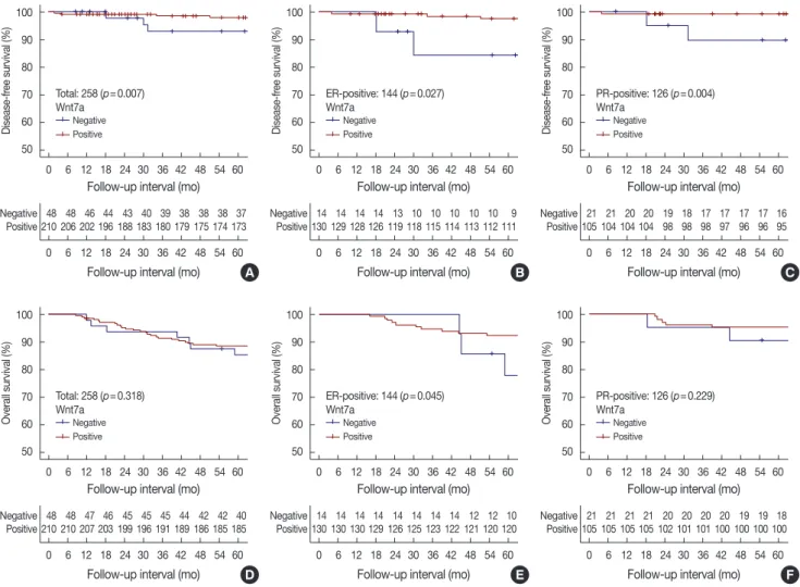
Table 3. Correlations between disease-free and overall survival and Wnt7a expression
Survival Univariate significance*
Multivariate
significance† HR 95% CI Disease-free survival
Total cases 0.007 0.008 9.12 1.80–46.09 ER-negative group 0.050 0.227 5.96 0.33–107.98 ER-positive group 0.027 0.042 13.54 1.11–165.73 Overall survival
Total cases 0.318 0.247 1.53 0.75–3.15 ER-negative group 0.314 0.304 1.71 0.62–4.75 ER-positive group 0.045 0.019 4.76 1.29–17.61 Total cases, 258 patients; ER-negative group, 114 patients; ER-positive group, 144 patients.
HR=hazard ratio; CI=confidence interval; ER=estrogen receptor.
*Log-rank test; †Cox proportional hazard model: adjusted for age (≤55 years vs. >55 years), T stage (1 or 2 vs. 3 or 4), histological grade (1 vs. 2 or 3), lymphovascular/perineural invasion (absence vs. presence), hormone therapy and chemotherapy.
Figure 2. Expression of Wnt7a according to molecular subtype. Wnt7a negativity is frequently observed in the triple-negative group compared with other groups including luminal A, luminal B human epidermal growth factor receptor 2 (HER2)–, luminal B HER2+, and HER2 (p<0.001). Values in parentheses represent percentage.
Figure 3. Patients’ survival according to Wnt7a expression. Kaplan-Meier curves representing disease-free survival in all cases (A), the estrogen re- ceptor (ER)-positive group (B) and the progesterone receptor (PR)-positive group (C) and overall survival in all cases (D), the ER-positive group (E) and the PR-positive group (F).
80
60
40
20
0
Luminal A Luminal B Luminal B HER2 Triple-
HER2− HER2+ negative
Molecular subtype
Wnt7a
(15.0) 15 (85.0) 85
Negative Positive
(13.3) 6 (86.7) 39
(9.5) 2 (90.5) 19
(15.4) 6 (84.6) 33
(35.8) 19 (64.2) 34
Count
100 90 80 70 60 50
100 90 80 70 60 50
100 90 80 70 60 50
100 90 80 70 60 50
100 90 80 70 60 50
100 90 80 70 60 50 0 6 12 18 24 30 36 42 48 54 60
0 6 12 18 24 30 36 42 48 54 60 0 6 12 18 24 30 36 42 48 54 60
0 6 12 18 24 30 36 42 48 54 60
0 6 12 18 24 30 36 42 48 54 60
0 6 12 18 24 30 36 42 48 54 60
0 6 12 18 24 30 36 42 48 54 60
0 6 12 18 24 30 36 42 48 54 60 48 48 46 44 43 40 39 38 38 38 37
210 206 202 196 188 183 180 179 175 174 173
48 48 47 46 45 45 45 44 42 42 40 210 210 207 203 199 196 191 189 186 185 185
14 14 14 14 13 10 10 10 10 10 9 130 129 128 126 119 118 115 114 113 112 111
14 14 14 14 14 14 14 14 12 12 10 130 130 130 129 126 125 123 122 121 120 120
21 21 20 20 19 18 17 17 17 17 16 105 104 104 104 98 98 98 97 96 96 95
21 21 21 21 20 20 20 20 19 19 18 105 105 105 105 102 101 101 100 100 100 100 Negative
Positive
Negative Positive
Negative Positive
Negative Positive
Negative Positive
Negative Positive 0 6 12 18 24 30 36 42 48 54 60
0 6 12 18 24 30 36 42 48 54 60
0 6 12 18 24 30 36 42 48 54 60
0 6 12 18 24 30 36 42 48 54 60 Follow-up interval (mo)
Follow-up interval (mo) Follow-up interval (mo)
Follow-up interval (mo)
Follow-up interval (mo)
Follow-up interval (mo)
Follow-up interval (mo)
Follow-up interval (mo) Follow-up interval (mo)
Follow-up interval (mo)
Follow-up interval (mo)
Follow-up interval (mo) Total: 258 (p=0.007)
Total: 258 (p=0.318)
ER-positive: 144 (p=0.027)
ER-positive: 144 (p=0.045)
PR-positive: 126 (p=0.004)
PR-positive: 126 (p=0.229)
Disease-free survival (%)Overall survival (%) Disease-free survival (%)Overall survival (%) Disease-free survival (%)Overall survival (%)
A
D
B
E
C
F Wnt7a
Wnt7a
Wnt7a
Wnt7a
Wnt7a
Wnt7a Negative
Negative
Negative
Negative
Negative
Negative Positive
Positive
Positive
Positive
Positive
Positive
nificantly associated with poor DFS (hazard ratio [HR], 4.63;
95% CI, 1.34–15.98; p=0.007), but not with poor OS. Other clinicopathological parameters such as age (HR, 2.46; 95% CI, 1.33–4.54; p=0.004), T stage (HR, 5.41; 95% CI, 1.14–25.56;
p=0.033), lymphovascular invasion (HR, 2.16; 95% CI, 1.12–
4.19; p=0.021), ER (HR, 1.93; 95% CI, 1.03–3.59; p=0.039), PR (HR, 2.86; 95% CI, 1.43–5.70; p=0.003), and hormone therapy (HR, 4.27; 95% CI, 2.09–8.72; p<0.001) were corre- lated with OS, whereas only T stage (HR, 5.16; 95% CI, 2.38–
11.19; p<0.001) was related to DFS. After adjusting for con- founding factors such as T criteria, histological grade, lym- phovascular/perineural invasion, hormone therapy, and che- motherapy, Wnt7a negativity (HR, 9.12; 95% CI, 1.80–46.09;
p=0.008) and old age (HR, 7.17; 95% CI, 1.19–43.09; p=
0.031) were still correlated with worse DFS, whereas advanced T stage (HR, 6.72; 95% CI, 2.89–15.66; p<0.001) and old age (HR, 3.23; 95% CI, 1.62–6.44; p=0.001) were associated with poor OS (Table 3, Figure 3).
We also evaluated DFS and OS in the groups based on ER, PR, and HER status. In the ER-positive group, Wnt7a negativ- ity had a significant correlation with poor DFS (multivariate HR, 13.54; 95% CI, 1.11–165.73; p=0.042) and OS (multivar- iate HR, 4.76; 95% CI, 1.29–17.61; p=0.019). In the PR-posi- tive group, Wnt7a negativity tended to be associated with worse DFS (multivariate HR, 12.29; 95% CI, 0.68–223.49; p=
0.090) (Figure 3). Regarding molecular subtypes, Wnt7a nega- tivity was significantly associated with poor DFS in the lumi- nal A group (HR, 6.04; 95% CI, 1.22–29.99; p=0.028). Mean- while, a significant relationship existed between Wnt7a ex- pression and worse OS in the luminal B HER2+ group (HR, 30.15; 95% CI, 2.67–340.66; p=0.006). However, no statistical significance was shown on multivariate analyses.
DISCUSSION
We showed here that Wnt7a expression was correlated with several clinicopathological factors including old age, ER posi- tivity, and DFS in 258 patients with IDC. Particularly, Wnt7a negativity was associated with worse DFS and OS, especially in the ER-positive group. Therefore, Wnt7a could play an im- portant role in promoting tumor progression, and its expres- sion may be helpful in predicting outcomes and improving prognostic models.
The Wnt signaling family includes glycoproteins that are highly conserved signaling molecules important to develop- ment and tissue homeostasis. Wnt ligands bind to the Frizzled family of receptors and activate β-catenin-dependent (canoni- cal) and β-catenin-independent (noncanonical) pathways.
These signaling pathways are fundamental to control cell pro-
liferation, cell polarity, and fate determination [5]. Wnt7a is known to induce cellular senescence via inactivation of S- phase kinase-associated protein 2, an important regulator of cellular senescence [11].
Previous studies on different types of malignancies have re- ported different roles of Wnt7a expression, which are either oncogenic or suppressive with respect to cancer progression.
In studies where Wnt7a promoted cancer progression, Wnt7a expression was associated with aggressive behavior in renal cell carcinomas [13], mesotheliomas [14], and uterine cervical carcinomas [10]. In studies where Wnt7a suppressed cancer progression, loss of Wnt7a expression might contribute to lung cancer progression through the loss of E-cadherin via the Wnt/β-catenin signaling pathway [17]. Moreover, methylation of Wnt7a was correlated with advanced AJCC stage and high histological grade in renal cell carcinoma [13]. Given these two paradoxical actions of oncogenic function and tumor suppression, Wnt7a may play a pivotal role in the crossroads of physiological pathways in several malignancies.
In studies of hormone-dependent organ cancers such as ovarian cancer [9,18] and breast cancer [7], Wnt7a expression was linked to oncogenic action and had a low survival rate.
However, in endometrial cancer, Wnt7a expression revealed two conflicting results [8,15]. In a study by Liu et al. [8], high Wnt7a expression was associated with lower DFS and OS rates. In contrast, another study showed a significant correla- tion between loss of Wnt7a expression and worse survival [15]. Based on experimental results [11], we hypothesized that loss of Wnt7a expression reduces tumor-suppressive cellular senescence such as apoptosis and autophagy, which increases carcinogen-induced breast tumorigenesis. However, the mechanism is limited to ER-positive breast cancer and is not related to other types of breast cancer. Here, Wnt7a expression was associated with ER positivity and a high survival rate, dis- similar to the findings of another study [7]. Especially in ER- or PR-positive groups, Wnt7a expression was significantly negatively correlated with worse DFS. A significant relation- ship between OS and Wnt7a occurred only in the ER-positive group.
In different types of malignancy, the Wnt signaling pathway has yet to be completely explained. Wnt7a has been shown to activate both canonical and noncanonical pathways according to the cellular environment [19-22]. In the complex molecular mechanism of the Wnt signaling pathway, contrary functions such as oncogenic action and tumor suppression are compli- cated by mutational heterogeneity in cancer progression. In- terestingly, previously published data and our results revealed that Wnt7a negativity is associated with poor clinical out- comes in ER-dependent malignancies such as endometrial
and breast cancer [15]. This is because synergistic biological effects such as inhibition of cellular senescence by ER and Wnt7a facilitate cancer progression [23]. However, some ex- ceptions to biological behaviors by Wnt7a and ER exist. Con- trary to our results, other hormone-dependent cancers did not exhibit tumor-suppressive effects by Wnt7a [7,9,18]. The functional role of Wnt7a may not apply in some parts of the senescence pathway and may depend on several environmen- tal factors and other multiple functions in breast cancer. Fur- thermore, the discrepancy in correlation between Wnt7a ex- pression and prognosis can be explained by various factors such as study design, cancer type, ethnic factors, and sample size.
Some limitations should be considered in this study. First, this cross-sectional study did not show continuous relation- ships over time, making it difficult to confirm a definite con- clusion. Second, representative areas may not have been eval- uated, because Wnt7a expression was only analyzed in one 2-mm sized core for each tumor specimen. Lastly, alteration in Wnt7a expression according to adjuvant therapeutic effects could not be evaluated, because the Wnt7a level after treat- ment was not investigated in fresh tumor tissue [24].
In summary, there was a significant relationship between Wnt7a expression and ER positivity in breast cancer. More- over, Wnt7a negativity was associated with poor DFS. In the ER-positive group, Wnt7a negativity was related to worse DFS and OS, whereas in the PR-positive group, there was a rela- tionship between Wnt7a and DFS. Therefore, Wnt7a expres- sion may have clinical utility as a prognostic marker in hor- mone-dependent cancer. Relevant larger-scale studies would be useful to confirm the relationships between Wnt7a expres- sion and prognosis in ER-positive breast cancer.
CONFLICT OF INTEREST
The authors declare that they have no competing interests.
REFERENCES
1. Jemal A, Bray F, Center MM, Ferlay J, Ward E, Forman D. Global cancer statistics. CA Cancer J Clin 2011;61:69-90.
2. Dixon JM, Page DL, Anderson TJ, Lee D, Elton RA, Stewart HJ, et al.
Long-term survivors after breast cancer. Br J Surg 1985;72:445-8.
3. Jezierska A, Motyl T. Matrix metalloproteinase-2 involvement in breast cancer progression: a mini-review. Med Sci Monit 2009;15:RA32-40.
4. Goldhirsch A, Wood WC, Coates AS, Gelber RD, Thürlimann B, Senn HJ, et al. Strategies for subtypes: dealing with the diversity of breast cancer: highlights of the St. Gallen International Expert Consensus on the Primary Therapy of Early Breast Cancer 2011. Ann Oncol 2011;22:
1736-47.
5. Logan CY, Nusse R. The Wnt signaling pathway in development and disease. Annu Rev Cell Dev Biol 2004;20:781-810.
6. Kirikoshi H, Katoh M. Expression of WNT7A in human normal tissues and cancer, and regulation of WNT7A and WNT7B in human cancer.
Int J Oncol 2002;21:895-900.
7. Avgustinova A, Iravani M, Robertson D, Fearns A, Gao Q, Klingbeil P, et al. Tumour cell-derived Wnt7a recruits and activates fibroblasts to promote tumour aggressiveness. Nat Commun 2016;7:10305.
8. Liu Y, Meng F, Xu Y, Yang S, Xiao M, Chen X, et al. Overexpression of Wnt7a is associated with tumor progression and unfavorable prognosis in endometrial cancer. Int J Gynecol Cancer 2013;23:304-11.
9. Zhang XL, Peng CJ, Peng J, Jiang LY, Ning XM, Zheng JH. Prognostic role of Wnt7a expression in ovarian carcinoma patients. Neoplasma 2010;57:545-51.
10. Ramos-Solano M, Meza-Canales ID, Torres-Reyes LA, Alvarez-Zavala M, Alvarado-Ruíz L, Rincon-Orozco B, et al. Expression of WNT genes in cervical cancer-derived cells: implication of WNT7A in cell prolifer- ation and migration. Exp Cell Res 2015;335:39-50.
11. Bikkavilli RK, Avasarala S, Van Scoyk M, Arcaroli J, Brzezinski C, Zhang W, et al. Wnt7a is a novel inducer of beta-catenin-independent tumor-suppressive cellular senescence in lung cancer. Oncogene 2015;
34:5317-28.
12. Ochoa-Hernández AB, Ramos-Solano M, Meza-Canales ID, García- Castro B, Rosales-Reynoso MA, Rosales-Aviña JA, et al. Peripheral T- lymphocytes express WNT7A and its restoration in leukemia-derived lymphoblasts inhibits cell proliferation. BMC Cancer 2012;12:60.
13. Kondratov AG, Kvasha SM, Stoliar LA, Romanenko AM, Zgonnyk YM, Gordiyuk VV, et al. Alterations of the WNT7A gene in clear cell renal cell carcinomas. PLoS One 2012;7:e47012.
14. Hirata T, Zheng Q, Chen Z, Kinoshita H, Okamoto J, Kratz J, et al.
Wnt7A is a putative prognostic and chemosensitivity marker in human malignant pleural mesothelioma. Oncol Rep 2015;33:2052-60.
15. Peng C, Zhang X, Wang Y, Li L, Wang Q, Zheng J. Expression and prognostic significance of Wnt7a in human endometrial carcinoma.
Obstet Gynecol Int 2012;2012:134962.
16. Remmele W, Stegner HE. Recommendation for uniform definition of an immunoreactive score (IRS) for immunohistochemical estrogen receptor detection (ER-ICA) in breast cancer tissue. Pathologe 1987;8:
138-40.
17. Ohira T, Gemmill RM, Ferguson K, Kusy S, Roche J, Brambilla E, et al.
WNT7a induces E-cadherin in lung cancer cells. Proc Natl Acad Sci U S A 2003;100:10429-34.
18. Yoshioka S, King ML, Ran S, Okuda H, MacLean JA 2nd, McAsey ME, et al. WNT7A regulates tumor growth and progression in ovarian cancer through the WNT/beta-catenin pathway. Mol Cancer Res 2012;10: 469-82.
19. Hirabayashi Y, Itoh Y, Tabata H, Nakajima K, Akiyama T, Masuyama N, et al. The Wnt/beta-catenin pathway directs neuronal differentiation of cortical neural precursor cells. Development 2004;131:2791-801.
20. Lucas FR, Salinas PC. WNT-7a induces axonal remodeling and in- creases synapsin I levels in cerebellar neurons. Dev Biol 1997;192:31- 44.
21. Lyu J, Joo CK. Wnt-7a up-regulates matrix metalloproteinase-12 expression and promotes cell proliferation in corneal epithelial cells during wound healing. J Biol Chem 2005;280:21653-60.
22. Winn RA, Marek L, Han SY, Rodriguez K, Rodriguez N, Hammond M, et al. Restoration of Wnt-7a expression reverses non-small cell lung cancer cellular transformation through frizzled-9-mediated growth inhibition and promotion of cell differentiation. J Biol Chem 2005;280:
19625-34.
23. Liu Z, Wang L, Yang J, Bandyopadhyay A, Kaklamani V, Wang S, et al.
Estrogen receptor alpha inhibits senescence-like phenotype and facili-
tates transformation induced by oncogenic Ras in human mammary epithelial cells. Oncotarget 2016;7:39097-107.
24. Choi M, Park YH, Ahn JS, Im YH, Nam SJ, Cho SY, et al. Evaluation of pathologic complete response in breast cancer patients treated with neoadjuvant chemotherapy: experience in a single institution over a 10- year period. J Pathol Transl Med 2017;51:69-78.