Delayed paresthesia of inferior alveolar nerve after dental surgery: case report and related pathophysiology
Re-Mee Doh, Sooil Shin, Tae Min You
Department of Advanced General Dentistry, School of Dentistry, Dankook University, Cheonan, Korea
Paresthesia is an altered sensation of the skin, manifesting as numbness, partial loss of local sensitivity, burning, or tingling. The inferior alveolar nerve (IAN) is the third branch of the trigeminal nerve and is very important in dental treatment. IAN paresthesia may occur after various dental procedures such as simple anesthetic injections, surgical procedures, and endodontic treatment, and is reported to range from 0.35% to 8.4%. The altered sensation usually follows immediately after the procedure, and reports of late onset of nerve involvement are rare. This report presents a rare case of delayed paresthesia after dental surgery and discusses the pathophysiology of IAN delayed paresthesia.
Keywords: Delayed Paresthesia, Inferior Alveolar Nerve, Pathophysiology.
This is an Open Access article distributed under the terms of the Creative Commons Attribution Non-Commercial License (http://creativecommons.org/licenses/by-nc/4.0/) which permits unrestricted non-commercial use, distribution, and reproduction in any medium, provided the original work is properly cited.
Received: May 29, 2018•Revised: June 4, 2018•Accepted: June 5, 2018
Corresponding Author:Tae Min You, Department of Advanced General Dentistry, School of Dentistry, Dankook University, 119 Dandae-ro, Dongnam-gu, Cheonan-si, Chungnam, 31116, Korea
Tel: +82.41.550.0281 Fax: +82.41.550.0117 E-mail: bestdenmin@naver.com Copyrightⓒ 2018 Journal of Dental Anesthesia and Pain Medicine
Paresthesia is an altered sensation of the skin, manifest- ing as numbness, partial loss of local sensitivity, burning, or tingling [1]. Facial paresthesia has a known etiology in 83% of cases, and 48% of these have been attributed to a dental procedure [2]. In paresthesia resulting from dental procedures, the inferior alveolar nerve (IAN) and lingual nerves are the most commonly implicated nerves [1,3].
The IAN is the third branch of the trigeminal nerve and is a very important nerve in dental treatment. After branching off from the trigeminal nerve, the IAN enters the mandibular foramen of the mandibular ramus and travels to the mandibular molars. After this nerve exits the mental foramen of the mandible, it controls sensation of the lower teeth, lips, chin, and cheek [4]. IAN paresthesia may occur after various dental procedures such as simple anesthetic injections, surgical procedures,
and endodontic treatment, and can manifest as altered sensation to the lips, skin of the cheek and chin, tongue, intraoral mucosa, and teeth [5].
IAN paresthesia occurs in 0.35% to 8.4% of patients, and the neurologic symptom duration varies greatly from days or weeks to several months [6,7]. In general, neurosensory deficits after third molar surgery sponta- neously recover in the first 6 postoperative months and the incidence of permanent sensory disturbance was reported as 0.12% [6,8]. Direct trauma to the IAN during dental procedures and indirect trauma from edema or hematoma are reported mechanisms of IAN paresthesia [1,9].
The altered sensation is usually noted by the patient on the day of surgery, once the effects of any local anesthetic have resolved [5,7,9]. However, on rare occasions, patients report onset of paresthesia a few days
(A) (B) (C)
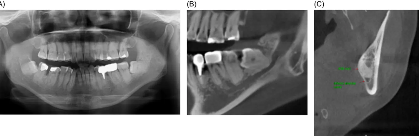
Fig. 1. Preoperative radiographs demonstrating left lower third molar, which was very close to the inferior alveolar canal. (A) Panoramic radiograph, (B) Cone beam computed tomography: panoramic view, (C) Cone beam computed tomography: cross-sectional view.
Fig. 2. Two weeks postoperatively (first day of paresthesia), the extraction site showed no swelling or redness of the soft tissue but partial loss of blood clot was observed.
to months after the procedure [10-14]. Delayed pares- thesia was represented by only 5% of the 60 cases of paresthesia reported in a study of 1477 third molar surgeries [15]. The biggest difference between classic paresthesia and delayed paresthesia is that the former begins immediately after the procedure and healing is not guaranteed, while the latter occurs later, with restoration to original condition [10].
Here we present a rare case of delayed paresthesia after dental surgery and discuss the pathophysiology of IAN-related delayed paresthesia.
CASE REPORT
A 53-year-old woman presented to the department of Advanced General Dentistry at Dankook University College of Dentistry in 2017 for dental treatment. She reported occasional discomfort of the left lower third molar, and her medical history was otherwise unremark- able.
The patient had slight swelling of the pericoronal tissue around the left lower third molar. The left lower third molar was very close to the inferior alveolar canal on radiography (Fig. 1). The patient was diagnosed with chronic pericoronitis and surgical removal was planned.
After informed consent was provided by the patient, IAN block anesthesia was performed using two ampoules of
2% lidocaine with 1:100000 epinephrine (Huons, Sung- namsi, Korea). After confirming efficacy of local anesthe- sia (absence of lip and chin sensation), surgical extraction and suturing were performed. No intraoperative com- plications were encountered, and the IAN was not visualized during surgery.
The early postoperative period was uneventful. How- ever, 2 weeks postoperatively, the patient returned complaining of recent onset of numbness of her lower lip, chin, and lower teeth on the left side, as well as increased discomfort of the extraction site. She reported no specific precipitating event, but sudden pain at the extraction site and a dulled sensitivity of the left side over the previous 24 h. On clinical examination, the extraction site showed no swelling and redness of soft tissue but
Fig. 3. Skin mapping of the affected area outlining abnormal sensation.
Right side Left side
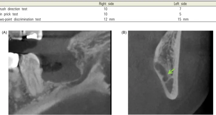
Brush direction test 10 7
Pin prick test 10 5
Two-point discrimination test 12 mm 15 mm
Table 1. The results of neurosensory tests performed 14 days after extraction (first day of paresthesia)
(A) (B)
Fig. 4. Postoperative radiographs demonstrating the discontinuity of upper cortical layer of the inferior alveolar canal. (A) Cone beam computed tomography:
panoramic view, (B) Cone beam computed tomography: cross-sectional view.
partial loss of blood clot was observed (Fig. 2).
Submandibular lymphadenopathy was not present. We performed careful subjective and objective assessments.
The patient did not have a pin prick sensation, but reported an overall sensation of dullness. Skin mapping of the affected area showed abnormal sensation in the lower lip and chin approximately 20 mm in width and spreading from the vermilion mucosal border of the lip down to the chin (Fig. 3). Neurosensory testing was performed according to the recommendations of Poort et
al. [16]. Three sensory tests, including the brush direction test, pin point test based on a visual analog scale, and two-point discrimination test were performed on the left chin with the unaffected right side as a control site [16].
The results showed that left side sensation was decreased (it was relatively lower than 10 when the control site sensation was 10) and the two-point discrimination test was 15 mm (normal right side was 12 mm) (Table 1).
Postoperative cone beam computed tomography revealed that continuity of the upper cortical layer of the inferior alveolar canal was partially destroyed (Fig. 4). The patient was diagnosed with delayed paresthesia of IAN, and anti-inflammatory drugs and steroids were prescribed.
The patient returned to the clinic one week later. The pain at the extraction site had disappeared, but there was no improvement in the sensory dullness of the lips and chin. The patient was instructed to take the remaining prescription medicines and to come in for follow-up in 1 month. One month later (6 weeks of paresthesia), the paresthesia had resolved completely by both patient report and subjective testing. We finally diagnosed this patient
with delayed paresthesia of neuropraxia of IAN.
DISCUSSION
This patient presented with typical symptoms of IAN paresthesia occurring approximately 2 weeks postopera- tively. The paresthesia lasted for about 5 weeks, after which the patient reported a complete recovery. We diagnosed this patient with delayed paresthesia by neuropraxia of IAN.
Seddon classified nerve injuries based on the severity of the injury as neurapraxia, axonotmesis, and neurotmesis [17]. Neurapraxia is the mildest classification of peripheral nerve injury, characterized by a temporary loss of sensory function due to blockage of nerve conduction, usually lasting an average of 6 to 8 weeks before full recovery. This condition is typically caused by a blunt neural injury due to nerve compression in which external pressure causes decreased blood flow to the nerve and deformation of the nerve fibers.
Neurapraxia results in temporary damage to the myelin sheath but leaves the nerve (axon) intact and is an impermanent condition. The thinning of the myelin sheath or focal demyelination are the main consequences of the injury that lead to conduction blockage. In order for the condition to be considered neurapraxia, there must be a complete and relatively rapid recovery of sensory function once nerve conduction has been restored;
otherwise, the injury would be classified as axonotmesis or neurotmesis [18,19].
Therefore, pressure from surrounding tissue edema may be the pathophysiology of delayed paresthesia. Dahli et al. [20] showed that rabbit tibial nerves compressed at 50 mmHg for 2 h had normal afferent and motor conduction velocity, whereas the nerves compressed at 200 mmHg for 2 h exhibited reduction of conduction velocity only at the area of compression. At 400 mmHg for 2 h, conduction velocity was reduced both at the level of compression and distal to the compressed segment.
Borgonovo et al. [10] reported three cases of delayed
paresthesia after third molar extraction, and considered compression caused by the clot, fibrous organization, and bone fragments as possible etiologies of delayed paresthesia. They indicated that all three may promote inflammation onset along the nerve trunk, and the paresthesia was induced by inflammatory edema [10]. In our case, however, neuropraxia due to compression is not applicable since the clot was lost rather than organized, and insertion of bone fragments was not observed on cone beam computed tomography.
Other papers have described the pathophysiology of delayed paresthesia from different perspectives.
Flanagan [11] commented in his article that hemo- globin has been associated with delayed neuropathy.
Hemoglobin degrades and liberates iron, which generates free radicals, which in turn degrade type I collagen and other molecules. Neuropathy from a hematoma may be related to the presence of iron compounds in the presence of the involved nerve. Goldberg and Galbraith [12]
commented in their study that the pathophysiologic mechanism of delayed paresthesia may include direct bacterial invasion of the neural sheath or inflammation of the nerve, as well as pressure secondary to the edema of the inflammatory process, as in inflammatory neuritis of peripheral nerves.
In our case, when the patient revisited due to dulled sensation of the left side, she also reported pain at the extraction site in which the soft tissue healing was normal but partial loss of blood clot was observed, similar to alveolar osteitis. Alveolar osteitis is described as post- operative pain originating from the extraction socket, which peaks approximately 1 to 3 days after tooth ex- traction and is associated with partial or total loss of the blood clot from the socket, with or without halitosis.
Clinical and laboratory studies have shown the signifi- cance of locally increased fibrinolytic activity in the pathogenesis of alveolar osteitis. Direct tissue activators after trauma and indirect activators produced by bacteria cleave other plasminogen molecules to plasmin, resulting in the breakup of the clot by disintegrating the fibrin [21].
Therefore, in our case, hemoglobin may be released after
fibrinolysis, and it may degrades and liberates iron, generating free radicals. The free radicals may damage the myelin sheath or affect nerve conduction. Ac- tinomyces viscosus, Streptococcus mutans, and anaerobic organisms (also the predominant organisms in pericoronitis) in the alveolar osteitis socket are regarded to have possible significance in the etiology of alveolar osteitis. Therefore, it can be suggested that the delayed paresthesia in our case may be caused by bacterial invasion [21]. However, there were no other signs of infection such as lymphadenopathy, swelling, or redness of extraction site, and bacterial invasion is unlikely to be a major factor.
In conclusion, the pathophysiology of delayed pares- thesia in our patient is thought to be a temporary conduction blockage due to degradation from free radicals in fibrinolysis and, partially, bacterial invasion of the neural sheath.
AUTHOR ORCIDs
Re-Mee Doh: https://orcid.org/0000-0001-8512-9134 Sooil Shin: https://orcid.org/0000-0002-7203-749X Tae Min You: https://orcid.org/0000-0001-7994-7608
NOTE: The authors have no conflicts of interest.
REFERENCES
1. Ahmad M. The Anatomical Nature of Dental Paresthesia:
A Quick Review. Open Dent J 2018; 12: 155-9.
2. Shadmehr E, Shekarchizade N. Endodontic periapical lesion-induced mental nerve paresthesia. Dent Res J 2015;
12: 192-6.
3. Smith MH, Lung KE. Nerve injuries after dental injection:
a review of the literature. J Can Dent Assoc 2006; 72:
559-64.
4. Lvovsky A, Bachrach S, Kim HC, Pawar A, Levinzon O, Ben Itzhak J, et al. Relationship between Root Apices and the Mandibular Canal: A Cone-beam Computed Tomographic Comparison of 3 Populations. J Endod 2018;
44: 555-8.
5. Devine M, Hirani M, Durham J, Nixdorf DR, Renton T. Identifying criteria for diagnosis of post-traumatic pain and altered sensation of the maxillary and mandibular branches of the trigeminal nerve: a systematic review. Oral Surg Oral Med Oral Pathol Oral Radiol 2018; 125: 526-40.
6. Sarikov R, Juodzbalys G. Inferior alveolar nerve injury after mandibular third molar extraction: a literature review.
J Oral Maxillofac Res 2014; 5: e1.
7. Shavit I, Juodzbalys G. Inferior alveolar nerve injuries following implant placement - importance of early diagnosis and treatment: a systematic review. J Oral Maxillofac Res 2014; 5: e2.
8. Cheung LK, Leung YY, Chow LK, Wong MC, Chan EK, Fok YH. Incidence of neurosensory deficits and recovery after lower third molar surgery: a prospective clinical study of 4338 cases. Int J Oral Maxillofac Surg 2010; 39: 320-6.
9. Charan Babu HS, Reddy PB, Pattathan RK, Desai R, Shubha AB. Factors influencing lingual nerve paraesthesia following third molar surgery: a prospective clinical study.
J Maxillofac Oral Surg 2013; 12: 168-72.
10. Borgonovo A, Bianchi A, Marchetti A, Censi R, Maiorana C. An uncommon clinical feature of IAN injury after third molar removal: a delayed paresthesia case series and literature review. Quintessence Int 2012; 43: 353-9.
11. Flanagan D. Delayed onset of altered sensation following dental implant placement and mental block local anesthesia:
a case report. Implant Dent 2002; 11: 324-30.
12. Goldberg MH, Galbraith DA. Late onset of mandibular and lingual dysesthesia secondary to postextraction infection. Oral Surg Oral Med Oral Pathol 1984; 58: 269-71.
13. Tolstunov L, Pogrel MA. Delayed paresthesia of inferior alveolar nerve after extraction of mandibular third molar:
case report and possible etiology. J Oral Maxillofac Surg 2009; 67: 1764-6.
14. Smyth J, Marley J. An unusual delayed complication of inferior alveolar nerve block. Br J Oral Maxillofac Surg 2010; 48: 51-2.
15. Kipp DP, Goldstein BH, Weiss WW, Jr. Dysesthesia after mandibular third molar surgery: a retrospective study and analysis of 1,377 surgical procedures. J Am Dent Assoc
1980; 100: 185-92.
16. Poort LJ, van Neck JW, van der Wal KG. Sensory testing of inferior alveolar nerve injuries: a review of methods used in prospective studies. J Oral Maxillofac Surg 2009;
67: 292-300.
17. Seddon HJ. Three types of nerve injury. Brain 1943; 66:
237-88.
18. Donoff RB. Nerve regeneration: basic and applied aspects.
Crit Rev Oral Biol Med 1995; 6: 18-24.
19. Campbell WW. Evaluation and management of peripheral nerve injury. Clin Neurophysiol 2008; 119: 1951-65.
20. Dahlin LB, Danielsen N, Ehira T, Lundborg G, Rydevik B. Mechanical effects of compression of peripheral nerves.
J Biomech Eng 1986; 108: 120-2.
21. Blum IR. Contemporary views on dry socket (alveolar osteitis): a clinical appraisal of standardization, aetiopatho- genesis and management: a critical review. Int J Oral Maxillofac Surg 2002; 31: 309-17.