Received February 24, 2011, Revised July 14, 2011, Accepted for publication August 9, 2011
Corresponding author: Jong Hee Lee, M.D., Ph.D., Department of Dermatology, Samsung Medical Center, 50 Ilwon-dong, Gangnam-gu, Seoul 135-710, Korea. Tel: 82-2-3410-3549, Fax: 82-2-3410-6578, E-mail: bell711@hanmail.net
This is an Open Access article distributed under the terms of the Creative Commons Attribution Non-Commercial License (http://
creativecommons.org/licenses/by-nc/3.0) which permits unrestricted non-commercial use, distribution, and reproduction in any medium, provided the original work is properly cited.
ORIGINAL ARTICLE
Intense Pulsed Light and Low-Fluence Q-Switched Nd:YAG Laser Treatment in Melasma Patients
Se Young Na, M.D., Soyun Cho, M.D., Ph.D., Jong Hee Lee, M.D., Ph.D.1
Department of Dermatology, Seoul National University Boramae Hospital, 1Samsung Medical Center, Sungkyunkwan University School of Medicine, Seoul, Korea
Background: Recently, low fluence collimated Q-switched (QS) Nd:YAG laser has drawn attention for the treatment of melasma. However, it needs a lot of treatment sessions for the substantial results and repetitive laser exposures may end up with unwanted depigmentation. Objective: We evaluated the clinical effects and safety of the combinational treatment, using intense pulsed light (IPL) and low fluence QS Nd:YAG laser. Methods: Retrospective case series of 20 female patients, with mixed type melasma, were analyzed using medical records. They were treated with IPL one time, and 4 times of weekly successive low fluence Nd:YAG laser treatments. At each visit, digital photographs were taken under the same condition. Melanin index (MI) and erythema index (EI) were measured on the highest point on the cheekbones. Modified melasma area and severity index (MASI) scores were calculated by two investigators using digital photographs. Results: The mean values of MI and EI decreased significantly after treatments. The modified MASI score has decreased by 59.35%, on average. Sixty percents of the participants did not require any more treatments, and no clinical aggravations were observed during the follow-up period (mean 5.9 months). Conclusion: IPL and low fluence laser may elicit a clinical resolution in the mixed type melasma with long term benefits. (Ann Dermatol 24(3) 267
∼273, 2012)
-Keywords-
IPL, Laser, Melasma treatment
INTRODUCTION
Melasma is a common, hyperpigmentary disorder, and may be the most concerning issue among the young to middle-aged Asian women. It is defined as a light to dark brown, irregular hypermelanosis of the face, which develops slowly, and is usually symmetrical1,2. Among the three histological patterns of melasma, the mixed type one, with hyperactive epidermal melanocytes and dermal melano- phages, is the most common in Korean women3.
Various treatment protocols for melasma have been suggested in the previous literature, and applied in various clinical settings1. The treatment efficacy and safety varies, according to the reports2. Generally, the epidermal com- ponent of melasma responds well to topical depigmenting agents3, as well as to other conventional treatments1. However, most Asian patients visiting the dermatologic clinic complain of mixed type melasma (having epidermal and dermal components), and mixed type melasma is hard to treat, due to its recurring nature and easy tendency of postinflammatory hypo- or hyperpigmentation after the treatments4,5.
During mid 2000s, intense pulsed light (IPL) was studied vigorously, and applied on various clinical conditions, including melasma. IPL emits a broad spectrum of light, from 500 nm upto 1,200 nm, with long pulse width of milliseconds unlike the other conventional pigment- targeting lasers. Its emitting spectrum includes lights absorbable to all of the skin chromophores (melanin, hemoglobin, and water) and thus, it was accepted that IPL could solve every cosmetic issue very successfully.
However, it has turned out that IPL was effective in clear-
ing the epidermal pigmentary and vascular issues, but not satisfactory enough in the mixed or dermal type mela- sma6. Some investigators even reported the experience of occurring subtle melasma after IPL treatment7.
Low-dose collimated 1,064 nm Q-switched (QS) Nd:YAG laser has drawn considerable attention for the treatment of melasma, since 20088. Several reports on the therapeutic results, using this method on melasma patients, pose substantial and long term beneficial effects. It delivers subthreshold dose of laser at a time and therefore, this treatment modality definitely requires a lot of treatment sessions4,9. Weekly repeated multiple treatment sessions of Nd:YAG laser can be a burden to patients and sometimes result in confetti-like hypopigmentation which is cosmetically unacceptable to patients10,11.
Considering merits and limitations of IPL and QS Nd:YAG laser, we thought the combination treatment will result in better clinical response. There have been no studies with the combination of IPL and low fluence QS-Nd:YAG laser in the treatment of mixed type melasma so far. The primary aim of this study is to investigate the efficacy and safety of the treatment method.
MATERIALS AND METHODS
Study patients
We carefully selected melasma patients visiting our laser clinic between March 2009 and December 2009 and cases were reviewed retrospectively by medical records under the approval of Institutional Review Board of Seoul National University Boramae Hospital. The diagnosis of melasma is based upon clinical appearance. On the basis of Wood's light examination, an epidermal, a dermal, and a mixed type were identified. We analyzed the patients who were treated with one time of IPL and 4 times of successive low fluence Nd:YAG laser treatments weekly (total of 5 treatments including IPL).
A total of twenty Korean female patients (Fitzpatrick skin type III and IV) with mixed-type melasma were included in this study. None of the patients used bleaching cream containing hydroquinone, and were treated with any other lasers on their faces for at least 6 months prior to this treatment.
Laser treatment
The patients were treated first with IPL (Ellipse Flex, Danish Dermatologic Development, Hoersholm, Denmark;
emitting 555∼950 nm), without topical anesthesia. The treatment parameters were as follows: fluence 10∼10.5 J/cm2, pulse width 2.5 ms, a delay time 10 ms between pulses, and double pulses. The clinical end point of IPL
treatment was mild erythema vanishing less than 30 minutes with ice pack application. After two weeks, the patients returned to visit the clinic for further treatments.
Several passes (mean 7∼8 passes) of low fluence of 1,064 nm QS Nd:YAG laser (MedLite C6, Hoya ConBio, Fre- mont, CA, USA) were applied on the whole face (6 mm spot size, using energy fluency ranging from 2.0 to 2.5 J/cm2 at 10 Hz). The notice of mild erythema without petechia on the perilesional area of melasma was the sign of stopping laser passes. A total of four times of QS Nd:YAG laser were applied on the melasma patients, at one-week interval, consecutively.
The patients were instructed to apply the ice packs for 30 minutes, right after either the IPL or the laser treatments.
They were banned to use any bleaching cream or topical agents containing retinoic acid, and its derivatives or alpha hydroxyl acid during the treatments. They were instructed to avoid sun exposure and to put on a broad spectrum sunscreen, during and after treatments.
Evaluation
Before every treatment session, digital photographic documentation was obtained, under the same circum- stances. Melanin index (MI) and erythema index (EI) were measured on the most prominent area of both zygomata, using the skin color-measuring device (Mexameter, MX18, Courage & Khazaka, Electronic GmbH, Clogne, Germany) at each visit. Modified melasma area and severity index (MASI) scores12 were evaluated by the two blinded investigators, using the photographs taken before the treatments and a week after the last treatment. Modified MASI score counted only a confined portion of the mala area (cheek), and it was calculated based on the percentage of the involved area (A=0∼6: 0=0%, 1≤
10%, 2=10∼29%, 3=30∼49%, 4=50∼69%, 5=70∼
89%, 6=90∼100%); darkness of pigment (D=0∼4: 0=
absent or normal skin color without evidence of hyper- pigmentation, 1=slight visible hyperpigmentation, 2=
mild visible, 3=marked, 4=severe), and homogenicity or density of the hyperpigmentation (number of pigmented lesions per unit facial area (H=0∼4: 0=minimal, 1=
slight, 2=mild, 3=marked, 4=severe).
Modified MASI score = (D+H)×A Safety assessments
At each visit, all possible side effects and complications, such as erythema, pain, edema, and postinflammatory hypopigmentation, were recorded.
Statistical analysis
To evaluate the treatment response, statistical analysis was
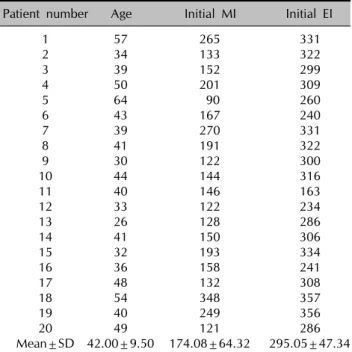
Table 1. Patients’ demographic data
Patient number Age Initial MI Initial EI
1 57 265 331
2 34 133 322
3 39 152 299
4 50 201 309
5 64 90 260
6 43 167 240
7 39 270 331
8 41 191 322
9 30 122 300
10 44 144 316
11 40 146 163
12 33 122 234
13 26 128 286
14 41 150 306
15 32 193 334
16 36 158 241
17 48 132 308
18 54 348 357
19 40 249 356
20 49 121 286
Mean±SD 42.00±9.50 174.08±64.32 295.05±47.34 MI: melanin index, EI: erythema index, SD: standard deviation.
Table 3. Spearman correlation analysis between the changes after intense pulsed light (IPL) and changes after full treatment sessions
Total ΔEI Total ΔMI
ΔEI after IPL 0.544* 0.093
ΔMI after IPL −0.048 0.694†
Total amount of erythema index change (ΔEI) and melanin index (MI) means changes of erythema index (EI) and MI in between baseline and after the last treatments. ΔEI and amount of MI change (ΔMI) after IPL indicates changes of EI and MI before IPL and 2 weeks after IPL. More reduction in EI and MI before and after IPL results in more decrease in EI and MI after all 5 times treatments (*p<0.05, †p<0.01).
Table 2. Melanin index (MI) and erythema index (EI) changes
Initial Final p-value
MI 174.08±64.32 128.65±41.36 <0.001 EI 295.05±47.34 238.40±48.67 <0.001 MI and EI declines significantly after treatments. This data imply clinical improvement in melasma patients.
Table 4. Mean modified melasma area and severity index after treatments
Initial mMASI Final mMASI % reduction p-value (Initial vs. final)
8.54 3.52 59.35±14.94† <0.001
mMASI: modified melasma area and severity index. †p<0.001.
carried out (SPSS Version 12.0.1, SPSS Inc., Chicago, IL, USA). All tests were assessed at an α=0.05 significance level. p<0.05 was considered to be statistically signi- ficant. Values were documented as the means±standard deviations.
RESULTS
Demographic characteristics
Demographic analysis is shown in Table 1. A total of twenty patients were all female. The mean age of all the patients was 42.00±9.50 years. The initial MI and EI was 174.08±64.32 and 295.05±47.34, respectively.
Treatment efficacy assessments
MI decreased significantly from 174.08±64.32 to 128.65±
41.36, and EI dropped from 295.05±47.34 to 238.40±
48.67 (p<0.001) (Table 2). The amount of MI change (ΔMI) between the before and after IPL showed positive correlations with significance to that of the ΔMI between the baseline and 1 week after the last treatment (R=
0.694). The amount of EI change (ΔEI) between the before and after IPL correlated well to that of the ΔEI between the baseline and 1 week after the last treatment (R=0.544) (Table 3). The mean modified melasma area and severity index (MASI) score decreased significantly, by 59.35% (from 8.54 to 3.52)(Table 4).
Safety assessments and follow ups
No serious adverse events occurred during the treatment period. Nineteen out of 20 patients experienced a mild erythema and prickling sensation, right after the treatment, which disappeared after a simple ice pack application.
Postinflammatory hypo- or hyperpigmentation was not observed in any of the patients during the 5 treatment sessions. Twelve out of 20 patients (60%) were very pleased with the results, and did not want any more treatment. Those 12 patients were instructed to use regular based cosmetics and put on broad spectrum sunscreen during the day. All these 12 patients revisited the clinic between 2 months to 1 year, after the last laser treatment, according to the medical records. The MI and EI were checked at the same area (on the highest point of both zygomata) and slight increment of EI and MI was noticed, which was not statistically significant (Table 5). The rest of the patients (8 patients) were recommended to use a topical retinoic acid/hydroquinone cream for better clinical results (Fig. 1).
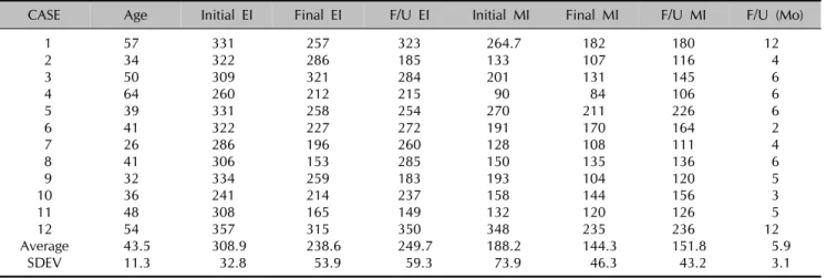
Table 5. Follow-up results in patients
CASE Age Initial EI Final EI F/U EI Initial MI Final MI F/U MI F/U (Mo)
1 57 331 257 323 264.7 182 180 12
2 34 322 286 185 133 107 116 4
3 50 309 321 284 201 131 145 6
4 64 260 212 215 90 84 106 6
5 39 331 258 254 270 211 226 6
6 41 322 227 272 191 170 164 2
7 26 286 196 260 128 108 111 4
8 41 306 153 285 150 135 136 6
9 32 334 259 183 193 104 120 5
10 36 241 214 237 158 144 156 3
11 48 308 165 149 132 120 126 5
12 54 357 315 350 348 235 236 12
Average 43.5 308.9 238.6 249.7 188.2 144.3 151.8 5.9
SDEV 11.3 32.8 53.9 59.3 73.9 46.3 43.2 3.1
Twelve out of 20 patients did not want more treatment sessions and their follow-up erythema index (EI) and melanin index (MI) data are shown here. Average follow-ups are 5.9 months (2 to 12 months). They did not use any bleaching cream or medication during follow-ups. They used their regular base cosmetics (without arbutin or kojic acid) and were educated to use broad spectrum sunscreen. Slight increases in EI and MI at follow-ups are noticed, which are not statistically significant. F/U: follow-up, SDEV: standard deviation.
DISCUSSION
The successful treatment of melasma is one of the hot issues in the field of dermatology. However, the patho- genesis of melasma has not yet been clearly understood.
Kang et al.13 demonstrated that epidermal hyperpigmen- tation, possibly caused by both an increased number of melanocytes and an increased activity of melanogenic enzymes overlying dermal changes, were caused by solar radiation. Increased vascularity is also one of the major findings in melasma14. Considering these histopathological findings in melasma, the ideal treatment could be achieved by targeting these several issuing components.
From this point of view, IPL was considered as a very promising treatment modality. The previous studies on IPL in melasma patients usually showed good to excellent results6,15.
In the meticulous reading of the previous studies of IPL or other lasers on melasma patients, the most worrisome hyperpigmentation started to be noticed, 2 to 4 weeks after the treatment. This is also very compatible with the clinical experiences in the treatment, especially the mixed type melasma. After the IPL irradiation, the distribution of melanosomes is drastically changed. Melanin cap structures, in which the melanosomes are dispersed within the basal keratinocytes collapse and melanosome accumulation as the intraepidermal microcrust (IEMC), gradually desqua- mates from the skin by 5 to 7 days16. Afterwards, it appears that the melanosomes are replenished and mela- nocytes are reactivated to produce melanin pigment in patients who showed hyperpigmentation after the treat-
ment. The survived melanocytes and melanosomes get back in their cycle, resulting in pigment transfer to the epidermis and dermis4,9,16. Therefore, we treated the patients first with IPL, and after two weeks, the patients were led into a successive weekly exposure to low fluence QS Nd:YAG laser in order to deplete the survived melanosomes and inactivate melanocytes. After one time of IPL, EI and MI significantly decreased (data not shown).
This might be explained by the IEMC phenomenon, which appears to be the most prominent in the first session of IPL in almost all patients16. After 6 weeks, patients showed 26.10% (174.08±64.32 to 128.65±41.36, Table 2) reduction of melanin index, and 59.35% decrease of the modified MASI scores. Accordingly, we thought that the main- tenance of the relatively rapid and significant improve- ment, after the irradiation of IPL and further improvement could be obtained by several times of QS Nd:YAG laser.
Recently, so-called “laser toning” that uses collimated low-fluence 1,064 nm QS Nd:YAG laser has gained popularity for the treatment of melasma in Eastern Asia.
The repetitive treatments of QS Nd:YAG laser of the fluence was just enough to damage the melanosomes subcellularly, which can induce excellent clinical results without the high risk of rebounding hyperpigmen- tation17,18. The QS Nd:YAG laser treatment may also produce nonspecific dermal wound and induce inflamma- tion, facilitating a migration of melanophages4,8,17. There was no epidermal disruption when low fluence of the laser was used. However, the inflammation may cause increased epidermal turnover, which can remove the up dispersed melanin pigment in the epidermal keratinocytes.
Fig. 1. (A) baseline, (B) 1 week after the last treatment, (C) follow-up (6 months).
Overall, several studies have demonstrated that several weekly treatments, with the 1,064 nm QS-Nd:YAG laser at sub-threshold photothermolytic fluences (<5 J/cm2), show relatively high efficacy and less side effects, like the hypo- or hyperpigmentation3,4,9.
The drawbacks of QS Nd:YAG laser applications in melasma patients can be listed below. Relatively long
period of treatment is required and weekly repetitive treatments can be a burden to patients, and it might exhaust the melanosomes completely out, which shows clinically the confetti-like hypopigmentation10,11. Moreover, this treatment doesn’t have any downtime or pain, which can make some patients very addictive to the treatment.
Chan et al.10 reported case series of facial depigmentation,
associated with low fluence QS Nd:YAG laser, and they pointed out the risk of possible occurrence of punctuate leukoderma, which is not cosmetically acceptable.
However, the complications might have not occurred if they had treated the patients with longer off-treatment period and lower energy. Although the adverse effects can be avoided, we have to consider the possibility of these unwanted outcomes.
The combinational treatment with IPL and QS Nd:YAG laser can induce clinical improvement, which means that it does not need multiple weekly exposures (more than 10 times in practice generally) of QS Nd:YAG laser, and it may lower the risk of rebound hyperpigmentation, after IPL alone. We analyzed the correlation MI and EI changes between the before and after IPL, and total MI and EI changes between the baseline and after all sessions to evaluate the effects of IPL treatment in this study. It is worthy to mention that the strong positive correlations between the MI changes before and after IPL, as well as the total MI changes after all sessions were observed. Swift removal of epidermal pigments, using IPL and suppression of reactivating melanosomes with subsequent low fluence QS Nd:YAG laser, enables only 5 treatment sessions to produce significant clinical results. Moreover, 60% of patients involved in the study did not show any clinical aggravation, during the follow-up periods (average 5.9 months) without further medical treatments.
Most previous studies used QS-Nd:YAG laser at 3.0 to 4.0 J/cm2 4,8,10. There were side effects, such as punctate leucoderma and postinflammatory hypopigmentation in those reports10,11. These adverse events did not occur in our patients. It might be because we used lower fluence of 2.0 to 2.5 J/cm2 than the ones used in the previous studies.
This study has limitations in that it was a retrospective chart review of the treatments, and there was no control group. Longer follow-ups were available for 12 out of the 20 patients, and those were the patients who were satisfied with the initial results, which can be an element of bias. However, the 5 treatment sessions can induce the substantial clinical results without any side effects or recurrences during the treatment free periods (mean 5.9 months) in 60% of the patients, and thus, it can be cautiously said that IPL and subsequent QS Nd:YAG laser therapy work beautifully on the melasma patients with less treatment sessions, and without serious side effects.
In this study, we first reported the combinational treatment of IPL and QS Nd:YAG laser for the treatment of mixed type melasma. It could improve both epidermal and dermal pigmentation and vascular component with fewer treatment sessions to reach acceptable clinical outcomes.
Therefore, the combination of IPL and QS Nd:YAG laser may be a good therapeutic option in the treatment, especially, of the mixed type melasma.
REFERENCES
1. Gupta AK, Gover MD, Nouri K, Taylor S. The treatment of melasma: a review of clinical trials. J Am Acad Dermatol 2006;55:1048-1065.
2. Rendon M, Berneburg M, Arellano I, Picardo M. Treatment of melasma. J Am Acad Dermatol 2006;54:S272-281.
3. Jeong SY, Shin JB, Yeo UC, Kim WS, Kim IH. Low-fluence Q-switched neodymium-doped yttrium aluminum garnet laser for melasma with pre- or post-treatment triple combi- nation cream. Dermatol Surg 2010;36:909-918.
4. Wattanakrai P, Mornchan R, Eimpunth S. Low-fluence Q-switched neodymium-doped yttrium aluminum garnet (1,064 nm) laser for the treatment of facial melasma in Asians. Dermatol Surg 2010;36:76-87.
5. Haddad AL, Matos LF, Brunstein F, Ferreira LM, Silva A, Costa D Jr. A clinical, prospective, randomized, double-blind trial comparing skin whitening complex with hydroquinone vs. placebo in the treatment of melasma. Int J Dermatol 2003;42:153-156.
6. Wang CC, Hui CY, Sue YM, Wong WR, Hong HS. Intense pulsed light for the treatment of refractory melasma in Asian persons. Dermatol Surg 2004;30:1196-1200.
7. Negishi K, Kushikata N, Tezuka Y, Takeuchi K, Miyamoto E, Wakamatsu S. Study of the incidence and nature of "very subtle epidermal melasma" in relation to intense pulsed light treatment. Dermatol Surg 2004;30:881-886.
8. Polnikorn N. Treatment of refractory dermal melasma with the MedLite C6 Q-switched Nd:YAG laser: two case reports.
J Cosmet Laser Ther 2008;10:167-173.
9. Choi M, Choi JW, Lee SY, Choi SY, Park HJ, Park KC, et al.
Low-dose 1064-nm Q-switched Nd:YAG laser for the treatment of melasma. J Dermatolog Treat 2010;21:224-228.
10. Chan NP, Ho SG, Shek SY, Yeung CK, Chan HH. A case series of facial depigmentation associated with low fluence Q-switched 1,064 nm Nd:YAG laser for skin rejuvenation and melasma. Lasers Surg Med 2010;42:712-719.
11. Kim MJ, Kim JS, Cho SB. Punctate leucoderma after melasma treatment using 1064-nm Q-switched Nd:YAG laser with low pulse energy. J Eur Acad Dermatol Venereol 2009;23:
960-962.
12. Kang WH, Chun SC, Lee S. Intermittent therapy for melasma in Asian patients with combined topical agents (retinoic acid, hydroquinone and hydrocortisone): clinical and histo- logical studies. J Dermatol 1998;25:587-596.
13. Kang WH, Yoon KH, Lee ES, Kim J, Lee KB, Yim H, et al.
Melasma: histopathological characteristics in 56 Korean patients. Br J Dermatol 2002;146:228-237.
14. Kim EH, Kim YC, Lee ES, Kang HY. The vascular charac- teristics of melasma. J Dermatol Sci 2007;46:111-116.
15. Li YH, Chen JZ, Wei HC, Wu Y, Liu M, Xu YY, et al. Efficacy and safety of intense pulsed light in treatment of melasma in Chinese patients. Dermatol Surg 2008;34:693-700.
16. Yamashita T, Negishi K, Hariya T, Kunizawa N, Ikuta K, Yanai M, et al. Intense pulsed light therapy for superficial pigmented lesions evaluated by reflectance-mode confocal microscopy and optical coherence tomography. J Invest Dermatol 2006;126:2281-2286.
17. Jeong SY, Chang SE, Bak H, Choi JH, Kim IH. New melasma
treatment by collimated low fluence Q-switched Nd:YAG laser. Korean J Dermatol 2008;46:1163-1170.
18. Kim JH, Kim H, Park HC, Kim IH. Subcellular selective photothermolysis of melanosomes in adult zebrafish skin following 1064-nm Q-switched Nd:YAG laser irradiation. J Invest Dermatol 2010;130:2333-2335.